Movie
Movie Controller

[English] 日本語
 Yorodumi
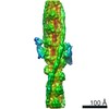
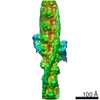
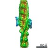
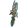
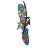
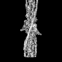
- EMDB-0729: Structure of human cardiac thin filament in the calcium bound state -
+ Open data

- Basic information
| Entry | Database: EMDB / ID: EMD-0729 | |||||||||
|---|---|---|---|---|---|---|---|---|---|---|
| Title | Structure of human cardiac thin filament in the calcium bound state | |||||||||
 Map data Map data | ||||||||||
 Sample Sample |
| |||||||||
 Keywords Keywords | Troponin / Tropomyosin / Actin / Thin filement / Muscle / CONTRACTILE PROTEIN-ACTIN BINDING PROTEIN complex | |||||||||
| Function / homology |  Function and homology information Function and homology informationpositive regulation of heart rate by epinephrine / muscle thin filament tropomyosin / regulation of systemic arterial blood pressure by ischemic conditions / troponin C binding / diaphragm contraction / regulation of muscle filament sliding speed / troponin T binding / cardiac Troponin complex / cardiac myofibril / negative regulation of ATP-dependent activity ...positive regulation of heart rate by epinephrine / muscle thin filament tropomyosin / regulation of systemic arterial blood pressure by ischemic conditions / troponin C binding / diaphragm contraction / regulation of muscle filament sliding speed / troponin T binding / cardiac Troponin complex / cardiac myofibril / negative regulation of ATP-dependent activity / troponin complex / regulation of muscle contraction / regulation of smooth muscle contraction / positive regulation of ATP-dependent activity / bleb / ruffle organization / transition between fast and slow fiber / Striated Muscle Contraction / muscle filament sliding / regulation of cardiac muscle contraction by calcium ion signaling / response to metal ion / sarcomere organization / cardiac muscle cell contraction / structural constituent of muscle / cytoskeletal motor activator activity / ventricular cardiac muscle tissue morphogenesis / myosin heavy chain binding / heart contraction / tropomyosin binding / negative regulation of vascular associated smooth muscle cell migration / regulation of heart contraction / actin filament bundle / troponin I binding / filamentous actin / mesenchyme migration / skeletal muscle myofibril / negative regulation of vascular associated smooth muscle cell proliferation / actin filament bundle assembly / striated muscle thin filament / skeletal muscle thin filament assembly / actin monomer binding / Smooth Muscle Contraction / vasculogenesis / calcium channel inhibitor activity / skeletal muscle contraction / skeletal muscle fiber development / cytoskeletal protein binding / Ion homeostasis / positive regulation of stress fiber assembly / cardiac muscle contraction / stress fiber / titin binding / actin filament polymerization / cytoskeleton organization / positive regulation of cell adhesion / negative regulation of cell migration / actin filament organization / sarcomere / filopodium / actin filament / wound healing / cellular response to reactive oxygen species / response to calcium ion / structural constituent of cytoskeleton / Hydrolases; Acting on acid anhydrides; Acting on acid anhydrides to facilitate cellular and subcellular movement / ruffle membrane / intracellular calcium ion homeostasis / calcium-dependent protein binding / actin filament binding / regulation of cell shape / lamellipodium / actin cytoskeleton / heart development / actin binding / cell body / cytoskeleton / protein heterodimerization activity / protein domain specific binding / hydrolase activity / calcium ion binding / positive regulation of gene expression / protein kinase binding / magnesium ion binding / protein homodimerization activity / ATP binding / identical protein binding / cytoplasm / cytosol Similarity search - Function | |||||||||
| Biological species |   Homo sapiens (human) Homo sapiens (human) | |||||||||
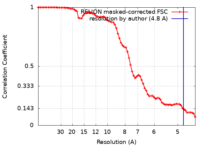
| Method | single particle reconstruction / cryo EM / Resolution: 4.8 Å | |||||||||
 Authors Authors | Fujii T / Yamada Y | |||||||||
 Citation Citation | Journal: Nat Commun / Year: 2020 Title: Cardiac muscle thin filament structures reveal calcium regulatory mechanism. Authors: Yurika Yamada / Keiichi Namba / Takashi Fujii /  Abstract: Contraction of striated muscles is driven by cyclic interactions of myosin head projecting from the thick filament with actin filament and is regulated by Ca released from sarcoplasmic reticulum. ...Contraction of striated muscles is driven by cyclic interactions of myosin head projecting from the thick filament with actin filament and is regulated by Ca released from sarcoplasmic reticulum. Muscle thin filament consists of actin, tropomyosin and troponin, and Ca binding to troponin triggers conformational changes of troponin and tropomyosin to allow actin-myosin interactions. However, the structural changes involved in this regulatory mechanism remain unknown. Here we report the structures of human cardiac muscle thin filament in the absence and presence of Ca by electron cryomicroscopy. Molecular models in the two states built based on available crystal structures reveal the structures of a C-terminal region of troponin I and an N-terminal region of troponin T in complex with the head-to-tail junction of tropomyosin together with the troponin core on actin filament. Structural changes of the thin filament upon Ca binding now reveal the mechanism of Ca regulation of muscle contraction. | |||||||||
| History |
|
- Structure visualization
| Movie |
Movie viewer |
|---|---|
| Structure viewer | EM map: SurfViewMolmilJmol/JSmol |
| Supplemental images |
- Downloads & links
-EMDB archive
| Map data | emd_0729.map.gz | 23.3 MB | EMDB map data format | |
|---|---|---|---|---|
| Header (meta data) | emd-0729-v30.xmlemd-0729.xml | 16.1 KB 16.1 KB | Display Display | EMDB header |
| FSC (resolution estimation) | emd_0729_fsc.xml | 7.1 KB | Display | FSC data file |
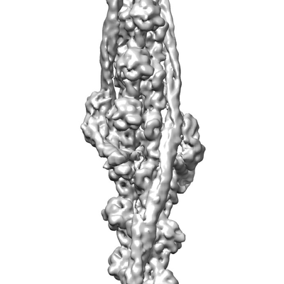
| Images |  emd_0729.png emd_0729.png | 45.6 KB | ||
| Filedesc metadata | emd-0729.cif.gz | 5.9 KB | ||
| Archive directory |  http://ftp.pdbj.org/pub/emdb/structures/EMD-0729ftp://ftp.pdbj.org/pub/emdb/structures/EMD-0729 http://ftp.pdbj.org/pub/emdb/structures/EMD-0729ftp://ftp.pdbj.org/pub/emdb/structures/EMD-0729 | HTTPS FTP |
-Related structure data
| Related structure data |  6kn8MC  7utiM  0728C  6kn7C M: atomic model generated by this map C: citing same article ( |
|---|---|
| Similar structure data |


-Links
| EMDB pages | EMDB (EBI/PDBe) / EMDataResource |
|---|---|
| Related items in Molecule of the Month |


-Map
| File | Download / File: emd_0729.map.gz / Format: CCP4 / Size: 30.5 MB / Type: IMAGE STORED AS FLOATING POINT NUMBER (4 BYTES) | ||||||||||||||||||||||||||||||||||||||||||||||||||||||||||||
|---|---|---|---|---|---|---|---|---|---|---|---|---|---|---|---|---|---|---|---|---|---|---|---|---|---|---|---|---|---|---|---|---|---|---|---|---|---|---|---|---|---|---|---|---|---|---|---|---|---|---|---|---|---|---|---|---|---|---|---|---|---|
| Projections & slices | Image control
Images are generated by Spider. | ||||||||||||||||||||||||||||||||||||||||||||||||||||||||||||
| Voxel size | X=Y=Z: 2.22 Å | ||||||||||||||||||||||||||||||||||||||||||||||||||||||||||||
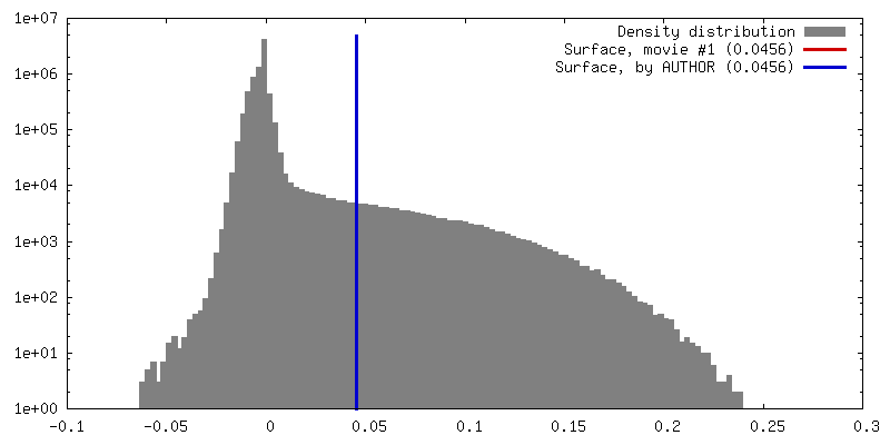
| Density |
| ||||||||||||||||||||||||||||||||||||||||||||||||||||||||||||
| Symmetry | Space group: 1 | ||||||||||||||||||||||||||||||||||||||||||||||||||||||||||||
| Details | EMDB XML:
CCP4 map header:
| ||||||||||||||||||||||||||||||||||||||||||||||||||||||||||||
 Z (Sec.)
 Y (Row.)
 X (Col.)


-Supplemental data
- Sample components
+Entire : Cardiac muscle thin filament in the calcium bound state
+Supramolecule #1: Cardiac muscle thin filament in the calcium bound state
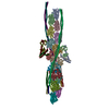
+Supramolecule #2: Actin, alpha skeletal muscle
+Supramolecule #3: Tropomyosin, Troponin
+Macromolecule #1: Actin, alpha skeletal muscle
+Macromolecule #2: Tropomyosin alpha-1 chain

+Macromolecule #3: Tropomyosin alpha-1 chain
+Macromolecule #4: Troponin T, cardiac muscle
+Macromolecule #5: Troponin I, cardiac muscle
+Macromolecule #6: Troponin C, slow skeletal and cardiac muscles
+Macromolecule #7: ADENOSINE-5'-DIPHOSPHATE

-Experimental details
-Structure determination
| Method | cryo EM |
|---|---|
 Processing Processing | single particle reconstruction |
| Aggregation state | filament |
-Sample preparation
| Concentration | 0.05 mg/mL |
|---|---|
| Buffer | pH: 7.5 |
| Vitrification | Cryogen name: ETHANE |
- Electron microscopy
| Microscope | JEOL CRYO ARM 200 |
|---|---|
| Image recording | Film or detector model: GATAN K2 SUMMIT (4k x 4k) / Average electron dose: 65.0 e/Å2 |
| Electron beam | Acceleration voltage: 200 kV / Electron source:  FIELD EMISSION GUN FIELD EMISSION GUN |
| Electron optics | Illumination mode: FLOOD BEAM / Imaging mode: BRIGHT FIELD |