 Movie
Movie Controller

[English] 日本語
 Yorodumi
- PDB-7uti: ALTERNATIVE MODELING OF TROPOMYOSIN IN HUMAN CARDIAC THIN FILAMEN... -
+ Open data

- Basic information
| Entry | Database: PDB / ID: 7uti | ||||||
|---|---|---|---|---|---|---|---|
| Title | ALTERNATIVE MODELING OF TROPOMYOSIN IN HUMAN CARDIAC THIN FILAMENT IN THE CALCIUM BOUND STATE | ||||||
 Components Components |
| ||||||
 Keywords Keywords | CONTRACTILE PROTEIN / TROPONIN / TROPOMYOSIN / ACTIN / THIN FILEMENT / MUSCLE / CONTRACTILE | ||||||
| Function / homology |  Function and homology information Function and homology informationpositive regulation of heart rate by epinephrine / muscle thin filament tropomyosin / regulation of systemic arterial blood pressure by ischemic conditions / troponin C binding / diaphragm contraction / regulation of muscle filament sliding speed / troponin T binding / cardiac Troponin complex / cardiac myofibril / negative regulation of ATP-dependent activity ...positive regulation of heart rate by epinephrine / muscle thin filament tropomyosin / regulation of systemic arterial blood pressure by ischemic conditions / troponin C binding / diaphragm contraction / regulation of muscle filament sliding speed / troponin T binding / cardiac Troponin complex / cardiac myofibril / negative regulation of ATP-dependent activity / troponin complex / regulation of muscle contraction / bleb / ruffle organization / positive regulation of ATP-dependent activity / Striated Muscle Contraction / muscle filament sliding / sarcomere organization / regulation of heart contraction / response to metal ion / regulation of cardiac muscle contraction by calcium ion signaling / regulation of smooth muscle contraction / cardiac muscle cell contraction / heart contraction / structural constituent of muscle / cytoskeletal motor activator activity / ventricular cardiac muscle tissue morphogenesis / negative regulation of vascular associated smooth muscle cell migration / myosin heavy chain binding / tropomyosin binding / actin filament bundle / troponin I binding / filamentous actin / vasculogenesis / mesenchyme migration / negative regulation of vascular associated smooth muscle cell proliferation / skeletal muscle myofibril / striated muscle thin filament / actin filament bundle assembly / skeletal muscle thin filament assembly / actin monomer binding / Smooth Muscle Contraction / positive regulation of stress fiber assembly / cardiac muscle contraction / calcium channel inhibitor activity / skeletal muscle fiber development / cytoskeleton organization / skeletal muscle contraction / cytoskeletal protein binding / actin filament polymerization / Ion homeostasis / stress fiber / titin binding / positive regulation of cell adhesion / negative regulation of cell migration / actin filament organization / wound healing / sarcomere / filopodium / actin filament / cellular response to reactive oxygen species / regulation of cell shape / response to calcium ion / intracellular calcium ion homeostasis / Hydrolases; Acting on acid anhydrides; Acting on acid anhydrides to facilitate cellular and subcellular movement / ruffle membrane / structural constituent of cytoskeleton / heart development / calcium-dependent protein binding / actin filament binding / lamellipodium / actin binding / cell body / cytoskeleton / hydrolase activity / protein heterodimerization activity / protein domain specific binding / positive regulation of gene expression / calcium ion binding / protein kinase binding / magnesium ion binding / protein homodimerization activity / ATP binding / identical protein binding / cytosol / cytoplasm Similarity search - Function | ||||||
| Biological species |  Homo sapiens (human) Homo sapiens (human)  Oryctolagus cuniculus (rabbit) Oryctolagus cuniculus (rabbit) | ||||||
| Method | ELECTRON MICROSCOPY / single particle reconstruction / cryo EM / Resolution: 4.8 Å | ||||||
 Authors Authors | Rynkiewicz, M.J. / Pavadai, E. / Lehman, W. | ||||||
| Funding support |  United States, 1items United States, 1items
| ||||||
 Citation Citation | Journal: Biophys J / Year: 2020 Title: Protein-Protein Docking Reveals Dynamic Interactions of Tropomyosin on Actin Filaments. Authors: Elumalai Pavadai / William Lehman / Michael J Rynkiewicz / Abstract: Experimental approaches such as fiber diffraction and cryo-electron microscopy reconstruction have defined regulatory positions of tropomyosin on actin but have not, as yet, succeeded at determining ...Experimental approaches such as fiber diffraction and cryo-electron microscopy reconstruction have defined regulatory positions of tropomyosin on actin but have not, as yet, succeeded at determining key atomic-level contacts between these proteins or fully substantiated the dynamics of their interactions at a structural level. To overcome this deficiency, we have previously employed computational approaches to deduce global dynamics of thin filament components by energy landscape determination and molecular dynamics simulations. Still, these approaches remain computationally challenging for any complex and large macromolecular assembly like the thin filament. For example, tropomyosin cable wrapping around actin of thin filaments features both head-to-tail polymeric interactions and local twisting, both of which depart from strict superhelical symmetry. This produces a complex energy surface that is difficult to model and thus to evaluate globally. Therefore, at this stage of our understanding, assessing global molecular dynamics can prove to be inherently impractical. As an alternative, we adopted a "divide and conquer" protocol to investigate actin-tropomyosin interactions at an atomistic level. Here, we first employed unbiased protein-protein docking tools to identify binding specificity of individual tropomyosin pseudorepeat segments over the actin surface. Accordingly, tropomyosin "ligand" segments were rotated and translated over potential "target" binding sites on F-actin where the corresponding interaction energetics of billions of conformational poses were ranked by the programs PIPER and ClusPro. These data were used to assess favorable interactions and then to rebuild models of seamless and continuous tropomyosin cables over the F-actin substrate, which were optimized further by flexible fitting routines and molecular dynamics. The models generated azimuthally distinct regulatory positions for tropomyosin cables along thin filaments on actin dominated by stereo-specific head-to-tail overlap linkage. The outcomes are in good agreement with current cryo-electron microscopy topology and consistent with long-thought residue-to-residue interactions between actin and tropomyosin. #1: Journal: Nat Commun / Year: 2020Title: Cardiac muscle thin filament structures reveal calcium regulatory mechanism. Authors: Yurika Yamada / Keiichi Namba / Takashi Fujii /  Abstract: Contraction of striated muscles is driven by cyclic interactions of myosin head projecting from the thick filament with actin filament and is regulated by Ca released from sarcoplasmic reticulum. ...Contraction of striated muscles is driven by cyclic interactions of myosin head projecting from the thick filament with actin filament and is regulated by Ca released from sarcoplasmic reticulum. Muscle thin filament consists of actin, tropomyosin and troponin, and Ca binding to troponin triggers conformational changes of troponin and tropomyosin to allow actin-myosin interactions. However, the structural changes involved in this regulatory mechanism remain unknown. Here we report the structures of human cardiac muscle thin filament in the absence and presence of Ca by electron cryomicroscopy. Molecular models in the two states built based on available crystal structures reveal the structures of a C-terminal region of troponin I and an N-terminal region of troponin T in complex with the head-to-tail junction of tropomyosin together with the troponin core on actin filament. Structural changes of the thin filament upon Ca binding now reveal the mechanism of Ca regulation of muscle contraction. | ||||||
| History |
|
- Structure visualization
| Structure viewer | Molecule: MolmilJmol/JSmol |
|---|
- Downloads & links
-Download
| PDBx/mmCIF format | 7uti.cif.gz | 2.5 MB | Display | PDBx/mmCIF format |
|---|---|---|---|---|
| PDB format | pdb7uti.ent.gz | Display | PDB format | |
| PDBx/mmJSON format | 7uti.json.gz | Tree view | PDBx/mmJSON format | |
| Others |  Other downloads Other downloads |
-Validation report
| Arichive directory | https://data.pdbj.org/pub/pdb/validation_reports/ut/7utiftp://data.pdbj.org/pub/pdb/validation_reports/ut/7uti | HTTPS FTP |
|---|
-Related structure data
| Related structure data | 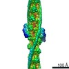 0729M  7utlC M: map data used to model this data C: citing same article ( |
|---|---|
| Similar structure data |
-Links
 PDBj
PDBj

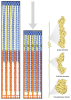




- Assembly
| Deposited unit | 
|
|---|---|
| 1 |
|
-Components
-Protein , 5 types, 32 molecules CDEFGHIJKLMNOPQRefWbijghXYcdVaUZ
| #1: Protein | Mass: 42109.973 Da / Num. of mol.: 16 / Source method: isolated from a natural source / Source: (natural) Oryctolagus cuniculus (rabbit) / References: UniProt: P68135#2: Protein | Mass: 32789.656 Da / Num. of mol.: 8 Source method: isolated from a genetically manipulated source Source: (gene. exp.) Homo sapiens (human) / Gene: TPM1, C15orf13, TMSA / Production host:   Escherichia coli (E. coli) / References: UniProt: P09493 Escherichia coli (E. coli) / References: UniProt: P09493#3: Protein | Mass: 34659.387 Da / Num. of mol.: 4 Source method: isolated from a genetically manipulated source Source: (gene. exp.) Homo sapiens (human) / Gene: TNNT2 / Production host: Escherichia coli (E. coli) / References: UniProt: P45379#4: Protein | Mass: 24058.686 Da / Num. of mol.: 2 Source method: isolated from a genetically manipulated source Source: (gene. exp.) Homo sapiens (human) / Gene: TNNI3, TNNC1 / Production host: Escherichia coli (E. coli) / References: UniProt: P19429#5: Protein | Mass: 18419.482 Da / Num. of mol.: 2 Source method: isolated from a genetically manipulated source Source: (gene. exp.) Homo sapiens (human) / Gene: TNNC1, TNNC / Production host: Escherichia coli (E. coli) / References: UniProt: P63316 |
|---|
-Non-polymers , 3 types, 38 molecules 


| #6: Chemical | ChemComp-ADP / 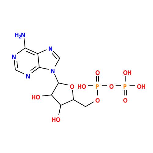 Mass: 427.201 Da / Num. of mol.: 16 / Source method: obtained synthetically / Formula: C10H15N5O10P2 / Comment: ADP, energy-carrying molecule*YM Mass: 427.201 Da / Num. of mol.: 16 / Source method: obtained synthetically / Formula: C10H15N5O10P2 / Comment: ADP, energy-carrying molecule*YM#7: Chemical | ChemComp-MG /  Mass: 24.305 Da / Num. of mol.: 16 / Source method: obtained synthetically / Formula: Mg Mass: 24.305 Da / Num. of mol.: 16 / Source method: obtained synthetically / Formula: Mg#8: Chemical | ChemComp-CA /  Mass: 40.078 Da / Num. of mol.: 6 / Source method: obtained synthetically / Formula: Ca Mass: 40.078 Da / Num. of mol.: 6 / Source method: obtained synthetically / Formula: Ca |
|---|
-Details
| Has ligand of interest | N |
|---|
-Experimental details
-Experiment
| Experiment | Method: ELECTRON MICROSCOPY |
|---|---|
| EM experiment | Aggregation state: FILAMENT / 3D reconstruction method: single particle reconstruction |
- Sample preparation
| Component | Name: HUMAN CARDIAC THIN FILAMENT IN THE CALCIUM BOUND STATE Type: COMPLEX / Entity ID: #1-#5 / Source: MULTIPLE SOURCES |
|---|---|
| Buffer solution | pH: 7.5 |
| Specimen | Conc.: 0.05 mg/ml / Embedding applied: NO / Shadowing applied: NO / Staining applied: NO / Vitrification applied: YES Details: CARDIAC MUSCLE THIN FILAMENT IN THE CALCIUM BOUND STATE; ACTIN, ALPHA SKELETAL MUSCLE; TROPOMYOSIN, TROPONIN |
| Specimen support | Grid material: MOLYBDENUM / Grid type: Quantifoil R1.2/1.3 |
| Vitrification | Instrument: LEICA EM GP / Cryogen name: ETHANE / Humidity: 80 % |
- Electron microscopy imaging
| Microscopy | Model: JEOL CRYO ARM 200 |
|---|---|
| Electron gun | Electron source:  FIELD EMISSION GUN / Accelerating voltage: 200 kV / Illumination mode: FLOOD BEAM FIELD EMISSION GUN / Accelerating voltage: 200 kV / Illumination mode: FLOOD BEAM |
| Electron lens | Mode: BRIGHT FIELD / Nominal defocus max: 1800 nm / Nominal defocus min: 700 nm |
| Image recording | Electron dose: 65 e/Å2 / Detector mode: COUNTING / Film or detector model: GATAN K2 SUMMIT (4k x 4k) / Num. of real images: 2880 |
- Processing
| EM software |
| |||||||||
|---|---|---|---|---|---|---|---|---|---|---|
| CTF correction | Type: PHASE FLIPPING AND AMPLITUDE CORRECTION | |||||||||
| 3D reconstruction | Resolution: 4.8 Å / Resolution method: FSC 0.143 CUT-OFF / Num. of particles: 23374 / Symmetry type: POINT |
