Movie
Movie Controller

[English] 日本語
 Yorodumi
- PDB-7wlm: The Cryo-EM structure of siphonaxanthin chlorophyll a/b type ligh... -
+ Open data

- Basic information
| Entry | Database: PDB / ID: 7wlm | ||||||
|---|---|---|---|---|---|---|---|
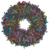
| Title | The Cryo-EM structure of siphonaxanthin chlorophyll a/b type light-harvesting complex II | ||||||
 Components Components | siphonaxanthin chlorophyll a/b binding light-harvesting complex II | ||||||
 Keywords Keywords | PHOTOSYNTHESIS / light-harvesting complex II / siphonaxanthin / Chlorophyll b replacement / blue-green utilization | ||||||
| Function / homology |  Function and homology information Function and homology informationChlorophyll a-b binding protein / Chlorophyll a/b binding protein domain / Orthogonal Bundle / Mainly Alpha Similarity search - Domain/homology | ||||||
| Biological species |  Codium fragile (dead man's fingers) Codium fragile (dead man's fingers) | ||||||
| Method | ELECTRON MICROSCOPY / single particle reconstruction / cryo EM / Resolution: 2.8 Å | ||||||
 Authors Authors | Seki, S. / Nakaniwa, T. / Castro-Hartmann, P. / Sader, K. / Kawamoto, A. / Tanaka, H. / Qian, P. / Kurisu, G. / Fujii, R. | ||||||
| Funding support | 1items
| ||||||
 Citation Citation | Journal: BBA Adv / Year: 2022 Title: Structural insights into blue-green light utilization by marine green algal light harvesting complex II at 2.78 Å. Authors: Soichiro Seki / Tetsuko Nakaniwa / Pablo Castro-Hartmann / Kasim Sader / Akihiro Kawamoto / Hideaki Tanaka / Pu Qian / Genji Kurisu / Ritsuko Fujii /   Abstract: Light-harvesting complex II (LHCII) present in plants and green algae absorbs solar energy to promote photochemical reactions. A marine green macroalga, , exhibits the unique characteristic of ...Light-harvesting complex II (LHCII) present in plants and green algae absorbs solar energy to promote photochemical reactions. A marine green macroalga, , exhibits the unique characteristic of absorbing blue-green light from the sun during photochemical reactions while being underwater owing to the presence of pigment-altered LHCII called siphonaxanthin-chlorophyll binding protein (SCP). In this study, we determined the structure of SCP at a resolution of 2.78 Å using cryogenic electron microscopy. SCP has a trimeric structure, wherein each monomer containing two lutein and two chlorophyll molecules in the plant-type LHCII are replaced by siphonaxanthin and its ester and two chlorophyll molecules, respectively. Siphonaxanthin occupies the binding site in SCP having a polarity in the trimeric inner core, and exhibits a distorted conjugated chain comprising a carbonyl group hydrogen bonded to a cysteine residue of apoprotein. These features suggest that the siphonaxanthin molecule is responsible for the characteristic green absorption of SCP. The replaced chlorophyll molecules extend the region of the stromal side chlorophyll cluster, spanning two adjacent monomers. | ||||||
| History |
|
- Structure visualization
| Structure viewer | Molecule: MolmilJmol/JSmol |
|---|
- Downloads & links
-Download
| PDBx/mmCIF format | 7wlm.cif.gz | 181.9 KB | Display | PDBx/mmCIF format |
|---|---|---|---|---|
| PDB format | pdb7wlm.ent.gz | 150.1 KB | Display | PDB format |
| PDBx/mmJSON format | 7wlm.json.gz | Tree view | PDBx/mmJSON format | |
| Others |  Other downloads Other downloads |
-Validation report
| Arichive directory | https://data.pdbj.org/pub/pdb/validation_reports/wl/7wlmftp://data.pdbj.org/pub/pdb/validation_reports/wl/7wlm | HTTPS FTP |
|---|
-Related structure data
| Related structure data |  32588MC M: map data used to model this data C: citing same article ( |
|---|---|
| Similar structure data |
-Links
 PDBj
PDBj


- Assembly
| Deposited unit | 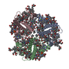
|
|---|---|
| 1 |
|
-Components
-Protein , 1 types, 3 molecules ABC
| #1: Protein | Mass: 24190.102 Da / Num. of mol.: 3 / Source method: isolated from a natural source / Source: (natural) Codium fragile (dead man's fingers) / Strain: KU-0654 |
|---|
-Non-polymers , 7 types, 105 molecules 


| #2: Chemical | ChemComp-CHL /  Mass: 907.472 Da / Num. of mol.: 24 / Source method: obtained synthetically / Formula: C55H70MgN4O6 / Feature type: SUBJECT OF INVESTIGATION Mass: 907.472 Da / Num. of mol.: 24 / Source method: obtained synthetically / Formula: C55H70MgN4O6 / Feature type: SUBJECT OF INVESTIGATION#3: Chemical | ChemComp-CLA /  Mass: 893.489 Da / Num. of mol.: 15 / Source method: obtained synthetically / Formula: C55H72MgN4O5 Mass: 893.489 Da / Num. of mol.: 15 / Source method: obtained synthetically / Formula: C55H72MgN4O5#4: Chemical |  Mass: 600.870 Da / Num. of mol.: 3 / Source method: obtained synthetically / Formula: C40H56O4 Mass: 600.870 Da / Num. of mol.: 3 / Source method: obtained synthetically / Formula: C40H56O4#5: Chemical |  Mass: 781.157 Da / Num. of mol.: 3 / Source method: obtained synthetically / Formula: C52H76O5 / Feature type: SUBJECT OF INVESTIGATION Mass: 781.157 Da / Num. of mol.: 3 / Source method: obtained synthetically / Formula: C52H76O5 / Feature type: SUBJECT OF INVESTIGATION#6: Chemical |  Mass: 600.870 Da / Num. of mol.: 3 / Source method: obtained synthetically / Formula: C40H56O4 / Feature type: SUBJECT OF INVESTIGATION Mass: 600.870 Da / Num. of mol.: 3 / Source method: obtained synthetically / Formula: C40H56O4 / Feature type: SUBJECT OF INVESTIGATION#7: Chemical |  Mass: 722.970 Da / Num. of mol.: 3 / Source method: obtained synthetically / Formula: C38H75O10P / Comment: phospholipid*YM Mass: 722.970 Da / Num. of mol.: 3 / Source method: obtained synthetically / Formula: C38H75O10P / Comment: phospholipid*YM#8: Water | ChemComp-HOH / | Mass: 18.015 Da / Num. of mol.: 54 / Source method: isolated from a natural source / Formula: H2O |
|---|
-Details
| Has ligand of interest | Y |
|---|
-Experimental details
-Experiment
| Experiment | Method: ELECTRON MICROSCOPY |
|---|---|
| EM experiment | Aggregation state: PARTICLE / 3D reconstruction method: single particle reconstruction |
- Sample preparation
| Component | Name: siphonaxanthin chlorophyll a/b-type light-harvesting complex II Type: COMPLEX / Entity ID: #1 / Source: NATURAL | |||||||||||||||
|---|---|---|---|---|---|---|---|---|---|---|---|---|---|---|---|---|
| Molecular weight | Value: 0.119 MDa / Experimental value: NO | |||||||||||||||
| Source (natural) | Organism: Codium fragile (dead man's fingers) | |||||||||||||||
| Buffer solution | pH: 8.2 | |||||||||||||||
| Buffer component |
| |||||||||||||||
| Specimen | Conc.: 10 mg/ml / Embedding applied: NO / Shadowing applied: NO / Staining applied: NO / Vitrification applied: YES | |||||||||||||||
| Specimen support | Grid material: COPPER | |||||||||||||||
| Vitrification | Cryogen name: ETHANE |
- Electron microscopy imaging
| Experimental equipment |  Model: Titan Krios / Image courtesy: FEI Company |
|---|---|
| Microscopy | Model: FEI TITAN KRIOS |
| Electron gun | Electron source:  FIELD EMISSION GUN / Accelerating voltage: 300 kV / Illumination mode: FLOOD BEAM FIELD EMISSION GUN / Accelerating voltage: 300 kV / Illumination mode: FLOOD BEAM |
| Electron lens | Mode: BRIGHT FIELD / Nominal magnification: 120000 X / Nominal defocus max: 2200 nm / Nominal defocus min: 800 nm / Alignment procedure: COMA FREE |
| Specimen holder | Cryogen: NITROGEN / Specimen holder model: FEI TITAN KRIOS AUTOGRID HOLDER |
| Image recording | Electron dose: 50.75 e/Å2 / Film or detector model: FEI FALCON IV (4k x 4k) / Num. of grids imaged: 2 / Num. of real images: 8935 |
| Image scans | Width: 4096 / Height: 4096 |
- Processing
| Software | Name: PHENIX / Version: 1.19.2_4158: / Classification: refinement | ||||||||||||||||||||||||||||||||||||||||
|---|---|---|---|---|---|---|---|---|---|---|---|---|---|---|---|---|---|---|---|---|---|---|---|---|---|---|---|---|---|---|---|---|---|---|---|---|---|---|---|---|---|
| EM software |
| ||||||||||||||||||||||||||||||||||||||||
| CTF correction | Type: PHASE FLIPPING AND AMPLITUDE CORRECTION | ||||||||||||||||||||||||||||||||||||||||
| Particle selection | Num. of particles selected: 2336076 | ||||||||||||||||||||||||||||||||||||||||
| Symmetry | Point symmetry: C3 (3 fold cyclic) | ||||||||||||||||||||||||||||||||||||||||
| 3D reconstruction | Resolution: 2.8 Å / Resolution method: FSC 0.143 CUT-OFF / Num. of particles: 250633 / Algorithm: FOURIER SPACE / Symmetry type: POINT | ||||||||||||||||||||||||||||||||||||||||
| Atomic model building | Protocol: RIGID BODY FIT / Space: REAL | ||||||||||||||||||||||||||||||||||||||||
| Atomic model building | PDB-ID: 1RWT Pdb chain-ID: B / Accession code: 1RWT / Pdb chain residue range: 14-231 / Source name: PDB / Type: experimental model | ||||||||||||||||||||||||||||||||||||||||
| Refine LS restraints |
|