 ムービー
ムービー コントローラー
 構造ビューア
 EMN検索について


-検索条件
-検索結果
検索 (著者・登録者: dolezal & m)の結果54件中、1から50件目までを表示しています

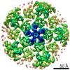
EMDB-19583: 
CryoEM structure of M. smegmatis GMP reductase in complex with GMP at pH 6.6, extended conformation.
手法: 単粒子 / : Dolezal M, Kouba T, Pichova I

EMDB-19584:
CryoEM structure of M. smegmatis GMP reductase in complex with GMP at pH 6.6, compressed conformation.
手法: 単粒子 / : Dolezal M, Kouba T, Pichova I

EMDB-19585:
CryoEM structure of M. smegmatis GMP reductase in complex with GMP and GTP at pH 6.6, extended conformation I.
手法: 単粒子 / : Dolezal M, Kouba T, Pichova I

EMDB-19586:
CryoEM structure of M. smegmatis GMP reductase in complex with GMP and GTP at pH 6.6, extended conformation II.
手法: 単粒子 / : Dolezal M, Kouba T, Pichova I

EMDB-19587:
CryoEM structure of M. smegmatis GMP reductase in complex with GMP and ATP at pH 6.6, compressed conformation.
手法: 単粒子 / : Dolezal M, Kouba T, Pichova I

EMDB-19588:
CryoEM structure of M. smegmatis GMP reductase apoform at pH 6.6, extended conformation I.
手法: 単粒子 / : Dolezal M, Kouba T, Pichova I

EMDB-19589:
CryoEM structure of M. smegmatis GMP reductase apoform at pH 6.6, extended conformation II.
手法: 単粒子 / : Dolezal M, Kouba T, Pichova I

EMDB-19590:
CryoEM structure of M. smegmatis GMP reductase apoform at pH 7.8, extended conformation I.
手法: 単粒子 / : Dolezal M, Kouba T, Svachova H, Pichova I

EMDB-19591:
CryoEM structure of M. smegmatis GMP reductase apoform at pH 7.8, extended conformation II.
手法: 単粒子 / : Dolezal M, Kouba T, Svachova H, Pichova I

EMDB-19592:
CryoEM structure of M. smegmatis GMP reductase apoform at pH 7.8, tetramer.
手法: 単粒子 / : Dolezal M, Kouba T, Svachova H, Pichova I

EMDB-19593:
CryoEM structure of M. smegmatis GMP reductase in complex with GMP at pH 7.8, extended conformation.
手法: 単粒子 / : Dolezal M, Tomas K, Filimonenko A, Pichova I

PDB-8ry0:
CryoEM structure of M. smegmatis GMP reductase in complex with GMP at pH 6.6, extended conformation.
手法: 単粒子 / : Dolezal M, Kouba T, Pichova I

PDB-8ry1:
CryoEM structure of M. smegmatis GMP reductase in complex with GMP at pH 6.6, compressed conformation.
手法: 単粒子 / : Dolezal M, Kouba T, Pichova I

PDB-8ry3:
CryoEM structure of M. smegmatis GMP reductase in complex with GMP and GTP at pH 6.6, extended conformation I.
手法: 単粒子 / : Dolezal M, Kouba T, Pichova I

PDB-8ry4:
CryoEM structure of M. smegmatis GMP reductase in complex with GMP and GTP at pH 6.6, extended conformation II.
手法: 単粒子 / : Dolezal M, Kouba T, Pichova I

PDB-8ry5:
CryoEM structure of M. smegmatis GMP reductase in complex with GMP and ATP at pH 6.6, compressed conformation.
手法: 単粒子 / : Dolezal M, Kouba T, Pichova I

PDB-8ry6:
CryoEM structure of M. smegmatis GMP reductase apoform at pH 6.6, extended conformation I.
手法: 単粒子 / : Dolezal M, Kouba T, Pichova I

PDB-8ry7:
CryoEM structure of M. smegmatis GMP reductase apoform at pH 6.6, extended conformation II.
手法: 単粒子 / : Dolezal M, Kouba T, Pichova I

PDB-8ry8:
CryoEM structure of M. smegmatis GMP reductase apoform at pH 7.8, extended conformation I.
手法: 単粒子 / : Dolezal M, Kouba T, Svachova H, Pichova I

PDB-8ry9:
CryoEM structure of M. smegmatis GMP reductase apoform at pH 7.8, extended conformation II.
手法: 単粒子 / : Dolezal M, Kouba T, Svachova H, Pichova I

PDB-8rya:
CryoEM structure of M. smegmatis GMP reductase apoform at pH 7.8, tetramer.
手法: 単粒子 / : Dolezal M, Kouba T, Svachova H, Pichova I

PDB-8ryb:
CryoEM structure of M. smegmatis GMP reductase in complex with GMP at pH 7.8, extended conformation.
手法: 単粒子 / : Dolezal M, Tomas K, Filimonenko A, Pichova I

EMDB-51463:
ApoF at 2.2A resolution imaged at 0.55A/pixel on Falcon C
手法: 単粒子 / : Koh FA, Karia D, Yang W, Yu L, Plitzko J, Aricescu R, Lander GC, Greber B, Kotecha A

EMDB-51481:
Apoferritin at 2.1A from Falcon C imaged at 0.7Apix
手法: 単粒子 / : Koh FA, Karia D, Yang W, Yu L, Kotecha A

EMDB-51483:
Apoferritin at 2.2A from Falcon C imaged at 0.9Apix
手法: 単粒子 / : Koh FA, Karia D, Yang W, Yu L, Kotecha A

EMDB-51487:
T20S Proteosome at 2.7A from Falcon C imaged at 0.7Apix
手法: 単粒子 / : Koh FA, Karia D, Yang W, Yu L, Khavnekar S, Kotecha A

EMDB-51503:
T20S Proteosome at 2.9A from Falcon C imaged at 0.9Apix
手法: 単粒子 / : Koh FA, Karia D, Yang W, Yu L, Khavnekar S, Kotecha A
EMDB-51506:
GABAA receptor at 2.8A from Falcon C imaged at 0.7Apix
手法: 単粒子 / : Koh FA, Karia D, Yang W, Yu L, Kotecha A

EMDB-51508:
GABAA receptor at 2.8A from Falcon C imaged at 0.9Apix
手法: 単粒子 / : Koh FA, Karia D, Yang W, Yu L, Kotecha A

EMDB-51511:
Rabbit muscle aldolase at 3A resolution imaged at 0.55A/pix
手法: 単粒子 / : Koh FA, Karia D, Yang W, Yu L, Kotecha A

EMDB-51512:
Rabbit muscle aldolase at 2.8A imaged at 0.7Apix
手法: 単粒子 / : Koh FA, Karia D, Yang W, Yu L, Kotecha A

EMDB-51519:
CAK complex at 4A imaged on Falcon C
手法: 単粒子 / : Koh FA, Karia D, Yang W, Yu L, Kotecha A, Greber B

EMDB-51525:
Human Haemoglobin at 5A imaged on Falcon C
手法: 単粒子 / : Koh FA, Karia D, Yang W, Yu L, Kotecha A

EMDB-51526:
Human Transthyretin at 3.5A imaged on Falcon C
手法: 単粒子 / : Koh FA, Karia D, Yang W, Yu L, Kotecha A, Lander GC
EMDB-51540:
Adeno-associated Virus 9 at 2.8A imaged on Falcon C
手法: 単粒子 / : Koh FA, Karia D, Yang W, Yu L, Kotecha A
EMDB-28189:
SARS-CoV-2 Spike in complex with biparatopic nanobody BP10
手法: 単粒子 / : Pymm PG, Glukhova A, Tham WH
EMDB-28190:
SARS-CoV-2 RBD in complex with biparatopic nanobody BP10 local refinement
手法: 単粒子 / : Pymm PG, Glukhova A, Tham WH
EMDB-25373:
Get3 bound to ADP and the transmembrane domain of the tail-anchored protein Bos1 - nucleotide binding domain map
手法: 単粒子 / : Fry MY, Maggiolo AO

EMDB-25374:
Get3 bound to ADP and the transmembrane domain of the tail-anchored protein Bos1 - overall map
手法: 単粒子 / : Fry MY, Maggiolo AO, Clemons Jr WM

EMDB-25375:
Nucleotide-free Get3 in the closed form
手法: 単粒子 / : Fry MY, Clemons Jr WM

EMDB-14249:
CryoEM structure of T20S proteasome at 3A from Tundra, 100kV microscope
手法: 単粒子 / : Karia D, Koh AF, Yu L, Kotecha A

EMDB-13816:
CryoEM structure of GABA(A)R-beta3 homopentamer at 3.4A from Tundra, 100kV microscope
手法: 単粒子 / : Karia D, Koh AF, Yu L, Kotecha A

EMDB-13769:
CryoEM structure of Apoferritin at 2.6A from Tundra, 100kV microscope
手法: 単粒子 / : Karia D, Hlavenkova H, Koh A, Malinsky M, Yu L, Kotecha A
EMDB-0290:
Immature M-PMV capsid hexamer structure in intact virus particles
手法: サブトモグラム平均 / : Qu K, Glass B
EMDB-0291:
Immature MLV capsid hexamer structure in intact virus particles
手法: サブトモグラム平均 / : Qu K, Glass B
EMDB-0292:
Mature MLV capsid hexamer structure in intact virus particles
手法: サブトモグラム平均 / : Qu K, Glass B
EMDB-0293:
Mature MLV capsid pentamer structure in intact virus particles
手法: サブトモグラム平均 / : Qu K, Glass B
EMDB-4419:
Representative tomogram of mature MLV particles
手法: トモグラフィー / : Qu K, Glass B, Dolezal M, Schur FKM, Rein A, Rumlova M, Ruml T, Kraeusslich HG, Briggs JAG
EMDB-4421:
Representative tomogram of immature M-PMV particles
手法: トモグラフィー / : Qu K, Glass B, Dolezal M, Schur FKM, Rein A, Rumlova M, Ruml T, Kraeusslich HG, Briggs JAG
EMDB-4422:
Representative tomogram of immature MLV particles
手法: トモグラフィー / : Qu K, Glass B, Dolezal M, Schur FKM, Rein A, Rumlova M, Ruml T, Kraeusslich HG, Briggs JAG
ページ:
 wwPDBはEMDBデータモデルのバージョン3へ移行します
