Journal: Structure / Year: 2025 Title: Sub-3 Å resolution protein structure determination by single-particle cryo-EM at 100 keV. Authors: Dimple Karia / Adrian F Koh / Wen Yang / Victoria I Cushing / Benjamin Basanta / Daniel B Mihaylov / Sagar Khavnekar / Ondřej Vyroubal / Miloš Malínský / Ondřej Sháněl / Vojtěch ...Authors: Dimple Karia / Adrian F Koh / Wen Yang / Victoria I Cushing / Benjamin Basanta / Daniel B Mihaylov / Sagar Khavnekar / Ondřej Vyroubal / Miloš Malínský / Ondřej Sháněl / Vojtěch Doležal / Jürgen Plitzko / Lingbo Yu / Gabriel C Lander / A Radu Aricescu / Basil J Greber / Abhay Kotecha / Abstract: Cryoelectron microscopy (cryo-EM) has transformed structural biology by providing high-resolution insights into biological macromolecules. We report sub-3 Å resolution structures using the 100 keV ...Cryoelectron microscopy (cryo-EM) has transformed structural biology by providing high-resolution insights into biological macromolecules. We report sub-3 Å resolution structures using the 100 keV Tundra cryo-TEM, equipped with the Falcon C direct electron detector (DED). This system combines advanced optics, extreme-brightness field emission gun (XFEG), and SP-TWIN lens to enhance coherence and resolution. The semi-automated loader reduced contamination and drift, enabling extended data collection, while the high detective quantum efficiency (DQE) of Falcon C improved signal-to-noise ratio. We validated performance by determining structures of biological samples, including apoferritin (2.1 Å), T20S proteasome (2.7 Å), GABA receptor (2.8 Å), hemoglobin (5.0 Å), transthyretin (3.5 Å), and AAV9 capsid (2.8 Å), spanning 50 kDa-3.9 MDa. This work highlights the potential of 100 keV transmission electron microscopes (TEMs) to make cryo-EM more accessible. It sets a precedent for using lower voltage TEMs not only for screening, but also for high-resolution protein structure determination.
History
Deposition
Nov 1, 2021
-
Header (metadata) release
Feb 2, 2022
-
Map release
Feb 2, 2022
-
Update
Aug 13, 2025
-
Current status
Aug 13, 2025
Processing site: PDBe / Status: Released
-
Structure visualization
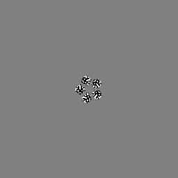
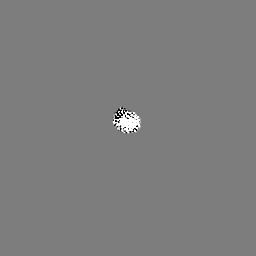
Movie
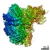
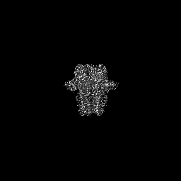
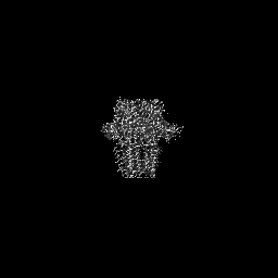
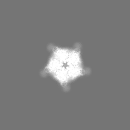
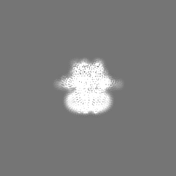
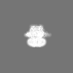
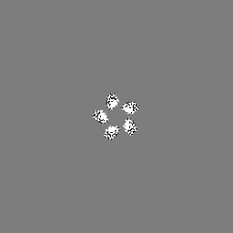
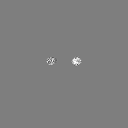
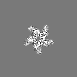
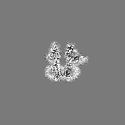
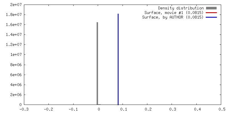
Surface view with section colored by density value
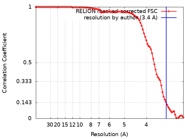
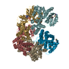
EMPIAR-10858 (Title: CryoEM structure of GABA(A)R-beta3 homopentamer at 3.4A from Tundra, 100kV microscope Data size: 2.2 TB Data #1: Unaligned multiframe movies of GABAA receptor from CETA-F camera [micrographs - multiframe])
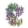
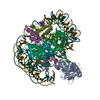
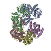
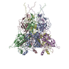
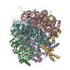
Supramolecule #1: Human GABA(A)R-beta3 homopentamer
Supramolecule
Name: Human GABA(A)R-beta3 homopentamer / type: complex / ID: 1 / Parent: 0 Details: Human GABA(A)R-beta3 homopentamer in complex with Megabody-25 in lipid nanodisc
Source (natural)
Organism: HEK 293S cells (E. coli)
Molecular weight
Theoretical: 200 KDa
-
Experimental details
-
Structure determination
Method
cryo EM
Processing
single particle reconstruction
Aggregation state
particle
-
Sample preparation
Buffer
pH: 7.6
Grid
Model: UltrAuFoil R2/2 / Material: GOLD / Mesh: 300 / Support film - Material: GOLD / Support film - topology: HOLEY / Pretreatment - Type: GLOW DISCHARGE / Pretreatment - Time: 30 sec.
In the structure databanks used in Yorodumi, some data are registered as the other names, "COVID-19 virus" and "2019-nCoV". Here are the details of the virus and the list of structure data.
Jan 31, 2019. EMDB accession codes are about to change! (news from PDBe EMDB page)
EMDB accession codes are about to change! (news from PDBe EMDB page)
The allocation of 4 digits for EMDB accession codes will soon come to an end. Whilst these codes will remain in use, new EMDB accession codes will include an additional digit and will expand incrementally as the available range of codes is exhausted. The current 4-digit format prefixed with “EMD-” (i.e. EMD-XXXX) will advance to a 5-digit format (i.e. EMD-XXXXX), and so on. It is currently estimated that the 4-digit codes will be depleted around Spring 2019, at which point the 5-digit format will come into force.
The EM Navigator/Yorodumi systems omit the EMD- prefix.
Related info.:Q: What is EMD? / ID/Accession-code notation in Yorodumi/EM Navigator
Yorodumi is a browser for structure data from EMDB, PDB, SASBDB, etc.
This page is also the successor to EM Navigator detail page, and also detail information page/front-end page for Omokage search.
The word "yorodu" (or yorozu) is an old Japanese word meaning "ten thousand". "mi" (miru) is to see.
Related info.:EMDB / PDB / SASBDB / Comparison of 3 databanks / Yorodumi Search / Aug 31, 2016. New EM Navigator & Yorodumi / Yorodumi Papers / Jmol/JSmol / Function and homology information / Changes in new EM Navigator and Yorodumi
 Movie
Movie Controller

 Yorodumi
 Open data
 Basic information
 Map data
 Sample
 Keywords
 Function and homology information
 Authors
 Netherlands, 1 items
  Citation


 Structure visualization
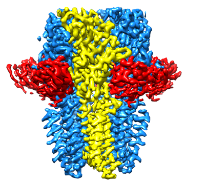
 Downloads & links
 emd_13816.png
 http://ftp.pdbj.org/pub/emdb/structures/EMD-13816


 Z (Sec.)
 Y (Row.)
 X (Col.)


 Sample components
 Processing
 Electron microscopy
 FIELD EMISSION GUN