 Movie
Movie Controller

[English] 日本語
 Yorodumi
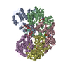
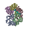
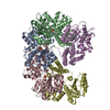
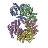
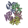
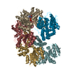
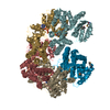
- PDB-3glh: Crystal Structure of the E. coli clamp loader bound to Psi Peptide -
+ Open data

- Basic information
| Entry | Database: PDB / ID: 3glh | ||||||
|---|---|---|---|---|---|---|---|
| Title | Crystal Structure of the E. coli clamp loader bound to Psi Peptide | ||||||
 Components Components |
| ||||||
 Keywords Keywords | TRANSFERASE / Clamp Loader / Gamma Complex / Replication / Psi / DNA replication / DNA-directed DNA polymerase / Nucleotidyltransferase / ATP-binding / Nucleotide-binding | ||||||
| Function / homology |  Function and homology information Function and homology informationDNA polymerase III, clamp loader complex / DNA clamp loader activity / DNA polymerase III complex / replisome / DNA polymerase processivity factor activity / 3'-5' exonuclease activity / DNA-templated DNA replication / ribonucleoside triphosphate phosphatase activity / DNA-directed DNA polymerase / DNA-directed DNA polymerase activity ...DNA polymerase III, clamp loader complex / DNA clamp loader activity / DNA polymerase III complex / replisome / DNA polymerase processivity factor activity / 3'-5' exonuclease activity / DNA-templated DNA replication / ribonucleoside triphosphate phosphatase activity / DNA-directed DNA polymerase / DNA-directed DNA polymerase activity / DNA replication / viral translational frameshifting / ATP hydrolysis activity / DNA binding / ATP binding / metal ion binding / identical protein binding Similarity search - Function | ||||||
| Biological species |  | ||||||
| Method |  X-RAY DIFFRACTION / SYNCHROTRON / MOLECULAR REPLACEMENT / Resolution: 3.891 Å X-RAY DIFFRACTION / SYNCHROTRON / MOLECULAR REPLACEMENT / Resolution: 3.891 Å | ||||||
 Authors Authors | Kazmirski, S.L. / Simonetta, K.R. / Kuriyan, J. | ||||||
 Citation Citation | Journal: Cell(Cambridge,Mass.) / Year: 2009 Title: The mechanism of ATP-dependent primer-template recognition by a clamp loader complex. Authors: Simonetta, K.R. / Kazmirski, S.L. / Goedken, E.R. / Cantor, A.J. / Kelch, B.A. / McNally, R. / Seyedin, S.N. / Makino, D.L. / O'Donnell, M. / Kuriyan, J. | ||||||
| History |
|
- Structure visualization
| Structure viewer | Molecule: MolmilJmol/JSmol |
|---|
- Downloads & links
-Download
| PDBx/mmCIF format | 3glh.cif.gz | 871.1 KB | Display | PDBx/mmCIF format |
|---|---|---|---|---|
| PDB format | pdb3glh.ent.gz | 699.9 KB | Display | PDB format |
| PDBx/mmJSON format | 3glh.json.gz | Tree view | PDBx/mmJSON format | |
| Others |  Other downloads Other downloads |
-Validation report
| Arichive directory | https://data.pdbj.org/pub/pdb/validation_reports/gl/3glhftp://data.pdbj.org/pub/pdb/validation_reports/gl/3glh | HTTPS FTP |
|---|
-Related structure data


-Links
 PDBj
PDBj
- Assembly
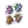
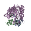
| Deposited unit | 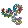
| ||||||||
|---|---|---|---|---|---|---|---|---|---|
| 1 | 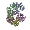
| ||||||||
| 2 | 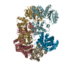
| ||||||||
| 3 | 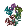
| ||||||||
| Unit cell |
|
-Components
| #1: Protein | Mass: 38745.574 Da / Num. of mol.: 3 Source method: isolated from a genetically manipulated source Source: (gene. exp.) #2: Protein | Mass: 41803.168 Da / Num. of mol.: 9 / Fragment: UNP residues 1-373 Source method: isolated from a genetically manipulated source Source: (gene. exp.) #3: Protein | Mass: 36980.484 Da / Num. of mol.: 3 Source method: isolated from a genetically manipulated source Source: (gene. exp.) |
|---|
-Experimental details
-Experiment
| Experiment | Method: X-RAY DIFFRACTION / Number of used crystals: 1 |
|---|
- Sample preparation
| Crystal | Density Matthews: 3.03 Å3/Da / Density % sol: 59.37 % |
|---|---|
| Crystal grow | Temperature: 293 K / Method: vapor diffusion, hanging drop / pH: 8.5 Details: 300 mM NaK Tartrate, 10% PEG 3350, 100 mM Tris, pH 8.5, VAPOR DIFFUSION, HANGING DROP, temperature 293K |
-Data collection
| Diffraction | Mean temperature: 100 K |
|---|---|
| Diffraction source | Source: SYNCHROTRON / Site: ALS  / Beamline: 8.2.2 / Wavelength: 0.9797 Å / Beamline: 8.2.2 / Wavelength: 0.9797 Å |
| Detector | Type: ADSC QUANTUM 315 / Detector: CCD / Date: Feb 9, 2004 |
| Radiation | Monochromator: KOHZU / Protocol: MAD / Monochromatic (M) / Laue (L): M / Scattering type: x-ray |
| Radiation wavelength | Wavelength: 0.9797 Å / Relative weight: 1 |
| Reflection | Resolution: 3.891→100 Å / Num. all: 67070 / Num. obs: 65729 / % possible obs: 98 % / Observed criterion σ(F): 1 / Observed criterion σ(I): 1 |
| Reflection shell | Resolution: 3.891→3.99 Å / % possible all: 94.2 |
- Processing
| Software |
| |||||||||||||||||||||||||||||||||||||||||||||||||||||||||||||||||||||||||||||||||||||||||||||||||||||||||||||||||||||||||||||||||||||||||||||||||||||||||||||||||||||||||||||||||||||||||||||||||||||||||||||||||||||||||
|---|---|---|---|---|---|---|---|---|---|---|---|---|---|---|---|---|---|---|---|---|---|---|---|---|---|---|---|---|---|---|---|---|---|---|---|---|---|---|---|---|---|---|---|---|---|---|---|---|---|---|---|---|---|---|---|---|---|---|---|---|---|---|---|---|---|---|---|---|---|---|---|---|---|---|---|---|---|---|---|---|---|---|---|---|---|---|---|---|---|---|---|---|---|---|---|---|---|---|---|---|---|---|---|---|---|---|---|---|---|---|---|---|---|---|---|---|---|---|---|---|---|---|---|---|---|---|---|---|---|---|---|---|---|---|---|---|---|---|---|---|---|---|---|---|---|---|---|---|---|---|---|---|---|---|---|---|---|---|---|---|---|---|---|---|---|---|---|---|---|---|---|---|---|---|---|---|---|---|---|---|---|---|---|---|---|---|---|---|---|---|---|---|---|---|---|---|---|---|---|---|---|---|---|---|---|---|---|---|---|---|---|---|---|---|---|---|---|---|
| Refinement | Method to determine structure: MOLECULAR REPLACEMENT / Resolution: 3.891→49.621 Å / Occupancy max: 1 / Occupancy min: 1 / SU ML: 0.87 / σ(F): 0.03 / Phase error: 41.25 / Stereochemistry target values: ML Details: There are several close contacts because this structure is only a 4A structure
| |||||||||||||||||||||||||||||||||||||||||||||||||||||||||||||||||||||||||||||||||||||||||||||||||||||||||||||||||||||||||||||||||||||||||||||||||||||||||||||||||||||||||||||||||||||||||||||||||||||||||||||||||||||||||
| Solvent computation | Shrinkage radii: 0.9 Å / VDW probe radii: 1.11 Å / Solvent model: FLAT BULK SOLVENT MODEL / Bsol: 14.525 Å2 / ksol: 0.309 e/Å3 | |||||||||||||||||||||||||||||||||||||||||||||||||||||||||||||||||||||||||||||||||||||||||||||||||||||||||||||||||||||||||||||||||||||||||||||||||||||||||||||||||||||||||||||||||||||||||||||||||||||||||||||||||||||||||
| Displacement parameters | Biso max: 291.12 Å2 / Biso mean: 111.018 Å2 / Biso min: 29.92 Å2
| |||||||||||||||||||||||||||||||||||||||||||||||||||||||||||||||||||||||||||||||||||||||||||||||||||||||||||||||||||||||||||||||||||||||||||||||||||||||||||||||||||||||||||||||||||||||||||||||||||||||||||||||||||||||||
| Refinement step | Cycle: LAST / Resolution: 3.891→49.621 Å
| |||||||||||||||||||||||||||||||||||||||||||||||||||||||||||||||||||||||||||||||||||||||||||||||||||||||||||||||||||||||||||||||||||||||||||||||||||||||||||||||||||||||||||||||||||||||||||||||||||||||||||||||||||||||||
| Refine LS restraints |
| |||||||||||||||||||||||||||||||||||||||||||||||||||||||||||||||||||||||||||||||||||||||||||||||||||||||||||||||||||||||||||||||||||||||||||||||||||||||||||||||||||||||||||||||||||||||||||||||||||||||||||||||||||||||||
| LS refinement shell |
|
