[English] 日本語
 Yorodumi
- PDB-8ry0: CryoEM structure of M. smegmatis GMP reductase in complex with GM... -
+ Open data
- Basic information
| Entry | Database: PDB / ID: 8ry0 | ||||||||||||||||||||||||||||||
|---|---|---|---|---|---|---|---|---|---|---|---|---|---|---|---|---|---|---|---|---|---|---|---|---|---|---|---|---|---|---|---|
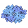
| Title | CryoEM structure of M. smegmatis GMP reductase in complex with GMP at pH 6.6, extended conformation. | ||||||||||||||||||||||||||||||
 Components Components | GMP reductase | ||||||||||||||||||||||||||||||
 Keywords Keywords | OXIDOREDUCTASE / GMP reductase / GuaB1 / CBS domain / Mycobacterium smegmatis | ||||||||||||||||||||||||||||||
| Function / homology |  Function and homology information Function and homology informationGMP reductase / GMP reductase activity / IMP salvage / IMP dehydrogenase activity / purine ribonucleoside salvage / cytosol Similarity search - Function | ||||||||||||||||||||||||||||||
| Biological species |  Mycolicibacterium smegmatis (bacteria) Mycolicibacterium smegmatis (bacteria) | ||||||||||||||||||||||||||||||
| Method | ELECTRON MICROSCOPY / single particle reconstruction / cryo EM / Resolution: 3.66 Å | ||||||||||||||||||||||||||||||
 Authors Authors | Dolezal, M. / Kouba, T. / Pichova, I. | ||||||||||||||||||||||||||||||
| Funding support | European Union, 1items
| ||||||||||||||||||||||||||||||
 Citation Citation |  Journal: To Be Published Journal: To Be PublishedTitle: Structural basis for allosteric regulation of mycobacterial guanosine 5'-monophosphate reductase with ATP and GTP. Authors: Dolezal, M. / Knejzlik, Z. / Kouba, T. / Filimonenko, A. / Svachova, H. / Klima, M. / Pichova, I. | ||||||||||||||||||||||||||||||
| History |
|
- Structure visualization
| Structure viewer | Molecule:  Molmil Molmil Jmol/JSmol Jmol/JSmol |
|---|
- Downloads & links
- Download
| PDBx/mmCIF format |  8ry0.cif.gz 8ry0.cif.gz | 707.8 KB | Display |  PDBx/mmCIF format PDBx/mmCIF format |
|---|---|---|---|---|
| PDB format |  pdb8ry0.ent.gz pdb8ry0.ent.gz | 496 KB | Display |  PDB format PDB format |
| PDBx/mmJSON format |  8ry0.json.gz 8ry0.json.gz | Tree view |  PDBx/mmJSON format PDBx/mmJSON format | |
| Others |  Other downloads Other downloads |
-Validation report
| Arichive directory |  https://data.pdbj.org/pub/pdb/validation_reports/ry/8ry0 https://data.pdbj.org/pub/pdb/validation_reports/ry/8ry0 ftp://data.pdbj.org/pub/pdb/validation_reports/ry/8ry0 ftp://data.pdbj.org/pub/pdb/validation_reports/ry/8ry0 | HTTPS FTP |
|---|
-Related structure data
| Related structure data |  19583MC  8ry1C  8ry3C  8ry4C  8ry5C  8ry6C  8ry7C  8ry8C  8ry9C  8ryaC  8rybC  9hfzC  9hg0C  9hg1C  9hg2C  9hg3C C: citing same article ( M: map data used to model this data |
|---|---|
| Similar structure data | Similarity search - Function & homology  F&H Search F&H Search |
- Links
Links
- Assembly
| Deposited unit | 
|
|---|---|
| 1 |
|
- Components
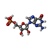
| #1: Protein | Mass: 51782.434 Da / Num. of mol.: 8 Source method: isolated from a genetically manipulated source Details: Guanosine 5'-monophosphate reductase / Source: (gene. exp.)  Mycolicibacterium smegmatis (bacteria) / Strain: ATCC 700084 / mc(2)155 / Gene: guaB1, MSMEG_3634, MSMEI_3548 / Plasmid: pTriex / Production host: Mycolicibacterium smegmatis (bacteria) / Strain: ATCC 700084 / mc(2)155 / Gene: guaB1, MSMEG_3634, MSMEI_3548 / Plasmid: pTriex / Production host:  #2: Chemical | ChemComp-5GP / Has ligand of interest | Y | Has protein modification | N | |
|---|
-Experimental details
-Experiment
| Experiment | Method: ELECTRON MICROSCOPY |
|---|---|
| EM experiment | Aggregation state: PARTICLE / 3D reconstruction method: single particle reconstruction |
- Sample preparation
| Component | Name: Msm GMPR with GMP at pH 6.6 / Type: COMPLEX / Entity ID: #1 / Source: RECOMBINANT | ||||||||||||||||||||
|---|---|---|---|---|---|---|---|---|---|---|---|---|---|---|---|---|---|---|---|---|---|
| Source (natural) | Organism:  Mycolicibacterium smegmatis (bacteria) / Strain: ATCC 700084 / mc(2)155 Mycolicibacterium smegmatis (bacteria) / Strain: ATCC 700084 / mc(2)155 | ||||||||||||||||||||
| Source (recombinant) | Organism:  | ||||||||||||||||||||
| Buffer solution | pH: 6.6 | ||||||||||||||||||||
| Buffer component |
| ||||||||||||||||||||
| Specimen | Embedding applied: NO / Shadowing applied: NO / Staining applied: NO / Vitrification applied: YES Details: 20 mg/ml Msm GMPR with 2 mM GMP in the storage buffer (50 mM Tris, pH 8.0, 2.5 mM TCEP) was diluted with the cryoEM buffer (50 mM HEPES, pH 6.6, 100 mM KCl, 2 mM MgCl2). | ||||||||||||||||||||
| Vitrification | Cryogen name: ETHANE |
- Electron microscopy imaging
| Experimental equipment |  Model: Talos Arctica / Image courtesy: FEI Company |
|---|---|
| Microscopy | Model: FEI TALOS ARCTICA |
| Electron gun | Electron source:  FIELD EMISSION GUN / Accelerating voltage: 200 kV / Illumination mode: FLOOD BEAM FIELD EMISSION GUN / Accelerating voltage: 200 kV / Illumination mode: FLOOD BEAM |
| Electron lens | Mode: BRIGHT FIELD / Nominal defocus max: 3000 nm / Nominal defocus min: 700 nm |
| Specimen holder | Cryogen: NITROGEN |
| Image recording | Electron dose: 40 e/Å2 / Film or detector model: GATAN K2 SUMMIT (4k x 4k) |
- Processing
| EM software |
| ||||||||||||||||||||||||||||||||
|---|---|---|---|---|---|---|---|---|---|---|---|---|---|---|---|---|---|---|---|---|---|---|---|---|---|---|---|---|---|---|---|---|---|
| CTF correction | Type: PHASE FLIPPING ONLY | ||||||||||||||||||||||||||||||||
| 3D reconstruction | Resolution: 3.66 Å / Resolution method: FSC 0.143 CUT-OFF / Num. of particles: 75197 / Symmetry type: POINT | ||||||||||||||||||||||||||||||||
| Atomic model building | Space: REAL Details: The initial structure was obtained by fitting an appropriate model into the cryo-EM map using MolRep and ChimeraX. The structure was then refined by iterative manual rebuilding in Coot and ...Details: The initial structure was obtained by fitting an appropriate model into the cryo-EM map using MolRep and ChimeraX. The structure was then refined by iterative manual rebuilding in Coot and Isolde, and automatic refinement in phenix.real_space_refine. | ||||||||||||||||||||||||||||||||
| Refine LS restraints |
|
 Movie
Movie Controller


 PDBj
PDBj


