 Movie
Movie Controller
 Structure viewers
 About EMN search


-Search query
-Search result
Showing 1 - 50 of 104 items for (author: cavadini & s)

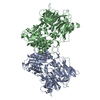
EMDB-53534: 
p53 bound to nucleosome at position SHL+5.9 (non-crosslinked sample, full map)

EMDB-53532:
p53 bound to nucleosome at position SHL+5.9 (non-crosslinked sample, composite map)

PDB-9r2m:
p53 bound to nucleosome at position SHL+5.9 (non-crosslinked sample, composite map)

EMDB-53478:
p53 bound to the nucleosome at position SHL-5.7 (crosslinked sample)

PDB-9r04:
p53 bound to the nucleosome at position SHL-5.7 (crosslinked sample)

EMDB-53517:
USP7 bound to a nucleosome/p53 complex

EMDB-53535:
p53 bound to nucleosome at position SHL+5.9 (non-crosslinked sample, focus refined map of p53)

EMDB-53537:
p53 bound to nucleosome at position SHL-5.7 (non-crosslinked sample)

PDB-9r2q:
p53 bound to nucleosome at position SHL-5.7 (non-crosslinked sample)

EMDB-53536:
p53 bound to nucleosome at position SHL+5.9 (crosslinked sample)

PDB-9r2p:
p53 bound to nucleosome at position SHL+5.9 (crosslinked sample)

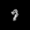
EMDB-17582:
Cryo-EM structure of Caenorhabditis elegans DPF-3 (apo)
PDB-8pba:
Cryo-EM structure of Caenorhabditis elegans DPF-3 (apo)

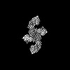
EMDB-19767:
Structure of a 2873 Scaffold Base DNA Origami V1

EMDB-19769:
Structure of a 2873 Scaffold Base DNA Origami V2

EMDB-19770:
Structure of a 2873 Scaffold Base DNA Origami V3

EMDB-19775:
Structure of a 1033 Scaffold Base DNA Origami Nanostructure V4 with Desalted Purified Staples

EMDB-19776:
Structure of a 1033 Scaffold Base DNA Origami Nanostructure V4 with HPLC Purified Staples

EMDB-19867:
Cryo-EM Structure of a 1033 Scaffold Base DNA Origami Nanostructure V4 and TBA

EMDB-19874:
Refinement Focused on the 1st Body of a 1033 Scaffold-Based DNA Origami Nanostructure V4 with TBA

EMDB-19875:
Refinement Focused on the 2nd Body of a 1033 Scaffold-Based DNA Origami Nanostructure V4 with TBA

EMDB-19876:
Refinement Focused on the 3rd Body of a 1033 Scaffold-Based DNA Origami Nanostructure V4 with TBA

PDB-9eoq:
Cryo-EM Structure of a 1033 Scaffold Base DNA Origami Nanostructure V4 and TBA
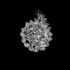
EMDB-17539:
Cryo-EM structure of dimeric UBR5

EMDB-17540:
Cryo-EM structure of full-length human UBR5 (homotetramer)

PDB-8p82:
Cryo-EM structure of dimeric UBR5

PDB-8p83:
Cryo-EM structure of full-length human UBR5 (homotetramer)
EMDB-17542:
Negative stain map of UBR5 (dimer) in complex with RARA/RXRA

EMDB-17154:
Cryo-EM structure of CLOCK-BMAL1 bound to a nucleosomal E-box at position SHL+5.8 (consensus and constituent map 1)

EMDB-17155:
Cryo-EM structure of CLOCK-BMAL1 bound to a nucleosomal E-box at position SHL-6.2 (DNA conformation 1)

EMDB-17156:
Cryo-EM structure of CLOCK-BMAL1 bound to a nucleosomal E-box at position SHL-6.2 (DNA conformation 2)

EMDB-17157:
Cryo-EM structure of CLOCK-BMAL1 bound to a nucleosomal E-box at position SHL+5.8 (composite map)

EMDB-17158:
Cryo-EM structure of CLOCK-BMAL1 bound to a nucleosomal E-box at position SHL+5.8 (constituent map 2 from additional focus classification on PAS domains)

EMDB-17159:
Cryo-EM map of MYC-MAX-OCT4-LIN28 complex

EMDB-17160:
Cryo-EM structure of CLOCK-BMAL1 bound to the native Por enhancer nucleosome (map 2, additional 3D classification and flexible refinement)

EMDB-17161:
Cryo-EM structure of CLOCK-BMAL1 bound to the native Por enhancer nucleosome (map 1)

EMDB-17162:
MAX-MAX bound to a nucleosome at SHL+5.1 and SHL-6.9.
EMDB-17183:
OCT4 and MYC-MAX co-bound to a nucleosome
EMDB-17184:
MYC-MAX bound to a nucleosome at SHL+5.8

PDB-8osj:
Cryo-EM structure of CLOCK-BMAL1 bound to a nucleosomal E-box at position SHL-6.2 (DNA conformation 1)

PDB-8osk:
Cryo-EM structure of CLOCK-BMAL1 bound to a nucleosomal E-box at position SHL+5.8 (composite map)

PDB-8osl:
Cryo-EM structure of CLOCK-BMAL1 bound to the native Por enhancer nucleosome (map 2, additional 3D classification and flexible refinement)
PDB-8ots:
OCT4 and MYC-MAX co-bound to a nucleosome
PDB-8ott:
MYC-MAX bound to a nucleosome at SHL+5.8

EMDB-15484:
Structure of human DDB1-DCAF12 in complex with the C-terminus of CCT5

EMDB-15485:
Structure of the human DDB1-DCAF12 complex

EMDB-15486:
Negative-stain electron microscopy structure of DDB1-DCAF12-CCT5

PDB-8ajm:
Structure of human DDB1-DCAF12 in complex with the C-terminus of CCT5

PDB-8ajn:
Structure of the human DDB1-DCAF12 complex

PDB-8ajo:
Negative-stain electron microscopy structure of DDB1-DCAF12-CCT5
Pages:
 wwPDB to switch to version 3 of the EMDB data model
