Loading
PDBj
MenuPDBj@FacebookPDBj@TwitterPDBj@YouTubewwPDB FoundationwwPDB
RCSB PDBPDBeBMRBAdv. SearchSearch help

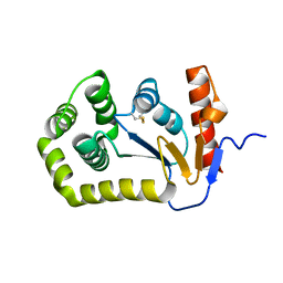
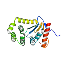
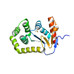
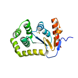
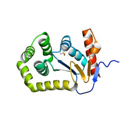
5QNT
DownloadVisualize
BU of 5qnt by Molmil
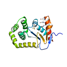
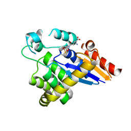
Group deposition of apo datasets for PANDDA analysis - Crystal Structure of apo EcDsbA after initial refinement (apo_dataset_2)
Descriptor: Thiol:disulfide interchange protein
Authors:Ilyichova, O.V, Bentley, M.R, Doak, B.C, Scanlon, M.J.
Deposit date:2019-01-27
Release date:2020-02-05
Last modified:2020-07-22
Method:X-RAY DIFFRACTION (1.747 Å)
Cite:Rapid Elaboration of Fragments into Leads by X-ray Crystallographic Screening of Parallel Chemical Libraries (REFiLX).
J.Med.Chem., 63, 2020
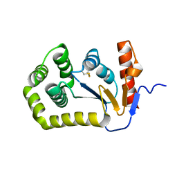
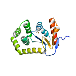
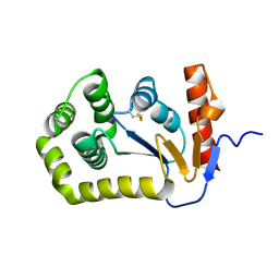
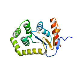
5QNV
DownloadVisualize
BU of 5qnv by Molmil
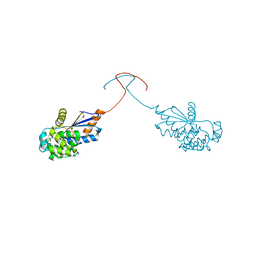
Group deposition of apo datasets for PANDDA analysis - Crystal Structure of apo EcDsbA after initial refinement (apo_dataset_21)
Descriptor: Thiol:disulfide interchange protein
Authors:Ilyichova, O.V, Bentley, M.R, Doak, B.C, Scanlon, M.J.
Deposit date:2019-01-27
Release date:2020-02-05
Last modified:2020-07-22
Method:X-RAY DIFFRACTION (1.75 Å)
Cite:Rapid Elaboration of Fragments into Leads by X-ray Crystallographic Screening of Parallel Chemical Libraries (REFiLX).
J.Med.Chem., 63, 2020
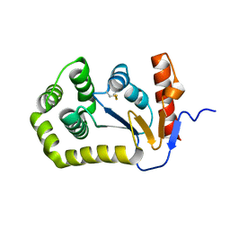
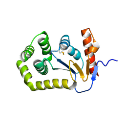
5QNL
DownloadVisualize
BU of 5qnl by Molmil
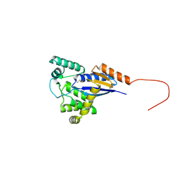
Group deposition of apo datasets for PANDDA analysis - Crystal Structure of apo EcDsbA after initial refinement (apo_dataset_12)
Descriptor: Thiol:disulfide interchange protein
Authors:Ilyichova, O.V, Bentley, M.R, Doak, B.C, Scanlon, M.J.
Deposit date:2019-01-27
Release date:2020-02-05
Last modified:2020-07-22
Method:X-RAY DIFFRACTION (1.752 Å)
Cite:Rapid Elaboration of Fragments into Leads by X-ray Crystallographic Screening of Parallel Chemical Libraries (REFiLX).
J.Med.Chem., 63, 2020
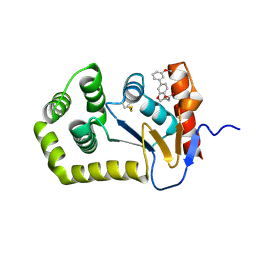
5QNR
DownloadVisualize
BU of 5qnr by Molmil
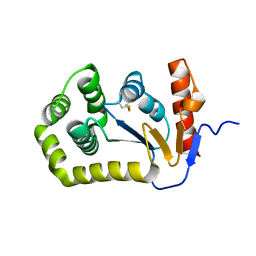
Group deposition of apo datasets for PANDDA analysis - Crystal Structure of apo EcDsbA after initial refinement (apo_dataset_18)
Descriptor: Thiol:disulfide interchange protein
Authors:Ilyichova, O.V, Bentley, M.R, Doak, B.C, Scanlon, M.J.
Deposit date:2019-01-27
Release date:2020-02-05
Last modified:2020-07-22
Method:X-RAY DIFFRACTION (1.752 Å)
Cite:Rapid Elaboration of Fragments into Leads by X-ray Crystallographic Screening of Parallel Chemical Libraries (REFiLX).
J.Med.Chem., 63, 2020
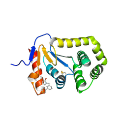
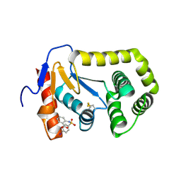
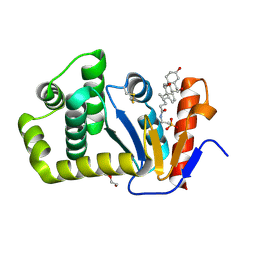
7S1F
DownloadVisualize
BU of 7s1f by Molmil
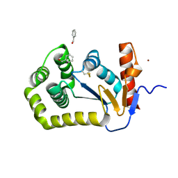
Crystal structure of E.coli DsbA in complex with compound MIPS-0001886 (compound 38)
Descriptor: 1-[(3-thiophen-3-ylphenyl)methyl]-3~{H}-pyrrol-2-one, COPPER (II) ION, GLYCEROL, ...
Authors:Heras, B, Scanlon, M.J, Martin, J.L, Caria, S.
Deposit date:2021-09-02
Release date:2023-02-08
Last modified:2024-04-03
Method:X-RAY DIFFRACTION (1.76 Å)
Cite:Fluoromethylketone-fragment conjugates designed as covalent modifiers of EcDsbA are atypical substrates
Chemrxiv, 2022
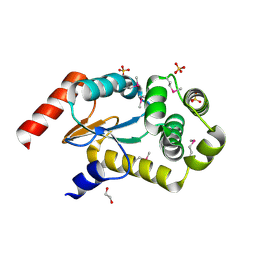
3GYK
DownloadVisualize
BU of 3gyk by Molmil
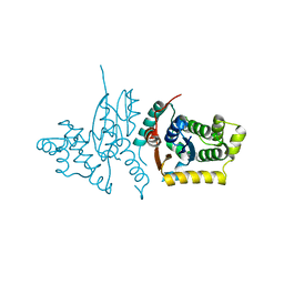
The crystal structure of a thioredoxin-like oxidoreductase from Silicibacter pomeroyi DSS-3
Descriptor: 1,2-ETHANEDIOL, 27kDa outer membrane protein, SULFATE ION
Authors:Fan, Y, Marshall, N, Keigher, L, Joachimiak, A, Midwest Center for Structural Genomics (MCSG)
Deposit date:2009-04-03
Release date:2009-06-09
Last modified:2011-07-13
Method:X-RAY DIFFRACTION (1.76 Å)
Cite:The crystal structure of a thioredoxin-like oxidoreductase from Silicibacter pomeroyi DSS-3
To be Published
4OCE
DownloadVisualize
BU of 4oce by Molmil
Crystal structure of the disulfide oxidoreductase DsbA from Proteus mirabilis
Descriptor: MALONATE ION, Thiol:disulfide interchange protein
Authors:Kurth, F, Martin, J.L.
Deposit date:2014-01-09
Release date:2014-05-28
Last modified:2023-09-20
Method:X-RAY DIFFRACTION (1.768 Å)
Cite:Crystal Structure of the Dithiol Oxidase DsbA Enzyme from Proteus Mirabilis Bound Non-covalently to an Active Site Peptide Ligand.
J.Biol.Chem., 289, 2014
5QO5
DownloadVisualize
BU of 5qo5 by Molmil
Group deposition of apo datasets for PANDDA analysis - Crystal Structure of apo EcDsbA after initial refinement (apo_dataset_34)
Descriptor: Thiol:disulfide interchange protein
Authors:Ilyichova, O.V, Bentley, M.R, Doak, B.C, Scanlon, M.J.
Deposit date:2019-01-27
Release date:2020-02-05
Last modified:2020-07-22
Method:X-RAY DIFFRACTION (1.769 Å)
Cite:Rapid Elaboration of Fragments into Leads by X-ray Crystallographic Screening of Parallel Chemical Libraries (REFiLX).
J.Med.Chem., 63, 2020
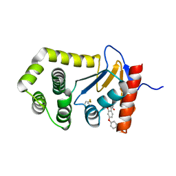
6POI
DownloadVisualize
BU of 6poi by Molmil
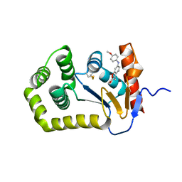
Crystal Structure of EcDsbA in complex phenyl ether 25
Descriptor: (6-phenoxy-1-benzofuran-3-yl)acetic acid, COPPER (II) ION, Thiol:disulfide interchange protein DsbA
Authors:Ilyichova, O.V, Scanlon, M.J.
Deposit date:2019-07-04
Release date:2019-11-06
Last modified:2023-10-11
Method:X-RAY DIFFRACTION (1.77 Å)
Cite:The Fragment-Based Development of a Benzofuran Hit as a New Class of Escherichia coli DsbA Inhibitors.
Molecules, 24, 2019
4ZIJ
DownloadVisualize
BU of 4zij by Molmil
Crystal structure of E.Coli DsbA in complex with 2-(4-iodophenylsulfonamido) benzoic acid
Descriptor: 1,2-ETHANEDIOL, 2-{[(4-iodophenyl)sulfonyl]amino}benzoic acid, Thiol:disulfide interchange protein DsbA
Authors:Vazirani, M, Ilyichova, O.V, Scanlon, M.J.
Deposit date:2015-04-28
Release date:2016-05-11
Last modified:2023-09-27
Method:X-RAY DIFFRACTION (1.78 Å)
Cite:Determination of ligand binding modes in weak protein-ligand complexes using sparse NMR data.
J.Biomol.Nmr, 66, 2016
5QO4
DownloadVisualize
BU of 5qo4 by Molmil
Group deposition of apo datasets for PANDDA analysis - Crystal Structure of apo EcDsbA after initial refinement (apo_dataset_32)
Descriptor: Thiol:disulfide interchange protein
Authors:Ilyichova, O.V, Bentley, M.R, Doak, B.C, Scanlon, M.J.
Deposit date:2019-01-27
Release date:2020-02-05
Last modified:2020-07-22
Method:X-RAY DIFFRACTION (1.78 Å)
Cite:Rapid Elaboration of Fragments into Leads by X-ray Crystallographic Screening of Parallel Chemical Libraries (REFiLX).
J.Med.Chem., 63, 2020
5QNS
DownloadVisualize
BU of 5qns by Molmil
Group deposition of apo datasets for PANDDA analysis - Crystal Structure of apo EcDsbA after initial refinement (apo_dataset_19)
Descriptor: Thiol:disulfide interchange protein
Authors:Ilyichova, O.V, Bentley, M.R, Doak, B.C, Scanlon, M.J.
Deposit date:2019-01-27
Release date:2020-02-05
Last modified:2020-07-22
Method:X-RAY DIFFRACTION (1.785 Å)
Cite:Rapid Elaboration of Fragments into Leads by X-ray Crystallographic Screening of Parallel Chemical Libraries (REFiLX).
J.Med.Chem., 63, 2020
7LSM
DownloadVisualize
BU of 7lsm by Molmil
Crystal structure of E.coli DsbA in complex with bile salt taurocholate
Descriptor: DI(HYDROXYETHYL)ETHER, TAUROCHOLIC ACID, Thiol:disulfide interchange protein DsbA
Authors:Wang, G, Heras, B.
Deposit date:2021-02-18
Release date:2021-12-29
Last modified:2023-10-18
Method:X-RAY DIFFRACTION (1.786 Å)
Cite:Selective Binding of Small Molecules to Vibrio cholerae DsbA Offers a Starting Point for the Design of Novel Antibacterials.
Chemmedchem, 17, 2022
5QOG
DownloadVisualize
BU of 5qog by Molmil
Group deposition of apo datasets for PANDDA analysis - Crystal Structure of apo EcDsbA after initial refinement (apo_dataset_9)
Descriptor: Thiol:disulfide interchange protein
Authors:Ilyichova, O.V, Bentley, M.R, Doak, B.C, Scanlon, M.J.
Deposit date:2019-01-27
Release date:2020-02-05
Last modified:2020-07-22
Method:X-RAY DIFFRACTION (1.787 Å)
Cite:Rapid Elaboration of Fragments into Leads by X-ray Crystallographic Screening of Parallel Chemical Libraries (REFiLX).
J.Med.Chem., 63, 2020
5QNN
DownloadVisualize
BU of 5qnn by Molmil
Group deposition of apo datasets for PANDDA analysis - Crystal Structure of apo EcDsbA after initial refinement (apo_dataset_14)
Descriptor: Thiol:disulfide interchange protein
Authors:Ilyichova, O.V, Bentley, M.R, Doak, B.C, Scanlon, M.J.
Deposit date:2019-01-27
Release date:2020-02-05
Last modified:2020-07-22
Method:X-RAY DIFFRACTION (1.787 Å)
Cite:Rapid Elaboration of Fragments into Leads by X-ray Crystallographic Screening of Parallel Chemical Libraries (REFiLX).
J.Med.Chem., 63, 2020
5QNZ
DownloadVisualize
BU of 5qnz by Molmil
Group deposition of apo datasets for PANDDA analysis - Crystal Structure of apo EcDsbA after initial refinement (apo_dataset_25)
Descriptor: Thiol:disulfide interchange protein
Authors:Ilyichova, O.V, Bentley, M.R, Doak, B.C, Scanlon, M.J.
Deposit date:2019-01-27
Release date:2020-02-05
Last modified:2020-07-22
Method:X-RAY DIFFRACTION (1.792 Å)
Cite:Rapid Elaboration of Fragments into Leads by X-ray Crystallographic Screening of Parallel Chemical Libraries (REFiLX).
J.Med.Chem., 63, 2020
5QO6
DownloadVisualize
BU of 5qo6 by Molmil
Group deposition of apo datasets for PANDDA analysis - Crystal Structure of apo EcDsbA after initial refinement (apo_dataset_36)
Descriptor: Thiol:disulfide interchange protein
Authors:Ilyichova, O.V, Bentley, M.R, Doak, B.C, Scanlon, M.J.
Deposit date:2019-01-27
Release date:2020-02-05
Last modified:2020-07-22
Method:X-RAY DIFFRACTION (1.794 Å)
Cite:Rapid Elaboration of Fragments into Leads by X-ray Crystallographic Screening of Parallel Chemical Libraries (REFiLX).
J.Med.Chem., 63, 2020
7L7C
DownloadVisualize
BU of 7l7c by Molmil
Crystal Structure of EcDsbA in a complex with 2-(6-(3-Methoxyphenyl)benzofuran-3-yl)acetic acid
Descriptor: COPPER (II) ION, Thiol:disulfide interchange protein DsbA, [6-(3-methoxyphenyl)-1-benzofuran-3-yl]acetic acid
Authors:Ilyichova, O.V, Scanlon, M.J.
Deposit date:2020-12-28
Release date:2021-08-11
Last modified:2023-10-18
Method:X-RAY DIFFRACTION (1.8 Å)
Cite:Elaboration of a benzofuran scaffold and evaluation of binding affinity and inhibition of Escherichia coli DsbA: A fragment-based drug design approach to novel antivirulence compounds.
Bioorg.Med.Chem., 45, 2021
8CZM
DownloadVisualize
BU of 8czm by Molmil
Crystal Structure of EcDsbA in a complex with 4-bromo-1H-pyrazole
Descriptor: 4-bromo-1H-pyrazole, TRIETHYLENE GLYCOL, Thiol:disulfide interchange protein DsbA
Authors:Whitehouse, R.L, Ilyichova, O.V, Taylor, A.J.
Deposit date:2022-05-25
Release date:2022-12-07
Last modified:2023-10-25
Method:X-RAY DIFFRACTION (1.8 Å)
Cite:Fragment screening libraries for the identification of protein hot spots and their minimal binding pharmacophores.
Rsc Med Chem, 14, 2023
8CXD
DownloadVisualize
BU of 8cxd by Molmil
Crystal Structure of EcDsbA in a complex with phenylmethanol
Descriptor: COPPER (II) ION, Thiol:disulfide interchange protein DsbA, phenylmethanol
Authors:Whitehouse, R.L, Ilyichova, O.V, Taylor, A.J.
Deposit date:2022-05-20
Release date:2022-12-07
Last modified:2023-10-25
Method:X-RAY DIFFRACTION (1.8 Å)
Cite:Fragment screening libraries for the identification of protein hot spots and their minimal binding pharmacophores.
Rsc Med Chem, 14, 2023
6POQ
DownloadVisualize
BU of 6poq by Molmil
Crystal Structure of EcDsbA in complex with anisidine 16
Descriptor: COPPER (II) ION, Thiol:disulfide interchange protein DsbA, {6-[(4-methoxyphenyl)amino]-1-benzofuran-3-yl}acetic acid
Authors:Ilyichova, O.V, Scanlon, M.J.
Deposit date:2019-07-04
Release date:2019-11-06
Last modified:2023-10-11
Method:X-RAY DIFFRACTION (1.8 Å)
Cite:The Fragment-Based Development of a Benzofuran Hit as a New Class of Escherichia coli DsbA Inhibitors.
Molecules, 24, 2019
3RPP
DownloadVisualize
BU of 3rpp by Molmil
Crystal structure of human kappa class glutathione transferase in apo form
Descriptor: Glutathione S-transferase kappa 1
Authors:Wang, B, Peng, Y, Zhang, T, Ding, J.
Deposit date:2011-04-27
Release date:2011-07-13
Last modified:2024-03-20
Method:X-RAY DIFFRACTION (1.8 Å)
Cite:Crystal structures and kinetic studies of human Kappa class glutathione transferase provide insights into the catalytic mechanism.
Biochem.J., 439, 2011
5HFI
DownloadVisualize
BU of 5hfi by Molmil
Cytosolic disulfide reductase DsbM from Pseudomonas aeruginosa with GSH
Descriptor: GLUTATHIONE, Uncharacterized protein, cytosolic disulfide reductase DsbM
Authors:Jo, I, Ha, N.-C.
Deposit date:2016-01-07
Release date:2016-10-26
Last modified:2024-04-03
Method:X-RAY DIFFRACTION (1.801 Å)
Cite:Crystal structures of the disulfide reductase DsbM from Pseudomonas aeruginosa
Acta Crystallogr D Struct Biol, 72, 2016
5CP1
DownloadVisualize
BU of 5cp1 by Molmil
Crystal structure of C239S mutant of a novel disulfide oxidoreductase from Deinococcus radiodurans
Descriptor: 1,2-ETHANEDIOL, ACETATE ION, FrnE protein, ...
Authors:Bihani, S.C, Panicker, L, Kumar, V.
Deposit date:2015-07-21
Release date:2016-07-20
Last modified:2023-11-08
Method:X-RAY DIFFRACTION (1.801 Å)
Cite:Crystal structure of a novel disulfide oxidoreductase from Deinococcus radiodurans
To Be Published
5COH
DownloadVisualize
BU of 5coh by Molmil
Crystal structure of a novel disulfide oxidoreductase from Deinococcus radiodurans crystallized in presence of beta-mercaptoethanol
Descriptor: FrnE protein
Authors:Bihani, S.C, Panicker, L, Kumar, V.
Deposit date:2015-07-20
Release date:2016-07-20
Last modified:2023-11-08
Method:X-RAY DIFFRACTION (1.801 Å)
Cite:drFrnE Represents a Hitherto Unknown Class of Eubacterial Cytoplasmic Disulfide Oxido-Reductases.
Antioxid. Redox Signal., 28, 2018

219140

PDB entries from 2024-05-01

PDB statisticsPDBj update infoContact PDBjnumon