Loading
PDBj
MenuPDBj@FacebookPDBj@TwitterPDBj@YouTubewwPDB FoundationwwPDB
RCSB PDBPDBeBMRBAdv. SearchSearch help

5UGJ
DownloadVisualize
BU of 5ugj by Molmil
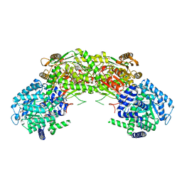
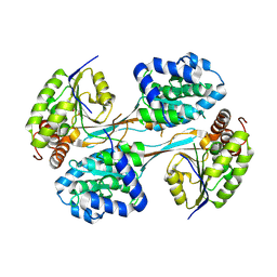
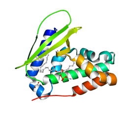
Crystal structure of HTPA Reductase from neisseria meningitidis
Descriptor: 4-hydroxy-tetrahydrodipicolinate reductase
Authors:Keown, J.K, Richards, E.W, Pearce, F.G, Goldstone, D.C.
Deposit date:2017-01-08
Release date:2017-12-13
Last modified:2023-10-04
Method:X-RAY DIFFRACTION (2.7 Å)
Cite:Plant DHDPR forms a dimer with unique secondary structure features that preclude higher-order assembly.
Biochem. J., 475, 2018
7VRR
DownloadVisualize
BU of 7vrr by Molmil
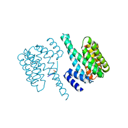
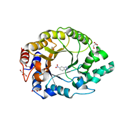
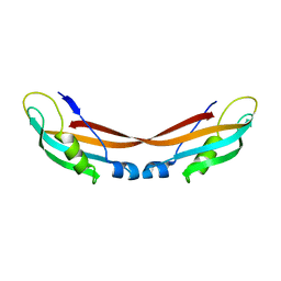
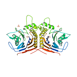
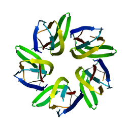
Crystal structure of Arabidopsis thaliana HDT1
Descriptor: Histone deacetylase HDT1
Authors:Kumar, A, Bobde, R.C, Vasudevan, D.
Deposit date:2021-10-23
Release date:2022-09-21
Last modified:2023-11-29
Method:X-RAY DIFFRACTION (2.95 Å)
Cite:Plant-specific HDT family histone deacetylases are nucleoplasmins.
Plant Cell, 34, 2022
7Q24
DownloadVisualize
BU of 7q24 by Molmil
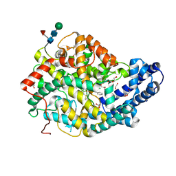
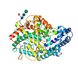
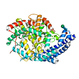
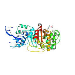
Crystal structure of Angiotensin-1 converting enzyme N-domain in complex with dual ACE/NEP inhibitor AD011
Descriptor: (2~{S})-2-[[(2~{S})-1-[[(2~{S})-3-(1~{H}-indol-3-yl)-1-oxidanyl-1-oxidanylidene-propan-2-yl]amino]-1-oxidanylidene-hexan-2-yl]amino]-4-phenyl-butanoic acid, 1,2-ETHANEDIOL, 2-acetamido-2-deoxy-beta-D-glucopyranose, ...
Authors:Cozier, G.E, Acharya, K.R.
Deposit date:2021-10-23
Release date:2022-02-16
Last modified:2024-01-31
Method:X-RAY DIFFRACTION (2 Å)
Cite:Probing the Requirements for Dual Angiotensin-Converting Enzyme C-Domain Selective/Neprilysin Inhibition.
J.Med.Chem., 65, 2022
5D3F
DownloadVisualize
BU of 5d3f by Molmil
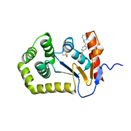
Crystal structure of human 14-3-3 zeta in complex with CFTR R-domain peptide pS753-pS768 and stabilizer fusicoccin-A
Descriptor: 14-3-3 protein zeta/delta, Cystic fibrosis transmembrane conductance regulator, FUSICOCCIN
Authors:Stevers, L.M, Leysen, S.F.R, Ottmann, C.
Deposit date:2015-08-06
Release date:2016-03-16
Last modified:2019-03-13
Method:X-RAY DIFFRACTION (2.74 Å)
Cite:Characterization and small-molecule stabilization of the multisite tandem binding between 14-3-3 and the R domain of CFTR.
Proc.Natl.Acad.Sci.USA, 113, 2016
7NA0
DownloadVisualize
BU of 7na0 by Molmil
Structure of Geobacter sulfurreducens proline utilization A (PutA) variant A206W
Descriptor: 1,2-ETHANEDIOL, Bifunctional protein PutA, FLAVIN-ADENINE DINUCLEOTIDE, ...
Authors:Tanner, J.J, Korasick, D.A.
Deposit date:2021-06-19
Release date:2021-09-22
Last modified:2023-10-18
Method:X-RAY DIFFRACTION (1.9 Å)
Cite:Probing the function of a ligand-modulated dynamic tunnel in bifunctional proline utilization A (PutA).
Arch.Biochem.Biophys., 712, 2021
7Q25
DownloadVisualize
BU of 7q25 by Molmil
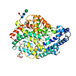
Crystal structure of Angiotensin-1 converting enzyme N-domain in complex with dual ACE/NEP inhibitor AD012
Descriptor: (2~{S})-2-[[(2~{S})-1-[[(2~{S})-3-(4-hydroxyphenyl)-1-oxidanyl-1-oxidanylidene-propan-2-yl]amino]-1-oxidanylidene-hexan-2-yl]amino]-4-phenyl-butanoic acid, 1,2-ETHANEDIOL, 2-acetamido-2-deoxy-beta-D-glucopyranose, ...
Authors:Cozier, G.E, Acharya, K.R.
Deposit date:2021-10-23
Release date:2022-02-16
Last modified:2024-01-31
Method:X-RAY DIFFRACTION (1.6 Å)
Cite:Probing the Requirements for Dual Angiotensin-Converting Enzyme C-Domain Selective/Neprilysin Inhibition.
J.Med.Chem., 65, 2022
8V9N
DownloadVisualize
BU of 8v9n by Molmil
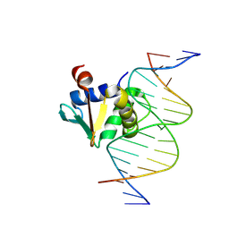
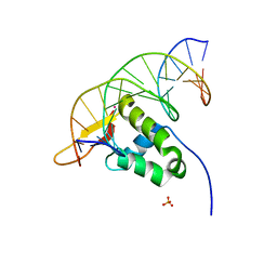
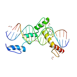
Human PU.1 ETS-Domain (165-270) Bound to d(AATAGAAGGAAGTGGG)
Descriptor: DNA (5'-D(*AP*AP*TP*AP*GP*AP*AP*GP*GP*AP*AP*GP*TP*GP*GP*G)-3'), DNA (5'-D(*TP*CP*CP*CP*AP*CP*TP*TP*CP*CP*TP*TP*CP*TP*AP*T)-3'), Transcription factor PU.1
Authors:Terrell, J.R, Ogbonna, E.N, Poon, G.M.K, Wilson, W.D.
Deposit date:2023-12-08
Release date:2024-10-02
Method:X-RAY DIFFRACTION (1.78 Å)
Cite:Structural Characterization of GC Base Pair Recognition by a Diamidine Small Molecule
To Be Published
5HK5
DownloadVisualize
BU of 5hk5 by Molmil
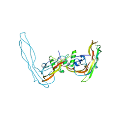
Structure of the Grem2-GDF5 Inhibitory Complex
Descriptor: Gremlin-2, Growth/differentiation factor 5
Authors:Nolan, K, Thompson, T.B, Read, R.J.
Deposit date:2016-01-13
Release date:2016-08-24
Last modified:2023-09-27
Method:X-RAY DIFFRACTION (2.9 Å)
Cite:Structure of Gremlin-2 in Complex with GDF5 Gives Insight into DAN-Family-Mediated BMP Antagonism.
Cell Rep, 16, 2016
8ONX
DownloadVisualize
BU of 8onx by Molmil
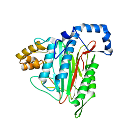
High resolution structure of Chaetomium thermophilum MAP2
Descriptor: MANGANESE (II) ION, Methionine aminopeptidase 2
Authors:Klein, M.A, Wild, K, Kisonaite, M, Sinning, I.
Deposit date:2023-04-04
Release date:2024-02-14
Method:X-RAY DIFFRACTION (1.3 Å)
Cite:Methionine aminopeptidase 2 and its autoproteolysis product have different binding sites on the ribosome.
Nat Commun, 15, 2024
7Q26
DownloadVisualize
BU of 7q26 by Molmil
Crystal structure of Angiotensin-1 converting enzyme N-domain in complex with dual ACE/NEP inhibitor AD013
Descriptor: (2~{S},5~{R})-5-(4-methylphenyl)-1-[2-[[(2~{S})-1-oxidanyl-1-oxidanylidene-4-phenyl-butan-2-yl]amino]ethanoyl]pyrrolidine-2-carboxylic acid, 1,2-ETHANEDIOL, 2-acetamido-2-deoxy-beta-D-glucopyranose, ...
Authors:Cozier, G.E, Acharya, K.R.
Deposit date:2021-10-23
Release date:2022-02-16
Last modified:2024-01-31
Method:X-RAY DIFFRACTION (1.7 Å)
Cite:Probing the Requirements for Dual Angiotensin-Converting Enzyme C-Domain Selective/Neprilysin Inhibition.
J.Med.Chem., 65, 2022
8V35
DownloadVisualize
BU of 8v35 by Molmil
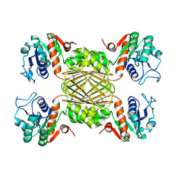
Crystal structure of HpsN from Cupriavidus pinatubonensis
Descriptor: 1,2-ETHANEDIOL, Sulfopropanediol 3-dehydrogenase, ZINC ION
Authors:Lee, M.
Deposit date:2023-11-27
Release date:2024-09-25
Last modified:2024-10-09
Method:X-RAY DIFFRACTION (1.94 Å)
Cite:Structural and kinetic insights into the stereospecific oxidation of R -2,3-dihydroxypropanesulfonate by DHPS-3-dehydrogenase from Cupriavidus pinatubonensis.
Chem Sci, 15, 2024
8RD5
DownloadVisualize
BU of 8rd5 by Molmil
Crystal structure of Kemp Eliminase HG3.R5 with bound transition state analog 6-nitrobenzotriazole
Descriptor: 6-NITROBENZOTRIAZOLE, ACETATE ION, Endo-1,4-beta-xylanase, ...
Authors:Schaub, D, Schwander, T, Hueppi, S, Buller, R.M.
Deposit date:2023-12-07
Release date:2024-09-04
Last modified:2024-10-23
Method:X-RAY DIFFRACTION (1.5 Å)
Cite:Enriching productive mutational paths accelerates enzyme evolution.
Nat.Chem.Biol., 2024
7Q27
DownloadVisualize
BU of 7q27 by Molmil
Crystal structure of Angiotensin-1 converting enzyme C-domain in complex with dual ACE/NEP inhibitor AD011
Descriptor: (2~{S})-2-[[(2~{S})-1-[[(2~{S})-3-(1~{H}-indol-3-yl)-1-oxidanyl-1-oxidanylidene-propan-2-yl]amino]-1-oxidanylidene-hexan-2-yl]amino]-4-phenyl-butanoic acid, 1,2-ETHANEDIOL, 2-acetamido-2-deoxy-beta-D-glucopyranose, ...
Authors:Cozier, G.E, Acharya, K.R.
Deposit date:2021-10-23
Release date:2022-02-16
Last modified:2024-01-31
Method:X-RAY DIFFRACTION (1.5 Å)
Cite:Probing the Requirements for Dual Angiotensin-Converting Enzyme C-Domain Selective/Neprilysin Inhibition.
J.Med.Chem., 65, 2022
8U59
DownloadVisualize
BU of 8u59 by Molmil
EcDsbA soaked with N-(2-fluorophenyl)-5-methylisoxazole-3-carboxamide and N-(4-(thiophen-3-yl)benzyl)cyclohexanamine
Descriptor: COPPER (II) ION, N-(2-fluorophenyl)-5-methyl-1,2-oxazole-3-carboxamide, Thiol:disulfide interchange protein DsbA, ...
Authors:Wang, G, Heras, B.
Deposit date:2023-09-12
Release date:2024-09-25
Method:X-RAY DIFFRACTION (1.77 Å)
Cite:Two-mode inhibition of DsbA: combination of two site-specific inhibitors enhances virulence inhibition in Salmonella enterica serovar Typhimurium
To Be Published
7ZC8
DownloadVisualize
BU of 7zc8 by Molmil
Crystal structure of the C-terminal domain of FusB, a TonB homologue
Descriptor: Protein TonB
Authors:Wojnowska, M, Walker, D, Yelland, T.
Deposit date:2022-03-25
Release date:2022-08-17
Last modified:2024-01-31
Method:X-RAY DIFFRACTION (2.08 Å)
Cite:Crystal structure of the C-terminal domain of FusB, a TonB homologue
To Be Published
8U9H
DownloadVisualize
BU of 8u9h by Molmil
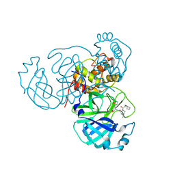
Structure of the SARS-CoV-2 main protease in complex with inhibitor MPI64
Descriptor: (1R,2S,5R)-3-[(cyclohexyloxy)acetyl]-N-{(2R)-1-hydroxy-3-[(3S)-2-oxopyrrolidin-3-yl]propan-2-yl}-6,6-dimethyl-3-azabicyclo[3.1.0]hexane-2-carboxamide, 3C-like proteinase nsp5
Authors:Blankenship, L.R, Liu, W.R.
Deposit date:2023-09-19
Release date:2024-09-25
Last modified:2024-10-23
Method:X-RAY DIFFRACTION (1.7 Å)
Cite:Structure of the SARS-CoV-2 main protease in complex with inhibitors
To Be Published
8OGE
DownloadVisualize
BU of 8oge by Molmil
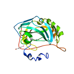
Structure of cobalt(II) substituted double mutant human carbonic anhydrase II bound to thiocyanate
Descriptor: 4-(HYDROXYMERCURY)BENZOIC ACID, COBALT (II) ION, Carbonic anhydrase 2, ...
Authors:Silva, J.M, Cerofolini, L, Carvalho, A.L, Ravera, E, Fragai, M, Parigi, G, Macedo, A.L, Geraldes, C.F.G.C, Luchinat, C.
Deposit date:2023-03-20
Release date:2024-02-07
Method:X-RAY DIFFRACTION (1.46 Å)
Cite:Elucidating the concentration-dependent effects of thiocyanate binding to carbonic anhydrase.
J.Inorg.Biochem., 244, 2023
8U9U
DownloadVisualize
BU of 8u9u by Molmil
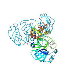
Structure of the SARS-CoV-2 main protease in complex with inhibitor MPI98
Descriptor: (1R,2S,5S)-3-[bis(4-chlorophenoxy)acetyl]-N-{(2S)-1-hydroxy-3-[(3R)-2-oxo-3,4-dihydro-2H-pyrrol-3-yl]propan-2-yl}-6,6-dimethyl-3-azabicyclo[3.1.0]hexane-2-carboxamide, 3C-like proteinase nsp5
Authors:Blankenship, L.R, Liu, W.R.
Deposit date:2023-09-20
Release date:2024-09-25
Method:X-RAY DIFFRACTION (1.85 Å)
Cite:Structure of the SARS-CoV-2 main protease in complex with inhibitors
To Be Published
7YZE
DownloadVisualize
BU of 7yze by Molmil
Crystal structure of the human FoxA2 bound to the TGTTTACT site (forkhead motif GTAAACA)
Descriptor: DNA (5'-D(*AP*GP*AP*TP*TP*GP*TP*TP*TP*AP*CP*TP*GP*AP*GP*A)-3'), DNA (5'-D(*TP*CP*TP*CP*AP*GP*TP*AP*AP*AP*CP*AP*AP*TP*CP*T)-3'), Hepatocyte nuclear factor 3-beta, ...
Authors:Pluta, R, Macias, M.J.
Deposit date:2022-02-19
Release date:2022-11-16
Last modified:2024-01-31
Method:X-RAY DIFFRACTION (1.99 Å)
Cite:Molecular basis for DNA recognition by the maternal pioneer transcription factor FoxH1.
Nat Commun, 13, 2022
5HKE
DownloadVisualize
BU of 5hke by Molmil
bile salt hydrolase from Lactobacillus salivarius
Descriptor: Bile salt hydrolase, PHOSPHATE ION
Authors:Hu, X.-J.
Deposit date:2016-01-14
Release date:2016-05-11
Last modified:2023-11-15
Method:X-RAY DIFFRACTION (1.9 Å)
Cite:Crystal structure of bile salt hydrolase from Lactobacillus salivarius.
Acta Crystallogr F Struct Biol Commun, 72, 2016
6MKI
DownloadVisualize
BU of 6mki by Molmil
Crystal structure of penicillin-binding protein 4 (PBP4) from Enterococcus faecalis in the ceftaroline-bound form
Descriptor: Ceftaroline, bound form, GLYCEROL, ...
Authors:D'Andrea, E.D, Moon, T.M, Peti, W, Page, R.
Deposit date:2018-09-25
Release date:2018-10-31
Last modified:2024-10-30
Method:X-RAY DIFFRACTION (2.984 Å)
Cite:The structures of penicillin-binding protein 4 (PBP4) and PBP5 fromEnterococciprovide structural insights into beta-lactam resistance.
J. Biol. Chem., 293, 2018
5HKK
DownloadVisualize
BU of 5hkk by Molmil
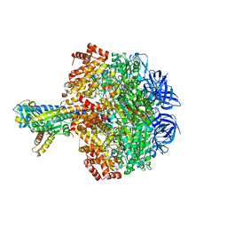
Caldalaklibacillus thermarum F1-ATPase (wild type)
Descriptor: ADENOSINE-5'-DIPHOSPHATE, ADENOSINE-5'-TRIPHOSPHATE, ATP synthase epsilon chain, ...
Authors:Ferguson, S.A, Cook, G.M, Montgomery, M.G, Leslie, A.G.W, Walker, J.E.
Deposit date:2016-01-14
Release date:2016-09-21
Last modified:2024-01-10
Method:X-RAY DIFFRACTION (3 Å)
Cite:Regulation of the thermoalkaliphilic F1-ATPase from Caldalkalibacillus thermarum.
Proc.Natl.Acad.Sci.USA, 113, 2016
7VR6
DownloadVisualize
BU of 7vr6 by Molmil
Crystal structure of MlaC from Escherichia coli in quasi-open state
Descriptor: 1,2-ETHANEDIOL, DI-PALMITOYL-3-SN-PHOSPHATIDYLETHANOLAMINE, Intermembrane phospholipid transport system binding protein MlaC
Authors:Dutta, A, Kanaujia, S.P.
Deposit date:2021-10-21
Release date:2022-09-21
Last modified:2023-11-29
Method:X-RAY DIFFRACTION (2.5 Å)
Cite:MlaC belongs to a unique class of non-canonical substrate-binding proteins and follows a novel phospholipid-binding mechanism.
J.Struct.Biol., 214, 2022
7Z5W
DownloadVisualize
BU of 7z5w by Molmil
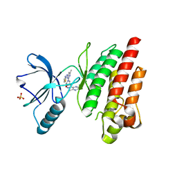
ROS1 with AstraZeneca ligand 1
Descriptor: Proto-oncogene tyrosine-protein kinase ROS, SULFATE ION, ~{N}-[6-methyl-2-[(2~{S})-2-[3-(3-methylpyrazin-2-yl)-1,2-oxazol-5-yl]pyrrolidin-1-yl]pyrimidin-4-yl]-1,3-thiazol-2-amine
Authors:Hargreaves, D.
Deposit date:2022-03-10
Release date:2022-08-17
Last modified:2024-01-31
Method:X-RAY DIFFRACTION (2.254 Å)
Cite:Virtual Screening in the Cloud Identifies Potent and Selective ROS1 Kinase Inhibitors.
J.Chem.Inf.Model., 62, 2022
6ML7
DownloadVisualize
BU of 6ml7 by Molmil
ZBTB24 Zinc Fingers 4-8 with 19+1mer DNA Oligonucleotide (Sequence 4 with a CpG 5mC Modification)
Descriptor: 1,2-ETHANEDIOL, DI(HYDROXYETHYL)ETHER, DNA (5'-D(*AP*CP*GP*CP*AP*GP*GP*TP*CP*CP*TP*GP*GP*AP*(5CM)P*GP*AP*AP*TP*T)-3'), ...
Authors:Horton, J.R, Cheng, X, Ren, R.
Deposit date:2018-09-26
Release date:2019-07-03
Last modified:2023-10-11
Method:X-RAY DIFFRACTION (1.75 Å)
Cite:Structural basis of specific DNA binding by the transcription factor ZBTB24.
Nucleic Acids Res., 47, 2019

226707

PDB entries from 2024-10-30

PDB statisticsPDBj update infoContact PDBjnumon