Loading
PDBj
MenuPDBj@FacebookPDBj@TwitterPDBj@YouTubewwPDB FoundationwwPDB
RCSB PDBPDBeBMRBAdv. SearchSearch help

3CLD
DownloadVisualize
BU of 3cld by Molmil
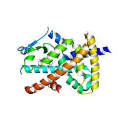
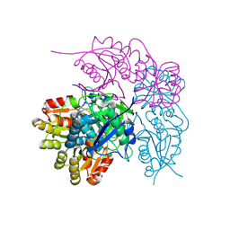
Ligand binding domain of the glucocorticoid receptor complexed with fluticazone furoate
Descriptor: (6alpha,11alpha,14beta,16alpha,17alpha)-6,9-difluoro-17-{[(fluoromethyl)sulfanyl]carbonyl}-11-hydroxy-16-methyl-3-oxoan drosta-1,4-dien-17-yl furan-2-carboxylate, Glucocorticoid receptor, Tif2 coactivator motif
Authors:Shewchuk, L.M, McLay, I, Stewart, E, Biggadike, K.B, Hassell, A.M, Bledsoe, R.K.
Deposit date:2008-03-18
Release date:2008-06-24
Last modified:2023-08-30
Method:X-RAY DIFFRACTION (2.84 Å)
Cite:X-ray crystal structure of the novel enhanced-affinity glucocorticoid agonist fluticasone furoate in the glucocorticoid receptor-ligand binding domain.
J.Med.Chem., 51, 2008
5F8U
DownloadVisualize
BU of 5f8u by Molmil
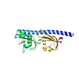
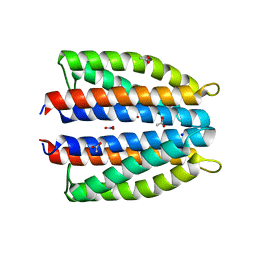
Ligand occupancy in crystal structure of beta1-adrenergic receptor previously submitted by Huang et al
Descriptor: 4-{[(2S)-3-(tert-butylamino)-2-hydroxypropyl]oxy}-3H-indole-2-carbonitrile, Beta-1 adrenergic receptor
Authors:Leslie, A.G.W, Warne, A, Tate, C.G.
Deposit date:2015-12-09
Release date:2015-12-23
Last modified:2024-01-10
Method:X-RAY DIFFRACTION (3.35 Å)
Cite:Ligand occupancy in crystal structure of Beta1 adrenergic G protein coupled receptor
Nat.Struct.Mol.Biol., 22, 2015
3U65
DownloadVisualize
BU of 3u65 by Molmil
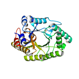
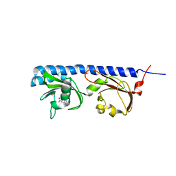
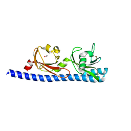
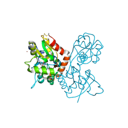
The Crystal Structure of Tat-P(T) (Tp0957)
Descriptor: 1,2-ETHANEDIOL, THIOCYANATE ION, Tp33 protein
Authors:Brautigam, C.A, Tomchick, D.R, Deka, R.K, Norgard, M.V.
Deposit date:2011-10-12
Release date:2012-02-22
Last modified:2024-02-28
Method:X-RAY DIFFRACTION (1.4 Å)
Cite:Structural, Bioinformatic, and In Vivo Analyses of Two Treponema pallidum Lipoproteins Reveal a Unique TRAP Transporter.
J.Mol.Biol., 416, 2012
6BEH
DownloadVisualize
BU of 6beh by Molmil
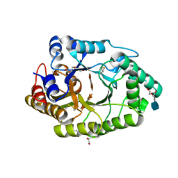
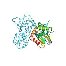
Crystal structure of VACV D13 in complex with Rifapentine
Descriptor: 1,2-ETHANEDIOL, FORMIC ACID, RIFAPENTINE, ...
Authors:Garriga, D, Accurso, C, Coulibaly, F.
Deposit date:2017-10-25
Release date:2018-07-18
Last modified:2023-10-04
Method:X-RAY DIFFRACTION (3 Å)
Cite:Structural basis for the inhibition of poxvirus assembly by the antibiotic rifampicin.
Proc. Natl. Acad. Sci. U.S.A., 115, 2018
3DRC
DownloadVisualize
BU of 3drc by Molmil
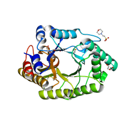
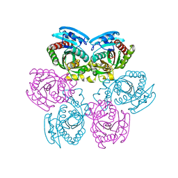
INVESTIGATION OF THE FUNCTIONAL ROLE OF TRYPTOPHAN-22 IN ESCHERICHIA COLI DIHYDROFOLATE REDUCTASE BY SITE-DIRECTED MUTAGENESIS
Descriptor: CALCIUM ION, CHLORIDE ION, DIHYDROFOLATE REDUCTASE, ...
Authors:Oatley, S.J, Kraut, J.
Deposit date:1992-09-22
Release date:1994-01-31
Last modified:2024-02-21
Method:X-RAY DIFFRACTION (1.9 Å)
Cite:Investigation of the functional role of tryptophan-22 in Escherichia coli dihydrofolate reductase by site-directed mutagenesis.
Biochemistry, 30, 1991
4GXS
DownloadVisualize
BU of 4gxs by Molmil
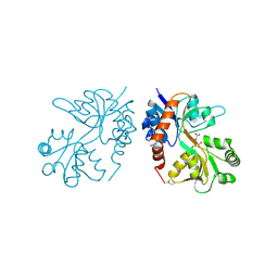
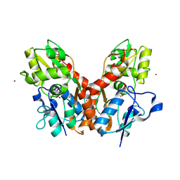
Ligand binding domain of GluA2 (AMPA/glutamate receptor) bound to (-)-kaitocephalin
Descriptor: (5R)-2-[(1S,2R)-2-amino-2-carboxy-1-hydroxyethyl]-5-{(2S)-2-carboxy-2-[(3,5-dichloro-4-hydroxybenzoyl)amino]ethyl}-L-proline, Glutamate receptor 2, ZINC ION
Authors:Ahmed, A.H, Oswald, R.E.
Deposit date:2012-09-04
Release date:2012-10-17
Last modified:2024-11-06
Method:X-RAY DIFFRACTION (1.9634 Å)
Cite:The structure of (-)-kaitocephalin bound to the ligand binding domain of the (S)-alpha-amino-3-hydroxy-5-methyl-4-isoxazolepropionic acid (AMPA)/glutamate receptor, GluA2.
J.Biol.Chem., 287, 2012
1JDL
DownloadVisualize
BU of 1jdl by Molmil
Structure of cytochrome c2 from Rhodospirillum Centenum
Descriptor: CYTOCHROME C2, ISO-2, PROTOPORPHYRIN IX CONTAINING FE
Authors:Camara-Artigas, A, Williams, J.C, Allen, J.P.
Deposit date:2001-06-14
Release date:2001-11-07
Last modified:2023-08-16
Method:X-RAY DIFFRACTION (1.7 Å)
Cite:Structure of cytochrome c2 from Rhodospirillum centenum.
Acta Crystallogr.,Sect.D, 57, 2001
1IUE
DownloadVisualize
BU of 1iue by Molmil
Crystal Structure Analysis of ferredoxin from Plasmodium falciparum
Descriptor: FE2/S2 (INORGANIC) CLUSTER, FERREDOXIN, SODIUM ION
Authors:Kimata-Ariga, Y, Kurisu, G, Hase, T.
Deposit date:2002-03-04
Release date:2003-09-30
Last modified:2023-12-27
Method:X-RAY DIFFRACTION (1.7 Å)
Cite:Redox power generator of a nonphotosynthetic plastid in malaria parasite
To be Published
1JH0
DownloadVisualize
BU of 1jh0 by Molmil
Photosynthetic Reaction Center Mutant With Glu L 205 Replaced to Leu
Descriptor: BACTERIOCHLOROPHYLL A, BACTERIOPHEOPHYTIN A, FE (III) ION, ...
Authors:Camara-Artigas, A, Magee, C.L, Williams, J.C, Allen, J.P.
Deposit date:2001-06-27
Release date:2001-09-05
Last modified:2023-08-16
Method:X-RAY DIFFRACTION (3.5 Å)
Cite:Individual interactions influence the crystalline order for membrane proteins.
Acta Crystallogr.,Sect.D, 57, 2001
1U3C
DownloadVisualize
BU of 1u3c by Molmil
Crystal Structure of the PHR domain of Cryptochrome 1 from Arabidopsis thaliana
Descriptor: CHLORIDE ION, Cryptochrome 1 apoprotein, ETHYL DIMETHYL AMMONIO PROPANE SULFONATE, ...
Authors:Brautigam, C.A, Smith, B.S, Ma, Z, Palnitkar, M, Tomchick, D.R, Machius, M, Deisenhofer, J.
Deposit date:2004-07-21
Release date:2004-08-24
Last modified:2023-08-23
Method:X-RAY DIFFRACTION (2.6 Å)
Cite:Structure of the photolyase-like domain of cryptochrome 1 from Arabidopsis thaliana.
Proc.Natl.Acad.Sci.USA, 101, 2004
2P81
DownloadVisualize
BU of 2p81 by Molmil
Engrailed homeodomain helix-turn-helix motif
Descriptor: Segmentation polarity homeobox protein engrailed
Authors:Religa, T.L.
Deposit date:2007-03-21
Release date:2007-06-12
Last modified:2024-05-22
Method:SOLUTION NMR
Cite:The helix-turn-helix motif as an ultrafast independently folding domain: The pathway of folding of Engrailed homeodomain.
Proc.Natl.Acad.Sci.Usa, 104, 2007
1U3D
DownloadVisualize
BU of 1u3d by Molmil
Crystal Structure of the PHR domain of Cryptochrome 1 from Arabidopsis thaliana with AMPPNP bound
Descriptor: CHLORIDE ION, Cryptochrome 1 apoprotein, ETHYL DIMETHYL AMMONIO PROPANE SULFONATE, ...
Authors:Brautigam, C.A, Smith, B.S, Ma, Z, Palnitkar, M, Tomchick, D.R, Machius, M, Deisenhofer, J.
Deposit date:2004-07-21
Release date:2004-08-24
Last modified:2023-08-23
Method:X-RAY DIFFRACTION (2.45 Å)
Cite:Structure of the photolyase-like domain of cryptochrome 1 from Arabidopsis thaliana.
Proc.Natl.Acad.Sci.USA, 101, 2004
3B3N
DownloadVisualize
BU of 3b3n by Molmil
Structure of neuronal NOS heme domain in complex with a inhibitor (+-)-N1-{cis-4'-[(6"-aminopyridin-2"-yl)methyl]pyrrolidin-3'-yl}ethane-1,2-diamine
Descriptor: 5,6,7,8-TETRAHYDROBIOPTERIN, ACETATE ION, N-{(3S,4S)-4-[(6-aminopyridin-2-yl)methyl]pyrrolidin-3-yl}ethane-1,2-diamine, ...
Authors:Igarashi, J, Li, H, Poulos, T.L.
Deposit date:2007-10-22
Release date:2008-07-15
Last modified:2024-02-21
Method:X-RAY DIFFRACTION (1.98 Å)
Cite:Minimal pharmacophoric elements and fragment hopping, an approach directed at molecular diversity and isozyme selectivity. Design of selective neuronal nitric oxide synthase inhibitors.
J.Am.Chem.Soc., 130, 2008
3B3M
DownloadVisualize
BU of 3b3m by Molmil
Structure of neuronal NOS heme domain in complex with a inhibitor (+-)-3-{cis-4'-[(6"-aminopyridin-2"-yl)methyl]pyrrolidin-3'-ylamino}propan-1-ol
Descriptor: 3-({(3S,4S)-4-[(6-aminopyridin-2-yl)methyl]pyrrolidin-3-yl}amino)propan-1-ol, 5,6,7,8-TETRAHYDROBIOPTERIN, ACETATE ION, ...
Authors:Igarashi, J, Li, H, Poulos, T.L.
Deposit date:2007-10-22
Release date:2008-07-15
Last modified:2024-02-21
Method:X-RAY DIFFRACTION (1.95 Å)
Cite:Minimal pharmacophoric elements and fragment hopping, an approach directed at molecular diversity and isozyme selectivity. Design of selective neuronal nitric oxide synthase inhibitors.
J.Am.Chem.Soc., 130, 2008
1CQF
DownloadVisualize
BU of 1cqf by Molmil
THE COMPLEX OF THE MUTATED SHIGA TOXIN B SUBUNIT AND GB3 TRISACCHARIDE
Descriptor: SHIGA TOXIN B-CHAIN, alpha-D-galactopyranose-(1-4)-beta-D-galactopyranose-(1-4)-beta-D-glucopyranose
Authors:Ling, H, Bast, D, Brunton, J.L, Read, R.J.
Deposit date:1999-08-06
Release date:2000-08-07
Last modified:2021-11-03
Method:X-RAY DIFFRACTION (2.2 Å)
Cite:The Complex of the Mutated Shiga Toxin B Subunit and Gb3 Trisaccharide
To be Published
1KFS
DownloadVisualize
BU of 1kfs by Molmil
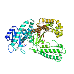
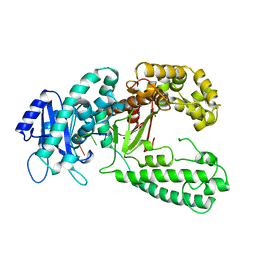
DNA POLYMERASE I KLENOW FRAGMENT (E.C.2.7.7.7) MUTANT/DNA COMPLEX
Descriptor: DNA (5'-D(*GP*CP*TP*TP*AP*CP*G)-3'), MAGNESIUM ION, PROTEIN (DNA POLYMERASE I KLENOW FRAGMENT (E.C.2.7.7.7)), ...
Authors:Brautigam, C.A, Steitz, T.A.
Deposit date:1997-08-18
Release date:1998-02-25
Last modified:2024-04-03
Method:X-RAY DIFFRACTION (2.1 Å)
Cite:Structural principles for the inhibition of the 3'-5' exonuclease activity of Escherichia coli DNA polymerase I by phosphorothioates.
J.Mol.Biol., 277, 1998
2DVJ
DownloadVisualize
BU of 2dvj by Molmil
phosphorylated Crk-II
Descriptor: V-crk sarcoma virus CT10 oncogene homolog, isoform a
Authors:Kobashigawa, Y, Inagaki, F.
Deposit date:2006-07-31
Release date:2007-05-08
Last modified:2022-03-09
Method:SOLUTION NMR
Cite:Structural basis for the transforming activity of human cancer-related signaling adaptor protein CRK.
Nat.Struct.Mol.Biol., 14, 2007
5CW0
DownloadVisualize
BU of 5cw0 by Molmil
Investigation of RNA structure in satellite panicum mosaic virus
Descriptor: Coat protein
Authors:Makino, D.L, Day, J, Larson, S.B, McPherson, A.
Deposit date:2015-07-27
Release date:2017-10-11
Last modified:2023-09-27
Method:X-RAY DIFFRACTION (4.6 Å)
Cite:Investigation of RNA structure in satellite panicum mosaic virus.
Virology, 351, 2006
7OL7
DownloadVisualize
BU of 7ol7 by Molmil
Crystal structure of Lysozyme in complex with trifluoroethanol: tetragonal form
Descriptor: CHLORIDE ION, Lysozyme, SODIUM ION, ...
Authors:Camara-Artigas, A, Salinas-Garcia, M.C, Plaza-Garrido, M.
Deposit date:2021-05-19
Release date:2022-06-01
Last modified:2024-01-31
Method:X-RAY DIFFRACTION (1.15 Å)
Cite:Crystal structure of Lysozyme in complex with TFE
To be published
7OL8
DownloadVisualize
BU of 7ol8 by Molmil
Crystal structure of Lysozyme in complex with trifluoroethanol: orthorhombic form
Descriptor: CHLORIDE ION, Lysozyme, SULFATE ION, ...
Authors:Camara-Artigas, A, Salinas-Garcia, M.C, Plaza-Garrido, M.
Deposit date:2021-05-19
Release date:2022-06-01
Last modified:2024-01-31
Method:X-RAY DIFFRACTION (1.15 Å)
Cite:Crystal structure of Lysozyme in complex with TFE
To be published
7OL6
DownloadVisualize
BU of 7ol6 by Molmil
Crystal structure of Lysozyme in complex with Imidazole
Descriptor: CHLORIDE ION, IMIDAZOLE, Lysozyme, ...
Authors:Camara-Artigas, A, Salinas-Garcia, M.C, Plaza-Garrido, M.
Deposit date:2021-05-19
Release date:2022-06-01
Last modified:2024-10-23
Method:X-RAY DIFFRACTION (1.1 Å)
Cite:Crystal structure of Lysozyme in complex with Imidazole
To be published
7OL5
DownloadVisualize
BU of 7ol5 by Molmil
Crystal structure of Lysozyme in complex with Hepes
Descriptor: 4-(2-HYDROXYETHYL)-1-PIPERAZINE ETHANESULFONIC ACID, ISOPROPYL ALCOHOL, Lysozyme
Authors:Camara-Artigas, A, Salinas-Garcia, M.C, Plaza-Garrido, M.
Deposit date:2021-05-19
Release date:2022-06-01
Last modified:2024-11-06
Method:X-RAY DIFFRACTION (0.94 Å)
Cite:Crystal structure of Lysozyme in complex with Hepes
To be published
7UJJ
DownloadVisualize
BU of 7ujj by Molmil
Stx2a and DARPin complex
Descriptor: 1,2-ETHANEDIOL, 3-PYRIDINIUM-1-YLPROPANE-1-SULFONATE, DARPin, ...
Authors:Jiang, M, Zhang, J.
Deposit date:2022-03-30
Release date:2023-04-12
Method:ELECTRON MICROSCOPY (6.5 Å)
Cite:A Multi-Specific DARPin Potently Neutralizes Shiga Toxin 2 via Simultaneous Modulation of Both Toxin Subunits.
Bioengineering (Basel), 9, 2022
3U9Q
DownloadVisualize
BU of 3u9q by Molmil
Ligand binding domain of PPARgamma complexed with Decanoic Acid and PGC-1a peptide
Descriptor: DECANOIC ACID, PGC-1a peptide, Peroxisome proliferator-activated receptor gamma
Authors:Malapaka, V.R, Xu, H.E.
Deposit date:2011-10-19
Release date:2011-11-09
Last modified:2024-02-28
Method:X-RAY DIFFRACTION (1.522 Å)
Cite:Identification and Mechanism of 10-Carbon Fatty Acid as Modulating Ligand of Peroxisome Proliferator-activated Receptors.
J.Biol.Chem., 287, 2012
5UN2
DownloadVisualize
BU of 5un2 by Molmil
Crystal Structure of Mouse Cadherin-23 EC19-21 with non-syndromic deafness (DFNB12) associated mutation R2029W
Descriptor: CALCIUM ION, Cadherin-23, POTASSIUM ION
Authors:Jaiganesh, A, Sotomayor, M.
Deposit date:2017-01-30
Release date:2018-02-07
Last modified:2023-10-04
Method:X-RAY DIFFRACTION (2.96 Å)
Cite:Zooming in on Cadherin-23: Structural Diversity and Potential Mechanisms of Inherited Deafness.
Structure, 26, 2018

227111

PDB entries from 2024-11-06

PDB statisticsPDBj update infoContact PDBjnumon