Loading
PDBj
MenuPDBj@FacebookPDBj@TwitterPDBj@YouTubewwPDB FoundationwwPDB
RCSB PDBPDBeBMRBAdv. SearchSearch help

3TX2
DownloadVisualize
BU of 3tx2 by Molmil
Structure of a Probable 6-phosphogluconolactonase from Mycobacterium abscessus
Descriptor: BENZOIC ACID, GLYCEROL, Probable 6-phosphogluconolactonase, ...
Authors:Seattle Structural Genomics Center for Infectious Disease (SSGCID)
Deposit date:2011-09-22
Release date:2011-10-05
Last modified:2023-09-13
Method:X-RAY DIFFRACTION (1.5 Å)
Cite:Increasing the structural coverage of tuberculosis drug targets.
Tuberculosis (Edinb), 95, 2015
3TRR
DownloadVisualize
BU of 3trr by Molmil
Crystal structure of a probable enoyl-CoA hydratase/isomerase from Mycobacterium abscessus
Descriptor: 1,2-ETHANEDIOL, DI(HYDROXYETHYL)ETHER, GLYCEROL, ...
Authors:Seattle Structural Genomics Center for Infectious Disease (SSGCID)
Deposit date:2011-09-09
Release date:2011-09-21
Last modified:2023-09-13
Method:X-RAY DIFFRACTION (2.09 Å)
Cite:Increasing the structural coverage of tuberculosis drug targets.
Tuberculosis (Edinb), 95, 2015
3QB0
DownloadVisualize
BU of 3qb0 by Molmil
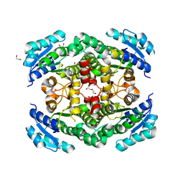
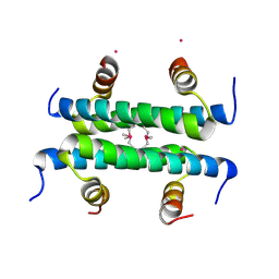
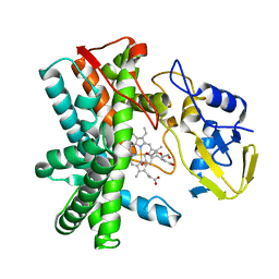
Crystal structure of Actin-related protein Arp4 from S. cerevisiae complexed with ATP
Descriptor: ADENOSINE-5'-TRIPHOSPHATE, Actin-related protein 4, CALCIUM ION
Authors:Fenn, S, Breitsprecher, D, Gerhold, C.B, Witte, G, Faix, J, Hopfner, K.P.
Deposit date:2011-01-12
Release date:2011-05-04
Last modified:2023-09-13
Method:X-RAY DIFFRACTION (3.404 Å)
Cite:Structural biochemistry of nuclear actin-related proteins 4 and 8 reveals their interaction with actin.
Embo J., 30, 2011
3QK8
DownloadVisualize
BU of 3qk8 by Molmil
Crystal structure of enoyl-coA hydratase EchA15 from Mycobacterium marinum in complex with an unknown ligand
Descriptor: 1,2-ETHANEDIOL, CHLORIDE ION, Enoyl-CoA hydratase EchA15, ...
Authors:Seattle Structural Genomics Center for Infectious Disease (SSGCID)
Deposit date:2011-01-31
Release date:2011-02-09
Last modified:2023-09-13
Method:X-RAY DIFFRACTION (1.6 Å)
Cite:Increasing the structural coverage of tuberculosis drug targets.
Tuberculosis (Edinb), 95, 2015
3QIV
DownloadVisualize
BU of 3qiv by Molmil
Crystal structure of a putative short-chain dehydrogenase or 3-oxoacyl-[acyl-carrier-protein] reductase from Mycobacterium paratuberculosis ATCC BAA-968 / K-10
Descriptor: short-chain dehydrogenase or 3-oxoacyl-[acyl-carrier-protein] reductase
Authors:Seattle Structural Genomics Center for Infectious Disease (SSGCID)
Deposit date:2011-01-27
Release date:2011-02-16
Last modified:2023-09-13
Method:X-RAY DIFFRACTION (2.25 Å)
Cite:Increasing the structural coverage of tuberculosis drug targets.
Tuberculosis (Edinb), 95, 2015
3Q8N
DownloadVisualize
BU of 3q8n by Molmil
Crystal structure of 4-aminobutyrate transaminase from Mycobacterium smegmatis
Descriptor: 4-aminobutyrate transaminase, 4-oxobutanoic acid
Authors:Seattle Structural Genomics Center for Infectious Disease (SSGCID)
Deposit date:2011-01-06
Release date:2011-02-09
Last modified:2023-09-13
Method:X-RAY DIFFRACTION (2.05 Å)
Cite:Increasing the structural coverage of tuberculosis drug targets.
Tuberculosis (Edinb), 95, 2015
3QK2
DownloadVisualize
BU of 3qk2 by Molmil
Structure-Based Analysis of the Interaction between the Simian Virus 40 T-Antigen Origin Binding Domain and Single-Stranded DNA
Descriptor: Large T antigen, THIOCYANATE ION
Authors:Meinke, G, Bullock, P.A, Bohm, A.
Deposit date:2011-01-31
Release date:2011-02-23
Last modified:2023-09-13
Method:X-RAY DIFFRACTION (1.643 Å)
Cite:Structure-based analysis of the interaction between the simian virus 40 T-antigen origin binding domain and single-stranded DNA.
J.Virol., 85, 2011
3QKA
DownloadVisualize
BU of 3qka by Molmil
Crystal structure of enoyl-CoA hydratase EchA5 from Mycobacterium marinum
Descriptor: Enoyl-CoA hydratase, EchA5
Authors:Seattle Structural Genomics Center for Infectious Disease (SSGCID)
Deposit date:2011-01-31
Release date:2011-02-09
Last modified:2023-09-13
Method:X-RAY DIFFRACTION (2.15 Å)
Cite:Increasing the structural coverage of tuberculosis drug targets.
Tuberculosis (Edinb), 95, 2015
3TZQ
DownloadVisualize
BU of 3tzq by Molmil
Crystal structure of a short-chain type dehydrogenase/reductase from Mycobacterium marinum
Descriptor: 1,2-ETHANEDIOL, DI(HYDROXYETHYL)ETHER, GLYCEROL, ...
Authors:Seattle Structural Genomics Center for Infectious Disease (SSGCID)
Deposit date:2011-09-27
Release date:2011-10-05
Last modified:2023-09-13
Method:X-RAY DIFFRACTION (2.5 Å)
Cite:Increasing the structural coverage of tuberculosis drug targets.
Tuberculosis (Edinb), 95, 2015
6QFU
DownloadVisualize
BU of 6qfu by Molmil
Human carbonic anhydrase II with bound IrCp* complex (cofactor 7) to generate an artificial transfer hydrogenase (ATHase)
Descriptor: 4-[2-(9-chloranyl-2',3',4',5',6'-pentamethyl-4-oxidanyl-7-oxidanylidene-spiro[1$l^{4},8-diaza-9$l^{8}-iridabicyclo[4.3.0]nona-1(6),2,4-triene-9,1'-1$l^{8}-iridapentacyclo[2.2.0.0^{1,3}.0^{1,5}.0^{2,6}]hexane]-8-yl)ethyl]benzenesulfonamide, Carbonic anhydrase 2, SULFATE ION, ...
Authors:Rebelein, J.G.
Deposit date:2019-01-10
Release date:2019-04-17
Last modified:2024-01-24
Method:X-RAY DIFFRACTION (1.8 Å)
Cite:Chemical Optimization of Whole-Cell Transfer Hydrogenation Using Carbonic Anhydrase as Host Protein.
Acs Catalysis, 9, 2019
6QFX
DownloadVisualize
BU of 6qfx by Molmil
Human carbonic anhydrase II with bound IrCp* complex (cofactor 10) to generate an artificial transfer hydrogenase (ATHase)
Descriptor: 2-(9-chloranyl-2',3',4',5',6'-pentamethyl-4-oxidanyl-7-oxidanylidene-spiro[1$l^{4},8-diaza-9$l^{8}-iridabicyclo[4.3.0]nona-1,3,5-triene-9,1'-1$l^{8}-iridapentacyclo[2.2.0.0^{1,3}.0^{1,5}.0^{2,6}]hexane]-8-yl)-~{N}-(4-sulfamoylphenyl)ethanamide, Carbonic anhydrase 2, SULFATE ION, ...
Authors:Rebelein, J.G.
Deposit date:2019-01-10
Release date:2019-04-17
Last modified:2024-05-15
Method:X-RAY DIFFRACTION (1.32 Å)
Cite:Chemical Optimization of Whole-Cell Transfer Hydrogenation Using Carbonic Anhydrase as Host Protein.
Acs Catalysis, 9, 2019
6QS5
DownloadVisualize
BU of 6qs5 by Molmil
Crystal Structure of maize CK2 in complex with tyrphostin AG99
Descriptor: (~{E})-3-[3,4-bis(oxidanyl)phenyl]-2-cyano-prop-2-enamide, Casein kinase II subunit alpha
Authors:Lolli, G, Mazzorana, M, Battistutta, R.
Deposit date:2019-02-20
Release date:2019-02-27
Last modified:2024-01-24
Method:X-RAY DIFFRACTION (1.961 Å)
Cite:Inhibition of protein kinase CK2 by flavonoids and tyrphostins. A structural insight.
Biochemistry, 51, 2012
6QZO
DownloadVisualize
BU of 6qzo by Molmil
Crystal structure of DyP-type peroxidase from Cellulomonas bogoriensis
Descriptor: PROTOPORPHYRIN IX CONTAINING FE, Peroxidase, TRIETHYLENE GLYCOL
Authors:Rozeboom, H.J, Fraaije, M.W.
Deposit date:2019-03-12
Release date:2019-04-03
Last modified:2024-01-24
Method:X-RAY DIFFRACTION (2.4 Å)
Cite:Characterization of a New DyP-Peroxidase from the Alkaliphilic Cellulomonad, Cellulomonas bogoriensis.
Molecules, 24, 2019
6QXD
DownloadVisualize
BU of 6qxd by Molmil
Crystal Structure of tyrosinase from Bacillus megaterium with JKB inhibitor in the active site.
Descriptor: (2,4-dinitrophenyl)-[4-[(4-fluorophenyl)methyl]piperazin-1-yl]methanone, COPPER (II) ION, Tyrosinase
Authors:Deri Zenaty, B, Gitto, R, Pazy, Y, Fishman, A.
Deposit date:2019-03-07
Release date:2019-06-19
Last modified:2024-01-24
Method:X-RAY DIFFRACTION (2.317 Å)
Cite:Exploiting the 1-(4-fluorobenzyl)piperazine fragment for the development of novel tyrosinase inhibitors as anti-melanogenic agents: Design, synthesis, structural insights and biological profile.
Eur.J.Med.Chem., 178, 2019
1BWW
DownloadVisualize
BU of 1bww by Molmil
BENCE-JONES IMMUNOGLOBULIN REI VARIABLE PORTION, T39K MUTANT
Descriptor: PROTEIN (IG KAPPA CHAIN V-I REGION REI)
Authors:Uson, I, Pohl, E, Schneider, T.R, Dauter, Z, Schmidt, A, Fritz, H.J, Sheldrick, G.M.
Deposit date:1998-09-29
Release date:1998-10-07
Last modified:2023-08-09
Method:X-RAY DIFFRACTION (1.7 Å)
Cite:1.7 A structure of the stabilized REIv mutant T39K. Application of local NCS restraints.
Acta Crystallogr.,Sect.D, 55, 1999
1BYQ
DownloadVisualize
BU of 1byq by Molmil
HSP90 N-TERMINAL DOMAIN BOUND TO ADP-MG
Descriptor: ADENOSINE-5'-DIPHOSPHATE, MAGNESIUM ION, PROTEIN (HEAT SHOCK PROTEIN 90)
Authors:Russo, A.A, Pavletich, N.P.
Deposit date:1998-10-19
Release date:1998-10-28
Last modified:2023-08-09
Method:X-RAY DIFFRACTION (1.5 Å)
Cite:In vivo function of Hsp90 is dependent on ATP binding and ATP hydrolysis.
J.Cell Biol., 143, 1998
4YT3
DownloadVisualize
BU of 4yt3 by Molmil
CYP106A2
Descriptor: ACETATE ION, Cytochrome P450(MEG), PROTOPORPHYRIN IX CONTAINING FE
Authors:janocha, S, carius, y, bernhardt, r, lancaster, c.r.d.
Deposit date:2015-03-17
Release date:2016-02-24
Last modified:2024-01-10
Method:X-RAY DIFFRACTION (1.8 Å)
Cite:Crystal Structure of CYP106A2 in Substrate-Free and Substrate-Bound Form.
Chembiochem, 17, 2016
4ZN8
DownloadVisualize
BU of 4zn8 by Molmil
Using molecular dynamics simulations to predict domain swapping of computationally designed protein variants
Descriptor: POTASSIUM ION, computationally modified engrailed homeodomain
Authors:Huang, P.-S, Thomas, L.M, Mayo, S.L.
Deposit date:2015-05-04
Release date:2015-05-27
Last modified:2019-11-27
Method:X-RAY DIFFRACTION (3 Å)
Cite:Using Molecular Dynamics Simulations as an Aid in the Prediction of Domain Swapping of Computationally Designed Protein Variants.
J.Mol.Biol., 427, 2015
5WQ6
DownloadVisualize
BU of 5wq6 by Molmil
Crystal Structure of hMNDA-PYD with MBP tag
Descriptor: 1,2-ETHANEDIOL, ACETATE ION, MBP tagged hMNDA-PYD, ...
Authors:Jin, T.C, Xiao, T.S.
Deposit date:2016-11-23
Release date:2017-02-15
Last modified:2023-11-08
Method:X-RAY DIFFRACTION (1.648 Å)
Cite:Design of an expression system to enhance MBP-mediated crystallization
Sci Rep, 7, 2017
5WDI
DownloadVisualize
BU of 5wdi by Molmil
Structure of Human Sts-2 histidine phosphatase domain
Descriptor: SULFATE ION, Ubiquitin-associated and SH3 domain-containing protein A
Authors:Zhou, W, Yin, Y, Weinheimer, A.W, Kaur, N, Carpino, N, French, J.B.
Deposit date:2017-07-05
Release date:2017-08-16
Last modified:2023-10-04
Method:X-RAY DIFFRACTION (2.43 Å)
Cite:Structural and Functional Characterization of the Histidine Phosphatase Domains of Human Sts-1 and Sts-2.
Biochemistry, 56, 2017
8SKQ
DownloadVisualize
BU of 8skq by Molmil
RNA oligonucleotide containing an alpha-(L)-threofuranosyl nucleic acid (TNA)
Descriptor: TNA-containing RNA oligonucleotide
Authors:Harp, J.M, Egli, M.
Deposit date:2023-04-20
Release date:2024-02-14
Method:X-RAY DIFFRACTION (1.35 Å)
Cite:Shorter Is Better: The alpha-(l)-Threofuranosyl Nucleic Acid Modification Improves Stability, Potency, Safety, and Ago2 Binding and Mitigates Off-Target Effects of Small Interfering RNAs.
J.Am.Chem.Soc., 145, 2023
8SWL
DownloadVisualize
BU of 8swl by Molmil
Substrate free structure of cytochrome P450 CYP105Q4 from mycobacterium marinum
Descriptor: Cytochrome P450 105Q4 Cyp105Q4, PROTOPORPHYRIN IX CONTAINING FE, TRIETHYLENE GLYCOL
Authors:Mohamed, H.A, Bruning, J.B, Bell, S.G.
Deposit date:2023-05-19
Release date:2024-03-27
Last modified:2024-04-17
Method:X-RAY DIFFRACTION (3.06 Å)
Cite:Structural determination and characterisation of the CYP105Q4 cytochrome P450 enzyme from Mycobacterium marinum.
Arch.Biochem.Biophys., 754, 2024
8SWH
DownloadVisualize
BU of 8swh by Molmil
Local refinement of SARS-CoV-2 (HP-GSAS-Mut7) spike NTD in complex with TXG-0078 Fab
Descriptor: 2-acetamido-2-deoxy-beta-D-glucopyranose, Spike glycoprotein, TXG-0078 Fab Heavy chain, ...
Authors:Bangaru, B, Ward, A.
Deposit date:2023-05-18
Release date:2024-05-22
Last modified:2024-06-26
Method:ELECTRON MICROSCOPY (3.88 Å)
Cite:Deep repertoire mining uncovers ultra-broad coronavirus neutralizing antibodies targeting multiple spike epitopes.
Cell Rep, 43, 2024
8T62
DownloadVisualize
BU of 8t62 by Molmil
Solution NMR structure of designed peptide BH21 (TMIEDPEAGHFHTSSA)
Descriptor: Designed peptide BH21
Authors:McShan, A.C, Torres, M.P.
Deposit date:2023-06-15
Release date:2023-06-28
Last modified:2024-05-15
Method:SOLUTION NMR
Cite:Generative beta-hairpin design using a residue-based physicochemical property landscape.
Biophys.J., 2024
8T63
DownloadVisualize
BU of 8t63 by Molmil
Solution NMR structure of designed peptide PH1 (WHMWNTVPNAKQVIAA)
Descriptor: Designed peptide PH1
Authors:McShan, A.C, Torres, M.P.
Deposit date:2023-06-15
Release date:2023-06-28
Last modified:2024-05-15
Method:SOLUTION NMR
Cite:Generative beta-hairpin design using a residue-based physicochemical property landscape.
Biophys.J., 2024

223790

PDB entries from 2024-08-14

PDB statisticsPDBj update infoContact PDBjnumon