Loading
PDBj
MenuPDBj@FacebookPDBj@TwitterPDBj@YouTubewwPDB FoundationwwPDB
RCSB PDBPDBeBMRBAdv. SearchSearch help

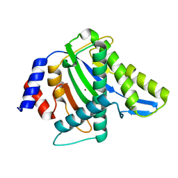
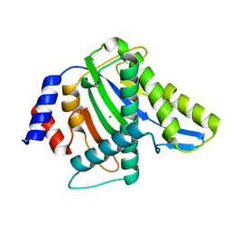
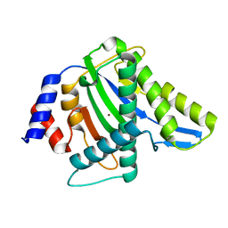
2F9G
DownloadVisualize
BU of 2f9g by Molmil
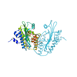
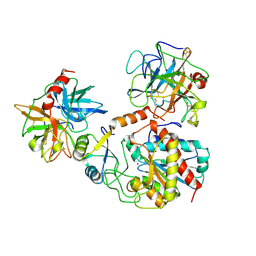
Crystal structure of Fus3 phosphorylated on Tyr182
Descriptor: ADENOSINE-5'-DIPHOSPHATE, MAGNESIUM ION, Mitogen-activated protein kinase FUS3
Authors:Bhattacharyya, R.P, Remenyi, A, Good, M.C, Bashor, C.J, Falick, A.M, Lim, W.A.
Deposit date:2005-12-05
Release date:2006-11-14
Last modified:2023-11-15
Method:X-RAY DIFFRACTION (2.1 Å)
Cite:The Ste5 scaffold allosterically modulates signaling output of the yeast mating pathway.
Science, 311, 2006
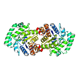
2K0U
DownloadVisualize
BU of 2k0u by Molmil
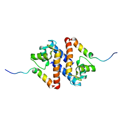
High Resolution Solution NMR Structures of Oxaliplatin-DNA Adduct
Descriptor: CYCLOHEXANE-1(R),2(R)-DIAMINE-PLATINUM(II), DNA (5'-D(*DCP*DCP*DTP*DCP*DTP*DGP*DGP*DTP*DCP*DTP*DCP*DC)-3'), DNA (5'-D(*DGP*DGP*DAP*DGP*DAP*DCP*DCP*DAP*DGP*DAP*DGP*DG)-3')
Authors:Bhattacharyya, D, King, C.L, Chaney, S.G, Campbell, S.L.
Deposit date:2008-02-15
Release date:2009-02-03
Last modified:2024-05-01
Method:SOLUTION NMR
Cite:Flanking Bases Influence the Nature of DNA Distortion by Platinum 1,2-Intrastrand (GG) Cross-Links.
Plos One, 6, 2011
2K0V
DownloadVisualize
BU of 2k0v by Molmil
High Resolution Solution NMR Structures of Undamaged DNA Dodecamer Duplex
Descriptor: DNA (5'-D(*DCP*DCP*DTP*DCP*DTP*DGP*DGP*DTP*DCP*DTP*DCP*DC)-3'), DNA (5'-D(*DGP*DGP*DAP*DGP*DAP*DCP*DCP*DAP*DGP*DAP*DGP*DG)-3')
Authors:Bhattacharyya, D, King, C.L, Chaney, S.G, Campbell, S.L.
Deposit date:2008-02-15
Release date:2009-02-03
Last modified:2024-05-01
Method:SOLUTION NMR
Cite:Flanking Bases Influence the Nature of DNA Distortion by Platinum 1,2-Intrastrand (GG) Cross-Links.
Plos One, 6, 2011
2K0T
DownloadVisualize
BU of 2k0t by Molmil
High Resolution Solution NMR Structures of Oxaliplatin-DNA Adduct
Descriptor: CYCLOHEXANE-1(R),2(R)-DIAMINE-PLATINUM(II), DNA (5'-D(*DCP*DCP*DTP*DCP*DTP*DGP*DGP*DTP*DCP*DTP*DCP*DC)-3'), DNA (5'-D(*DGP*DGP*DAP*DGP*DAP*DCP*DCP*DAP*DGP*DAP*DGP*DG)-3')
Authors:Bhattacharyya, D, King, C.L, Chaney, S.G, Campbell, S.L.
Deposit date:2008-02-14
Release date:2009-02-03
Last modified:2024-05-01
Method:SOLUTION NMR
Cite:Flanking Bases Influence the Nature of DNA Distortion by Platinum 1,2-Intrastrand (GG) Cross-Links.
Plos One, 6, 2011
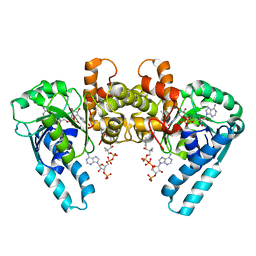
1D6N
DownloadVisualize
BU of 1d6n by Molmil
TERNARY COMPLEX STRUCTURE OF HUMAN HGPRTASE, PRPP, MG2+, AND THE INHIBITOR HPP REVEALS THE INVOLVEMENT OF THE FLEXIBLE LOOP IN SUBSTRATE BINDING
Descriptor: 1-O-pyrophosphono-5-O-phosphono-alpha-D-ribofuranose, 3H-PYRAZOLO[4,3-D]PYRIMIDIN-7-OL, MAGNESIUM ION, ...
Authors:Balendiran, G.K.
Deposit date:1999-10-14
Release date:1999-12-30
Last modified:2024-02-07
Method:X-RAY DIFFRACTION (2.7 Å)
Cite:Ternary complex structure of human HGPRTase, PRPP, Mg2+, and the inhibitor HPP reveals the involvement of the flexible loop in substrate binding.
Protein Sci., 8, 1999
1D15
DownloadVisualize
BU of 1d15 by Molmil
TERNARY INTERACTIONS OF SPERMINE WITH DNA: 4'-EPIADRIAMYCIN AND OTHER DNA: ANTHRACYCLINE COMPLEXES
Descriptor: 4'-EPIDOXORUBICIN, DNA (5'-D(*CP*GP*AP*TP*CP*G)-3'), SPERMINE
Authors:Williams, L.D, Frederick, C.A, Ughetto, G, Rich, A.
Deposit date:1990-07-03
Release date:1991-07-15
Last modified:2024-02-07
Method:X-RAY DIFFRACTION (1.5 Å)
Cite:Ternary interactions of spermine with DNA: 4'-epiadriamycin and other DNA: anthracycline complexes.
Nucleic Acids Res., 18, 1990
1X6G
DownloadVisualize
BU of 1x6g by Molmil
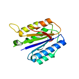
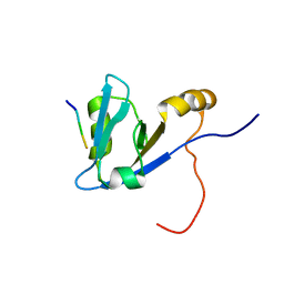
Solution structures of the SH3 domain of human megakaryocyte-associated tyrosine-protein kinase.
Descriptor: Megakaryocyte-associated tyrosine-protein kinase
Authors:Sato, M, Koshiba, S, Inoue, M, Kigawa, T, Yokoyama, S, RIKEN Structural Genomics/Proteomics Initiative (RSGI)
Deposit date:2005-05-17
Release date:2005-11-17
Last modified:2024-05-29
Method:SOLUTION NMR
Cite:Solution structures of the SH3 domain of human megakaryocyte-associated tyrosine-protein kinase.
To be Published
1M76
DownloadVisualize
BU of 1m76 by Molmil
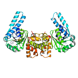
Crystal Structure of the S137C Mutant of L-3-HYDROXYACYL-COA Dehydrogenase in Complex with NAD and Acetoacetyl-COA
Descriptor: 3-HYDROXYACYL-COA DEHYDROGENASE, ACETOACETYL-COENZYME A, NICOTINAMIDE-ADENINE-DINUCLEOTIDE
Authors:Barycki, J.J, Banaszak, L.J.
Deposit date:2002-07-18
Release date:2004-02-03
Last modified:2024-02-14
Method:X-RAY DIFFRACTION (2.15 Å)
Cite:Crystal Structure of the S137C Mutant of L-3-Hydroxyacyl-COA Dehydrogenase in Complex with NAD and Acetoacetyl-COA
To be Published
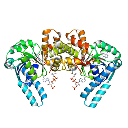
1M75
DownloadVisualize
BU of 1m75 by Molmil
Crystal Structure of the N208S Mutant of L-3-Hydroxyacyl-COA Dehydrogenase in Complex with NAD and Acetoacetyl-COA
Descriptor: 3-HYDROXYACYL-COA DEHYDROGENASE, ACETOACETYL-COENZYME A, NICOTINAMIDE-ADENINE-DINUCLEOTIDE
Authors:Barycki, J.J, Banaszak, L.J.
Deposit date:2002-07-18
Release date:2004-02-03
Last modified:2024-02-14
Method:X-RAY DIFFRACTION (2.3 Å)
Cite:Crystal Structure of the N208S Mutant of L-3-Hydroxyacyl-COA Dehydrogenase in Complex with NAD and Acetoacetyl-COA
To be Published
1LSO
DownloadVisualize
BU of 1lso by Molmil
Crystal Structure of the S137A mutant of L-3-Hydroxyacyl-CoA Dehydrogenase in Complex with NAD
Descriptor: 3-HYDROXYACYL-COA DEHYDROGENASE, NICOTINAMIDE-ADENINE-DINUCLEOTIDE
Authors:Barycki, J.J, Banaszak, L.J.
Deposit date:2002-05-17
Release date:2004-02-03
Last modified:2024-02-14
Method:X-RAY DIFFRACTION (2.6 Å)
Cite:Crystal Structure of the S137A mutant of L-3-Hydroxyacyl-CoA Dehydrogenase in Complex with NAD
To be Published
1LSJ
DownloadVisualize
BU of 1lsj by Molmil
Crystal Structure of the E110Q Mutant of L-3-Hydroxyacyl-CoA Dehydrogenase in Complex with NAD
Descriptor: 3-HYDROXYACYL-COA DEHYDROGENASE, NICOTINAMIDE-ADENINE-DINUCLEOTIDE
Authors:Barycki, J.J, Banaszak, L.J.
Deposit date:2002-05-17
Release date:2004-02-03
Last modified:2024-02-14
Method:X-RAY DIFFRACTION (2.5 Å)
Cite:Crystal Structure of the E110Q Mutant of L-3-Hydroxyacyl-CoA Dehydrogenase in Complex with NAD
To be Published
2MLF
DownloadVisualize
BU of 2mlf by Molmil
NMR structure of the dilated cardiomyopathy mutation G159D in troponin C bound to the anchoring region of troponin I
Descriptor: CALCIUM ION, Troponin C, slow skeletal and cardiac muscles
Authors:Baryshnikova, O.K, Robertson, I.M, Mercier, P, Sykes, B.D.
Deposit date:2014-02-26
Release date:2014-03-12
Last modified:2024-05-15
Method:SOLUTION NMR
Cite:The dilated cardiomyopathy G159D mutation in cardiac troponin C weakens the anchoring interaction with troponin I.
Biochemistry, 47, 2008
1JKU
DownloadVisualize
BU of 1jku by Molmil
Crystal Structure of Manganese Catalase from Lactobacillus plantarum
Descriptor: CALCIUM ION, HYDROXIDE ION, MANGANESE (III) ION, ...
Authors:Barynin, V.V, Whittaker, M.M, Antonyuk, S.V, Lamzin, V.S, Harrison, P.M, Artymiuk, P.J, Whittaker, J.W.
Deposit date:2001-07-13
Release date:2002-07-13
Last modified:2024-04-03
Method:X-RAY DIFFRACTION (1.84 Å)
Cite:Crystal structure of manganese catalase from Lactobacillus plantarum.
Structure, 9, 2001
1JKV
DownloadVisualize
BU of 1jkv by Molmil
Crystal Structure of Manganese Catalase from Lactobacillus plantarum complexed with azide
Descriptor: 1,2-ETHANEDIOL, AZIDE ION, CALCIUM ION, ...
Authors:Barynin, V.V, Whittaker, M.M, Antonyuk, S.V, Lamzin, V.S, Harrison, P.M, Artymiuk, P.J, Whittaker, J.W.
Deposit date:2001-07-13
Release date:2002-07-13
Last modified:2023-08-16
Method:X-RAY DIFFRACTION (1.39 Å)
Cite:Crystal structure of manganese catalase from Lactobacillus plantarum.
Structure, 9, 2001
1I11
DownloadVisualize
BU of 1i11 by Molmil
SOLUTION STRUCTURE OF THE DNA BINDING DOMAIN, SOX-5 HMG BOX FROM MOUSE
Descriptor: TRANSCRIPTION FACTOR SOX-5
Authors:Cary, P.D, Read, C.M, Davis, B, Driscoll, P.C, Crane-Robinson, C.
Deposit date:2001-01-30
Release date:2001-02-14
Last modified:2024-05-22
Method:SOLUTION NMR
Cite:Solution structure and backbone dynamics of the DNA-binding domain of mouse Sox-5.
Protein Sci., 10, 2001
1Y60
DownloadVisualize
BU of 1y60 by Molmil
Structure of the tetrahydromethanopterin dependent formaldehyde-activating enzyme (Fae) from Methylobacterium extorquens AM1 with bound 5,10-methylene tetrahydromethanopterin
Descriptor: 5,10-DIMETHYLENE TETRAHYDROMETHANOPTERIN, Formaldehyde-activating enzyme fae
Authors:Acharya, P, Goenrich, M, Hagemeier, C.H, Demmer, U, Vorholt, J.A, Thauer, R.K, Ermler, U.
Deposit date:2004-12-03
Release date:2005-01-11
Last modified:2023-08-23
Method:X-RAY DIFFRACTION (1.9 Å)
Cite:How an enzyme binds the C1-carrier tetrahydromethanopterin: Structure of the tetrahydromethanopterin dependent formaldehyde-activating enzyme (Fae) from Methylobacterium extorquens AM1
J.Biol.Chem., 280, 2005
1YSM
DownloadVisualize
BU of 1ysm by Molmil
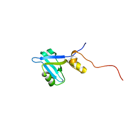
NMR Structure of N-terminal domain (Residues 1-77) of Siah-Interacting Protein.
Descriptor: Calcyclin-binding protein
Authors:Bhattacharya, S, Lee, Y.T, Michowski, W, Jastrzebska, B, Filipek, A, Kuznicki, J, Chazin, W.J.
Deposit date:2005-02-08
Release date:2005-07-26
Last modified:2024-05-22
Method:SOLUTION NMR
Cite:The Modular Structure of SIP Facilitates Its Role in Stabilizing Multiprotein Assemblies.
Biochemistry, 44, 2005
1Y5Y
DownloadVisualize
BU of 1y5y by Molmil
Structure of the tetrahydromethanopterin dependent formaldehyde-activating enzyme (Fae) from Methylobacterium extorquens AM1
Descriptor: CALCIUM ION, Formaldehyde-activating enzyme fae, SODIUM ION
Authors:Acharya, P, Goenrich, M, Hagemeier, C.H, Demmer, U, Vorholt, J.A, Thauer, R.K, Ermler, U.
Deposit date:2004-12-03
Release date:2005-01-11
Last modified:2024-02-14
Method:X-RAY DIFFRACTION (2 Å)
Cite:How an enzyme binds the C1-carrier tetrahydromethanopterin: Structure of the tetrahydromethanopterin dependent formaldehyde-activating enzyme (Fae) from Methylobacterium extorquens AM1
J.Biol.Chem., 280, 2005
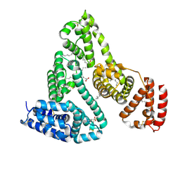
1E7A
DownloadVisualize
BU of 1e7a by Molmil
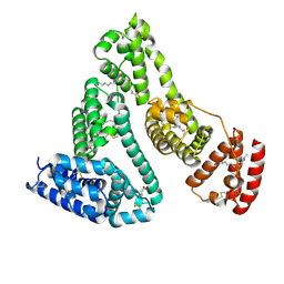
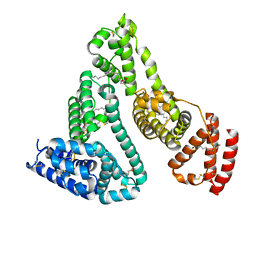
Crystal structure of human serum albumin complexed with the general anesthetic propofol
Descriptor: 2,6-BIS(1-METHYLETHYL)PHENOL, SERUM ALBUMIN
Authors:Bhattacharya, A.A, Curry, S, Franks, N.P.
Deposit date:2000-08-26
Release date:2001-01-17
Last modified:2023-12-13
Method:X-RAY DIFFRACTION (2.2 Å)
Cite:Binding of the General Anesthetics Propofol and Halothane to Human Serum Albumin. High Resolution Crystal Structures
J.Biol.Chem., 275, 2000
1E7C
DownloadVisualize
BU of 1e7c by Molmil
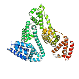
HUMAN SERUM ALBUMIN COMPLEXED WITH MYRISTIC ACID and the general anesthetic halothane
Descriptor: 2-BROMO-2-CHLORO-1,1,1-TRIFLUOROETHANE, MYRISTIC ACID, SERUM ALBUMIN
Authors:Bhattacharya, A.A, Curry, S, Franks, N.P.
Deposit date:2000-08-26
Release date:2001-01-17
Last modified:2023-12-13
Method:X-RAY DIFFRACTION (2.4 Å)
Cite:Binding of the General Anesthetics Propofol and Halothane to Human Serum Albumin. High Resolution Crystal Structures
J.Biol.Chem., 275, 2000
1E78
DownloadVisualize
BU of 1e78 by Molmil
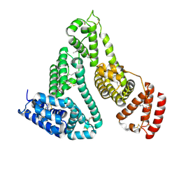
Crystal structure of human serum albumin
Descriptor: SERUM ALBUMIN
Authors:Bhattacharya, A.A, Curry, S, Franks, N.P.
Deposit date:2000-08-25
Release date:2001-03-23
Last modified:2023-12-13
Method:X-RAY DIFFRACTION (2.6 Å)
Cite:Binding of the General Anesthetics Propofol and Halothane to Human Serum Albumin. High Resolution Crystal Structures
J.Biol.Chem., 275, 2000
1E7B
DownloadVisualize
BU of 1e7b by Molmil
Crystal structure of human serum albumin complexed with the general anesthetic halothane
Descriptor: 2-BROMO-2-CHLORO-1,1,1-TRIFLUOROETHANE, SERUM ALBUMIN
Authors:Bhattacharya, A.A, Curry, S, Franks, N.P.
Deposit date:2000-08-26
Release date:2001-01-17
Last modified:2023-12-13
Method:X-RAY DIFFRACTION (2.38 Å)
Cite:Binding of the General Anesthetics Propofol and Halothane to Human Serum Albumin. High Resolution Crystal Structures
J.Biol.Chem., 275, 2000
7CK5
DownloadVisualize
BU of 7ck5 by Molmil
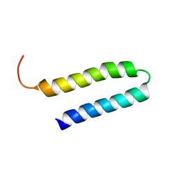
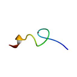
Solution structure of 28 amino acid polypeptide (354-381) in Plantago asiatica mosaic virus replicase bound to SDS micelle
Descriptor: PlAMV replicase peptide from RNA-dependent RNA polymerase
Authors:Komatsu, K, Sasaki, N, Yoshida, T, Suzuki, K, Masujima, Y, Hashimoto, M, Watanabe, S, Tochio, N, Kigawa, T, Yamaji, Y, Oshima, K, Namba, S, Nelson, R, Arie, T.
Deposit date:2020-07-15
Release date:2021-07-21
Last modified:2024-05-15
Method:SOLUTION NMR
Cite:Identification of a Proline-Kinked Amphipathic alpha-Helix Downstream from the Methyltransferase Domain of a Potexvirus Replicase and Its Role in Virus Replication and Perinuclear Complex Formation.
J.Virol., 95, 2021
3CE6
DownloadVisualize
BU of 3ce6 by Molmil
Crystal structure of Mycobacterium tuberculosis S-adenosyl-L-homocysteine hydrolase in ternary complex with NAD and adenosine
Descriptor: ADENOSINE, Adenosylhomocysteinase, NICOTINAMIDE-ADENINE-DINUCLEOTIDE
Authors:Reddy, M.C.M, Gokulan, K, Shetty, N.D, Owen, J.L, Ioerger, T.R, Sacchettini, J.C.
Deposit date:2008-02-28
Release date:2008-09-16
Last modified:2023-08-30
Method:X-RAY DIFFRACTION (1.6 Å)
Cite:Crystal structures of Mycobacterium tuberculosis S-adenosyl-L-homocysteine hydrolase in ternary complex with substrate and inhibitors.
Protein Sci., 17, 2008
3DHY
DownloadVisualize
BU of 3dhy by Molmil
Crystal Structures of Mycobacterium tuberculosis S-Adenosyl-L-Homocysteine Hydrolase in Ternary Complex with Substrate and Inhibitors
Descriptor: 5'-S-ethyl-5'-thioadenosine, Adenosylhomocysteinase, NICOTINAMIDE-ADENINE-DINUCLEOTIDE
Authors:Shetty, N.D, Ioerger, T.R, Gokulan, K, Reddy, M.C.M, Owen, J.L, Sacchettini, J.C, TB Structural Genomics Consortium (TBSGC)
Deposit date:2008-06-19
Release date:2008-09-09
Last modified:2023-08-30
Method:X-RAY DIFFRACTION (2 Å)
Cite:Crystal structures of Mycobacterium tuberculosis S-adenosyl-L-homocysteine hydrolase in ternary complex with substrate and inhibitors.
Protein Sci., 17, 2008

223532

數據於2024-08-07公開中

PDB statisticsPDBj update infoContact PDBjnumon