Loading
PDBj
MenuPDBj@FacebookPDBj@TwitterPDBj@YouTubewwPDB FoundationwwPDB
RCSB PDBPDBeBMRBAdv. SearchSearch help

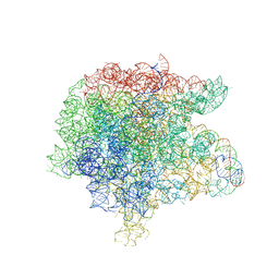
3JQ4
DownloadVisualize
BU of 3jq4 by Molmil
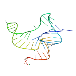
The structure of the complex of the large ribosomal subunit from D. Radiodurans with the antibiotic lankacidin
Descriptor: 23S ribosomal RNA, 5S ribosomal RNA, N-[(1S,2R,3E,5E,7S,9E,11E,13S,15R,19R)-7,13-dihydroxy-1,4,10,19-tetramethyl-17,18-dioxo-16-oxabicyclo[13.2.2]nonadeca-3,5,9,11-tetraen-2-yl]-2-oxopropanamide
Authors:Auerbach-Nevo, T, Mermershtain, I, Davidovich, C, Bashan, A, Rozenberg, H, Yonath, A.
Deposit date:2009-09-06
Release date:2010-09-08
Last modified:2023-11-01
Method:X-RAY DIFFRACTION (3.52 Å)
Cite:The structure of ribosome-lankacidin complex reveals ribosomal sites for synergistic antibiotics
Proc.Natl.Acad.Sci.USA, 107, 2010
1K8G
DownloadVisualize
BU of 1k8g by Molmil
Crystal Structure of the N-terminal domain of Oxytricha nova telomere end binding protein alpha subunit both uncomplexed and complexed with telomeric ssDNA
Descriptor: 5'-D(TP*TP*GP*GP*GP*G)-3', SULFATE ION, Telomere-Binding Protein alpha Subunit
Authors:Classen, S, Ruggles, J.A, Schultz, S.C.
Deposit date:2001-10-24
Release date:2001-12-20
Last modified:2023-08-16
Method:X-RAY DIFFRACTION (2.6 Å)
Cite:Crystal structure of the N-terminal domain of Oxytricha nova telomere end-binding protein alpha subunit both uncomplexed and complexed with telomeric ssDNA.
J.Mol.Biol., 314, 2001
3AU5
DownloadVisualize
BU of 3au5 by Molmil
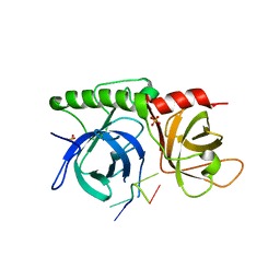
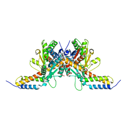
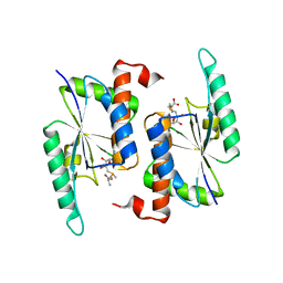
Structure of the human myosin-X MyTH4-FERM cassette
Descriptor: Myosin-X
Authors:Hirano, Y, Takahashi, A, Hakoshima, T.
Deposit date:2011-01-28
Release date:2011-07-13
Last modified:2023-11-01
Method:X-RAY DIFFRACTION (2.55 Å)
Cite:Structural basis of cargo recognition by the myosin-X MyTH4-FERM domain
Embo J., 30, 2011
3RVD
DownloadVisualize
BU of 3rvd by Molmil
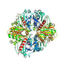
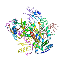
Crystal structure of the binary complex, obtained by soaking, of photosyntetic a4 glyceraldehyde 3-phosphate dehydrogenase (gapdh) with cp12-2, both from arabidopsis thaliana.
Descriptor: Glyceraldehyde-3-phosphate dehydrogenase A, chloroplastic, NICOTINAMIDE-ADENINE-DINUCLEOTIDE, ...
Authors:Fermani, S, Thumiger, A, Falini, G, Marri, L, Sparla, F, Trost, P.
Deposit date:2011-05-06
Release date:2012-04-25
Last modified:2024-03-13
Method:X-RAY DIFFRACTION (2.7 Å)
Cite:Conformational Selection and Folding-upon-binding of Intrinsically Disordered Protein CP12 Regulate Photosynthetic Enzymes Assembly.
J.Biol.Chem., 287, 2012
3AU4
DownloadVisualize
BU of 3au4 by Molmil
Structure of the human myosin-X MyTH4-FERM cassette bound to its specific cargo, DCC
Descriptor: Myosin-X, Netrin receptor DCC
Authors:Hirano, Y, Hatano, T, Hakoshima, T.
Deposit date:2011-01-28
Release date:2011-07-13
Last modified:2024-03-13
Method:X-RAY DIFFRACTION (1.9 Å)
Cite:Structural basis of cargo recognition by the myosin-X MyTH4-FERM domain
Embo J., 30, 2011
1Q89
DownloadVisualize
BU of 1q89 by Molmil
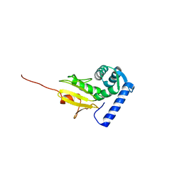
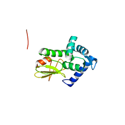
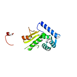
Crystal structure of the C-domain of the T.vaginalis Inr binding protein, IBP39 (cubic crystal form)
Descriptor: 39 kDa initiator binding protein
Authors:Schumacher, M.A, Johnson, P.J.
Deposit date:2003-08-20
Release date:2003-11-18
Last modified:2023-08-16
Method:X-RAY DIFFRACTION (2.75 Å)
Cite:Structural Basis of Core Promoter Recognition in a Primitive Eukaryote
Cell(Cambridge,Mass.), 115, 2003
1Q88
DownloadVisualize
BU of 1q88 by Molmil
Crystal structure of the C-domain of the T.vaginalis Inr binding protein, IBP39 (monoclinic form)
Descriptor: 39 kDa initiator binding protein
Authors:Schumacher, M.A, Johnson, P.J.
Deposit date:2003-08-20
Release date:2003-11-18
Last modified:2024-02-14
Method:X-RAY DIFFRACTION (2.42 Å)
Cite:Structural Basis of Core Promoter Recognition in a Primitive Eukaryote
Cell(Cambridge,Mass.), 115, 2003
1Q87
DownloadVisualize
BU of 1q87 by Molmil
Crystal structure of the C-domain of the T.vaginalis Inr binding protein, IBP39 (tetragonal form)
Descriptor: 39 kDa initiator binding protein
Authors:Schumacher, M.A, Johnson, P.J.
Deposit date:2003-08-20
Release date:2003-11-18
Last modified:2023-08-16
Method:X-RAY DIFFRACTION (2.32 Å)
Cite:Structural Basis of Core Promoter Recognition in a Primitive Eukaryote
Cell(Cambridge,Mass.), 115, 2003
2FCC
DownloadVisualize
BU of 2fcc by Molmil
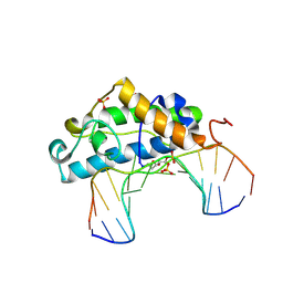
Crystal Structure of T4 Pyrimidine Dimer Glycosylase (T4-Pdg) Covalently Complexed with a DNA Substrate Containing Abasic Site
Descriptor: DNA (5'-D(*CP*CP*AP*GP*GP*AP*(PED)P*GP*AP*AP*GP*CP*C)-3'), DNA (5'-D(*GP*GP*CP*(BRU)P*(BRU)P*CP*AP*(BRU)P*CP*CP*(BRU)P*GP*G)-3'), Endonuclease V, ...
Authors:Golan, G, Zharkov, D.O, Fernandes, A.S, Dodson, M.L, McCullough, A.K, Grollman, A.P, Lloyd, R.S, Shoham, G.
Deposit date:2005-12-12
Release date:2006-10-03
Last modified:2023-08-30
Method:X-RAY DIFFRACTION (2.3 Å)
Cite:Structure of T4 Pyrimidine Dimer Glycosylase in a Reduced Imine Covalent Complex with Abasic Site-containing DNA.
J.Mol.Biol., 362, 2006
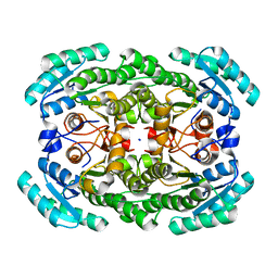
3GDF
DownloadVisualize
BU of 3gdf by Molmil
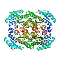
Crystal structure of the NADP-dependent mannitol dehydrogenase from Cladosporium herbarum.
Descriptor: Probable NADP-dependent mannitol dehydrogenase, ZINC ION
Authors:Nuess, D, Goettig, P, Magler, I, Denk, U, Breitenbach, M, Schneider, P.B, Brandstetter, H, Simon-Nobbe, B.
Deposit date:2009-02-24
Release date:2010-05-26
Last modified:2023-09-06
Method:X-RAY DIFFRACTION (2.5 Å)
Cite:Crystal structure of the NADP-dependent mannitol dehydrogenase from Cladosporium herbarum: Implications for oligomerisation and catalysis.
Biochimie, 92, 2010
5J43
DownloadVisualize
BU of 5j43 by Molmil
CdiA-CT from uropathogenic Escherichia coli in complex with CysK
Descriptor: Cysteine synthase A, tRNA nuclease CdiA
Authors:Morse, R.P, Goulding, C.W, Johnson, P.M.
Deposit date:2016-03-31
Release date:2016-07-27
Last modified:2023-11-15
Method:X-RAY DIFFRACTION (2.7 Å)
Cite:Unraveling the essential role of CysK in CDI toxin activation.
Proc. Natl. Acad. Sci. U.S.A., 113, 2016
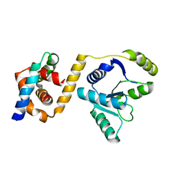
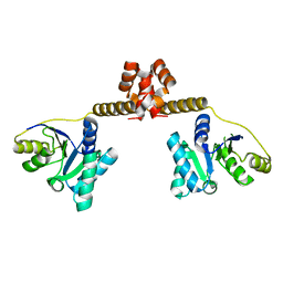
2QKM
DownloadVisualize
BU of 2qkm by Molmil
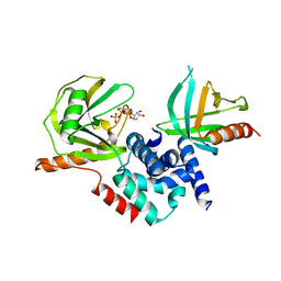
The crystal structure of fission yeast mRNA decapping enzyme Dcp1-Dcp2 complex
Descriptor: ADENOSINE-5'-TRIPHOSPHATE, SPAC19A8.12 protein, SPBC3B9.21 protein
Authors:She, M, Song, H.
Deposit date:2007-07-11
Release date:2008-04-15
Last modified:2023-08-30
Method:X-RAY DIFFRACTION (2.8 Å)
Cite:Structural basis of dcp2 recognition and activation by dcp1.
Mol.Cell, 29, 2008
6WRZ
DownloadVisualize
BU of 6wrz by Molmil
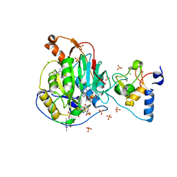
Crystal Structure of Nsp16-Nsp10 Heterodimer from SARS-CoV-2 with 7-methyl-GpppA and S-adenosyl-L-homocysteine in the Active Site and Sulfates in the mRNA Binding Groove.
Descriptor: 2'-O-methyltransferase, 7-METHYL-GUANOSINE-5'-TRIPHOSPHATE, CHLORIDE ION, ...
Authors:Minasov, G, Shuvalova, L, Rosas-Lemus, M, Brunzelle, J.S, Kiryukhina, O, Satchell, K.J.F, Center for Structural Genomics of Infectious Diseases (CSGID)
Deposit date:2020-04-30
Release date:2020-05-13
Last modified:2023-10-18
Method:X-RAY DIFFRACTION (2.25 Å)
Cite:High-resolution structures of the SARS-CoV-2 2'- O -methyltransferase reveal strategies for structure-based inhibitor design.
Sci.Signal., 13, 2020
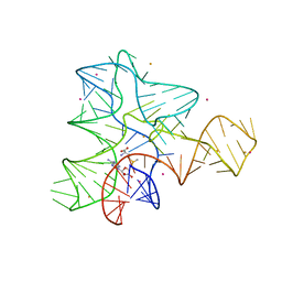
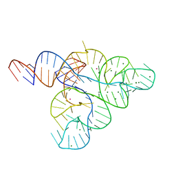
4ENC
DownloadVisualize
BU of 4enc by Molmil
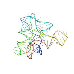
Crystal structure of fluoride riboswitch
Descriptor: FLUORIDE ION, Fluoride riboswitch, MAGNESIUM ION, ...
Authors:Ren, A.M, Rajashankar, K.R, Patel, D.J.
Deposit date:2012-04-12
Release date:2012-05-09
Last modified:2023-09-13
Method:X-RAY DIFFRACTION (2.272 Å)
Cite:Fluoride ion encapsulation by Mg2+ ions and phosphates in a fluoride riboswitch.
Nature, 486, 2012
4FXE
DownloadVisualize
BU of 4fxe by Molmil
Crystal structure of the intact E. coli RelBE toxin-antitoxin complex
Descriptor: Antitoxin RelB, SULFATE ION, mRNA interferase RelE
Authors:Brodersen, D.E, Boggild, A, Sofos, N.
Deposit date:2012-07-03
Release date:2012-08-29
Last modified:2024-02-28
Method:X-RAY DIFFRACTION (2.7503 Å)
Cite:The crystal structure of the intact E. coli RelBE toxin-antitoxin complex provides the structural basis for conditional cooperativity.
Structure, 20, 2012
5TWJ
DownloadVisualize
BU of 5twj by Molmil
Crystal Structure of RlmH in Complex with S-Adenosylmethionine
Descriptor: Ribosomal RNA large subunit methyltransferase H, S-ADENOSYLMETHIONINE
Authors:Koh, C.S, Madireddy, R, Beane, T.J, Zamore, P.D, Korostelev, A.A.
Deposit date:2016-11-14
Release date:2017-05-03
Last modified:2023-10-04
Method:X-RAY DIFFRACTION (2.299 Å)
Cite:Small methyltransferase RlmH assembles a composite active site to methylate a ribosomal pseudouridine.
Sci Rep, 7, 2017
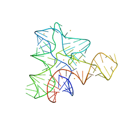
3IQR
DownloadVisualize
BU of 3iqr by Molmil
SAM-I riboswitch from T. tencongensis variant A94G bound with SAM
Descriptor: BARIUM ION, S-ADENOSYLMETHIONINE, SAM-I riboswitch
Authors:Montange, R.K, Batey, R.T.
Deposit date:2009-08-20
Release date:2010-08-04
Last modified:2023-09-06
Method:X-RAY DIFFRACTION (2.55 Å)
Cite:Free state conformational sampling of the SAM-I riboswitch aptamer domain.
Structure, 18, 2010
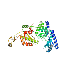
3EAR
DownloadVisualize
BU of 3ear by Molmil
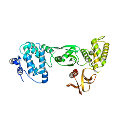
Novel dimerization motif in the DEAD box RNA helicase Hera: form 1, partial dimer
Descriptor: Hera
Authors:Klostermeier, D, Rudolph, M.G.
Deposit date:2008-08-26
Release date:2008-12-09
Last modified:2023-08-30
Method:X-RAY DIFFRACTION (2.3 Å)
Cite:A novel dimerization motif in the C-terminal domain of the Thermus thermophilus DEAD box helicase Hera confers substantial flexibility.
Nucleic Acids Res., 37, 2009
3IQN
DownloadVisualize
BU of 3iqn by Molmil
Free-state structural transitions of the SAM-I riboswitch
Descriptor: BARIUM ION, POTASSIUM ION, S-ADENOSYLMETHIONINE, ...
Authors:Montange, R.K, Batey, R.T.
Deposit date:2009-08-20
Release date:2010-08-04
Last modified:2023-09-06
Method:X-RAY DIFFRACTION (2.7 Å)
Cite:Free state conformational sampling of the SAM-I riboswitch aptamer domain.
Structure, 18, 2010
3IQP
DownloadVisualize
BU of 3iqp by Molmil
SAM-I riboswitch from T. tencongensis variant A94G apo form
Descriptor: BARIUM ION, SAM-I riboswitch
Authors:Montange, R.K, Batey, R.T.
Deposit date:2009-08-20
Release date:2010-08-04
Last modified:2023-09-06
Method:X-RAY DIFFRACTION (2.9 Å)
Cite:Free state conformational sampling of the SAM-I riboswitch aptamer domain.
Structure, 18, 2010
1UET
DownloadVisualize
BU of 1uet by Molmil
Divergent evolutions of trinucleotide polymerization revealed by an archaeal CCA-adding enzyme structure
Descriptor: ACETATE ION, CALCIUM ION, MAGNESIUM ION, ...
Authors:Nureki, O, RIKEN Structural Genomics/Proteomics Initiative (RSGI)
Deposit date:2003-05-21
Release date:2003-12-02
Last modified:2023-12-27
Method:X-RAY DIFFRACTION (2 Å)
Cite:Divergent evolutions of trinucleotide polymerization revealed by an archaeal CCA-adding enzyme structure.
Embo J., 22, 2003
3EAS
DownloadVisualize
BU of 3eas by Molmil
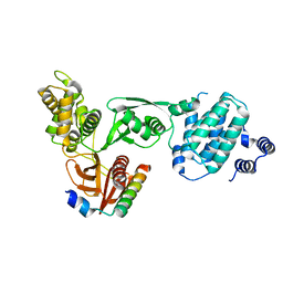
Novel dimerization motif in the DEAD box RNA helicase Hera: form 1, complete dimer, asymmetric
Descriptor: Hera
Authors:Klostermeier, D, Rudolph, M.G.
Deposit date:2008-08-26
Release date:2008-12-09
Last modified:2023-08-30
Method:X-RAY DIFFRACTION (2.8 Å)
Cite:A novel dimerization motif in the C-terminal domain of the Thermus thermophilus DEAD box helicase Hera confers substantial flexibility.
Nucleic Acids Res., 37, 2009
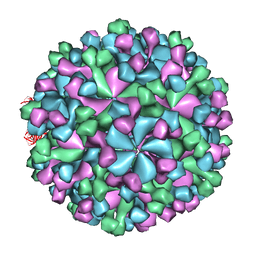
3IYO
DownloadVisualize
BU of 3iyo by Molmil
Cryo-EM model of virion-sized HEV virion-sized capsid
Descriptor: Capsid protein
Authors:Xing, L, Mayazaki, N, Li, T.C, Simons, M.N, Wall, J.S, Moore, M, Wang, C.Y, Takeda, N, Wakita, T, Miyamura, T, Cheng, R.H.
Deposit date:2010-03-19
Release date:2010-08-18
Last modified:2024-02-21
Method:ELECTRON MICROSCOPY (10.5 Å)
Cite:Structural basis for the RNA-dependent assembly pathway of hepatitis E virion-sized particles
J.Biol.Chem., 2010
2YIF
DownloadVisualize
BU of 2yif by Molmil
Crystal structure of a F. nucleatum FMN riboswitch - Free state
Descriptor: FMN RIBOSWITCH, MAGNESIUM ION, POTASSIUM ION
Authors:Vicens, Q, Mondragon, E, Batey, R.T.
Deposit date:2011-05-12
Release date:2011-08-31
Last modified:2023-12-20
Method:X-RAY DIFFRACTION (3.298 Å)
Cite:Molecular Sensing by the Aptamer Domain of the Fmn Riboswitch: A General Model for Ligand Binding by Conformational Selection.
Nucleic Acids Res., 39, 2011
3GDG
DownloadVisualize
BU of 3gdg by Molmil
Crystal structure of the NADP-dependent mannitol dehydrogenase from Cladosporium herbarum.
Descriptor: Probable NADP-dependent mannitol dehydrogenase, SODIUM ION
Authors:Nuess, D, Goettig, P, Magler, I, Denk, U, Breitenbach, M, Schneider, P.B, Brandstetter, H, Simon-Nobbe, B.
Deposit date:2009-02-24
Release date:2010-06-09
Last modified:2023-09-06
Method:X-RAY DIFFRACTION (2.3 Å)
Cite:Crystal structure of the NADP-dependent mannitol dehydrogenase from Cladosporium herbarum: Implications for oligomerisation and catalysis.
Biochimie, 92, 2010

226707

数据于2024-10-30公开中

PDB statisticsPDBj update infoContact PDBjnumon