Loading
PDBj
MenuPDBj@FacebookPDBj@TwitterPDBj@YouTubewwPDB FoundationwwPDB
RCSB PDBPDBeBMRBAdv. SearchSearch help

3DQ2
DownloadVisualize
BU of 3dq2 by Molmil
Structure of the Yellow Fluorescent Protein Citrine Frozen at 4000 Atmospheres Number 1: Structure 23 in a Series of 26 High Pressure Structures
Descriptor: Green fluorescent protein
Authors:Barstow, B, Kim, C.U.
Deposit date:2008-07-09
Release date:2008-09-23
Last modified:2023-11-15
Method:X-RAY DIFFRACTION (1.6 Å)
Cite:Alteration of citrine structure by hydrostatic pressure explains the accompanying spectral shift.
Proc.Natl.Acad.Sci.Usa, 105, 2008
3DQC
DownloadVisualize
BU of 3dqc by Molmil
Structure of the Yellow Fluorescent Protein Citrine Frozen at 1250 Atmospheres Number 3: Structure 13 in a Series of 26 High Pressure Structures
Descriptor: Green fluorescent protein
Authors:Barstow, B, Kim, C.U.
Deposit date:2008-07-09
Release date:2008-09-23
Last modified:2023-11-15
Method:X-RAY DIFFRACTION (1.49 Å)
Cite:Alteration of citrine structure by hydrostatic pressure explains the accompanying spectral shift.
Proc.Natl.Acad.Sci.Usa, 105, 2008
3DPW
DownloadVisualize
BU of 3dpw by Molmil
Structure of the Yellow Fluorescent Protein Citrine Frozen at 1 Atmosphere Number 1: Structure 1 in a Series of 26 High Pressure Structures
Descriptor: Green fluorescent protein
Authors:Barstow, B, Kim, C.U.
Deposit date:2008-07-09
Release date:2008-09-23
Last modified:2023-11-15
Method:X-RAY DIFFRACTION (1.59 Å)
Cite:Alteration of citrine structure by hydrostatic pressure explains the accompanying spectral shift.
Proc.Natl.Acad.Sci.Usa, 105, 2008
3DQ4
DownloadVisualize
BU of 3dq4 by Molmil
Structure of the Yellow Fluorescent Protein Citrine Frozen at 2000 Atmospheres Number 2: Structure 20 in a Series of 26 High Pressure Structures
Descriptor: Green fluorescent protein
Authors:Barstow, B, Kim, C.U.
Deposit date:2008-07-09
Release date:2008-09-23
Last modified:2023-11-15
Method:X-RAY DIFFRACTION (1.47 Å)
Cite:Alteration of citrine structure by hydrostatic pressure explains the accompanying spectral shift.
Proc.Natl.Acad.Sci.Usa, 105, 2008
3DQF
DownloadVisualize
BU of 3dqf by Molmil
Structure of the Yellow Fluorescent Protein Citrine Frozen at 1000 Atmospheres Number 6: Structure 10 in a Series of 26 High Pressure Structures
Descriptor: Green fluorescent protein
Authors:Barstow, B, Kim, C.U.
Deposit date:2008-07-09
Release date:2008-09-23
Last modified:2023-11-15
Method:X-RAY DIFFRACTION (1.46 Å)
Cite:Alteration of citrine structure by hydrostatic pressure explains the accompanying spectral shift.
Proc.Natl.Acad.Sci.Usa, 105, 2008
3DQO
DownloadVisualize
BU of 3dqo by Molmil
Structure of the Yellow Fluorescent Protein Citrine Frozen at 1 Atmosphere Number 2: Structure 2 in a Series of 26 High Pressure Structures
Descriptor: Green fluorescent protein
Authors:Barstow, B, Kim, C.U.
Deposit date:2008-07-09
Release date:2008-09-23
Last modified:2023-11-15
Method:X-RAY DIFFRACTION (1.5 Å)
Cite:Alteration of citrine structure by hydrostatic pressure explains the accompanying spectral shift.
Proc.Natl.Acad.Sci.Usa, 105, 2008
3DPX
DownloadVisualize
BU of 3dpx by Molmil
Structure of the Yellow Fluorescent Protein Citrine Frozen at 5000 Atmospheres: Structure 26 in a Series of 26 High Pressure Structures
Descriptor: Green fluorescent protein
Authors:Barstow, B, Kim, C.U.
Deposit date:2008-07-09
Release date:2008-09-23
Last modified:2023-11-15
Method:X-RAY DIFFRACTION (1.5 Å)
Cite:Alteration of citrine structure by hydrostatic pressure explains the accompanying spectral shift.
Proc.Natl.Acad.Sci.Usa, 105, 2008
3DQ5
DownloadVisualize
BU of 3dq5 by Molmil
Structure of the Yellow Fluorescent Protein Citrine Frozen at 1960 Atmospheres: Structure 19 in a Series of 26 High Pressure Structures
Descriptor: Green fluorescent protein
Authors:Barstow, B, Kim, C.U.
Deposit date:2008-07-09
Release date:2008-09-23
Last modified:2023-11-15
Method:X-RAY DIFFRACTION (1.5 Å)
Cite:Alteration of citrine structure by hydrostatic pressure explains the accompanying spectral shift.
Proc.Natl.Acad.Sci.Usa, 105, 2008
3DQE
DownloadVisualize
BU of 3dqe by Molmil
Structure of the Yellow Fluorescent Protein Citrine Frozen at 1250 Atmospheres Number 1: Structure 11 in a Series of 26 High Pressure Structures
Descriptor: Green fluorescent protein
Authors:Barstow, B, Kim, C.U.
Deposit date:2008-07-09
Release date:2008-09-23
Last modified:2023-11-15
Method:X-RAY DIFFRACTION (1.43 Å)
Cite:Alteration of citrine structure by hydrostatic pressure explains the accompanying spectral shift.
Proc.Natl.Acad.Sci.Usa, 105, 2008
3DQI
DownloadVisualize
BU of 3dqi by Molmil
Structure of the Yellow Fluorescent Protein Citrine Frozen at 1000 Atmospheres Number 4: Structure 8 in a Series of 26 High Pressure Structures
Descriptor: Green fluorescent protein
Authors:Barstow, B, Kim, C.U.
Deposit date:2008-07-09
Release date:2008-09-23
Last modified:2023-11-15
Method:X-RAY DIFFRACTION (1.42 Å)
Cite:Alteration of citrine structure by hydrostatic pressure explains the accompanying spectral shift.
Proc.Natl.Acad.Sci.Usa, 105, 2008
3DQU
DownloadVisualize
BU of 3dqu by Molmil
Structure of the Yellow Fluorescent Protein Citrine Frozen at 2000 Atmospheres Number 1: Structure 20 in a Series of 26 High Pressure Structures
Descriptor: Green fluorescent protein
Authors:Barstow, B, Kim, C.U.
Deposit date:2008-07-09
Release date:2008-09-23
Last modified:2023-11-15
Method:X-RAY DIFFRACTION (1.42 Å)
Cite:Alteration of citrine structure by hydrostatic pressure explains the accompanying spectral shift.
Proc.Natl.Acad.Sci.Usa, 105, 2008
3DQJ
DownloadVisualize
BU of 3dqj by Molmil
Structure of the Yellow Fluorescent Protein Citrine Frozen at 1000 Atmospheres Number 3: Structure 7 in a Series of 26 High Pressure Structures
Descriptor: Green fluorescent protein
Authors:Barstow, B, Kim, C.U.
Deposit date:2008-07-09
Release date:2008-09-23
Last modified:2023-11-15
Method:X-RAY DIFFRACTION (1.51 Å)
Cite:Alteration of citrine structure by hydrostatic pressure explains the accompanying spectral shift.
Proc.Natl.Acad.Sci.Usa, 105, 2008
3DQ7
DownloadVisualize
BU of 3dq7 by Molmil
Structure of the Yellow Fluorescent Protein Citrine Frozen at 1920 Atmospheres Number 1: Structure 17 in a Series of 26 High Pressure Structures
Descriptor: Green fluorescent protein
Authors:Barstow, B, Kim, C.U.
Deposit date:2008-07-09
Release date:2008-09-23
Last modified:2023-11-15
Method:X-RAY DIFFRACTION (1.23 Å)
Cite:Alteration of citrine structure by hydrostatic pressure explains the accompanying spectral shift.
Proc.Natl.Acad.Sci.Usa, 105, 2008
3DQK
DownloadVisualize
BU of 3dqk by Molmil
Structure of the Yellow Fluorescent Protein Citrine Frozen at 1000 Atmospheres Number 2: Structure 6 in a Series of 26 High Pressure Structures
Descriptor: Green fluorescent protein
Authors:Barstow, B, Kim, C.U.
Deposit date:2008-07-09
Release date:2008-09-23
Last modified:2023-11-15
Method:X-RAY DIFFRACTION (1.4 Å)
Cite:Alteration of citrine structure by hydrostatic pressure explains the accompanying spectral shift.
Proc.Natl.Acad.Sci.Usa, 105, 2008
3DPZ
DownloadVisualize
BU of 3dpz by Molmil
Structure of the Yellow Fluorescent Protein Citrine Frozen at 4000 Atmospheres Number 3: Structure 25 in a Series of 26 High Pressure Structures
Descriptor: Green fluorescent protein
Authors:Barstow, B, Kim, C.U.
Deposit date:2008-07-09
Release date:2008-09-23
Last modified:2023-11-15
Method:X-RAY DIFFRACTION (1.7 Å)
Cite:Alteration of citrine structure by hydrostatic pressure explains the accompanying spectral shift.
Proc.Natl.Acad.Sci.Usa, 105, 2008
3DQ8
DownloadVisualize
BU of 3dq8 by Molmil
Structure of the Yellow Fluorescent Protein Citrine Frozen at 1500 Atmospheres Number 2: Structure 16 in a Series of 26 High Pressure Structures
Descriptor: Green fluorescent protein
Authors:Barstow, B, Kim, C.U.
Deposit date:2008-07-09
Release date:2008-09-23
Last modified:2023-11-15
Method:X-RAY DIFFRACTION (1.51 Å)
Cite:Alteration of citrine structure by hydrostatic pressure explains the accompanying spectral shift.
Proc.Natl.Acad.Sci.Usa, 105, 2008
1LL9
DownloadVisualize
BU of 1ll9 by Molmil
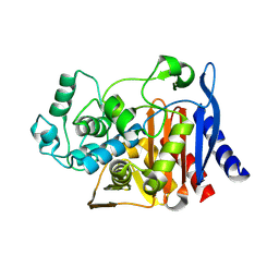
Crystal Structure Of AmpC beta-Lactamase From E. Coli In Complex With Amoxicillin
Descriptor: 2-{1-[2-AMINO-2-(4-HYDROXY-PHENYL)-ACETYLAMINO]-2-OXO-ETHYL}-5,5-DIMETHYL-THIAZOLIDINE-4-CARBOXYLIC ACID, beta-lactamase
Authors:Trehan, I, Morandi, F, Blaszczak, L.C, Shoichet, B.K.
Deposit date:2002-04-26
Release date:2002-10-02
Last modified:2023-08-16
Method:X-RAY DIFFRACTION (1.87 Å)
Cite:Using steric hindrance to design new inhibitors of class C beta-lactamases.
Chem.Biol., 9, 2002
2HGW
DownloadVisualize
BU of 2hgw by Molmil
Crystal structure of Cys318Ala mutant of human mitochondrial branched chain aminotransferase
Descriptor: 4-(2-HYDROXYETHYL)-1-PIPERAZINE ETHANESULFONIC ACID, ACETIC ACID, Branched-chain-amino-acid aminotransferase, ...
Authors:Yennawar, N.H, Hutson, S.M.
Deposit date:2006-06-27
Release date:2006-10-24
Last modified:2021-10-20
Method:X-RAY DIFFRACTION (1.98 Å)
Cite:Human Mitochondrial Branched Chain Aminotransferase Isozyme: STRUCTURAL ROLE OF THE CXXC CENTER IN CATALYSIS.
J.Biol.Chem., 281, 2006
2HDK
DownloadVisualize
BU of 2hdk by Molmil
Crystal Structure of Cys315Ala-Cys318Ala Mutant of Human Mitochondrial Branched Chain Aminotransferase
Descriptor: 2-OXO-4-METHYLPENTANOIC ACID, ACETIC ACID, Branched-chain-amino-acid aminotransferase, ...
Authors:Yennawar, N.H, Hutson, S.M.
Deposit date:2006-06-20
Release date:2006-10-24
Last modified:2021-10-20
Method:X-RAY DIFFRACTION (2.4 Å)
Cite:Human Mitochondrial Branched Chain Aminotransferase Isozyme: STRUCTURAL ROLE OF THE CXXC CENTER IN CATALYSIS.
J.Biol.Chem., 281, 2006
8CDQ
DownloadVisualize
BU of 8cdq by Molmil
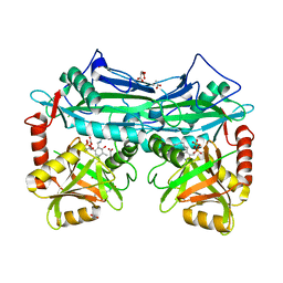
Plasmodium falciparum Myosin A full-length, post-rigor state complexed to the inhibitor KNX-002 and Mg.ATP-gamma-S
Descriptor: 1,2-ETHANEDIOL, 1-(4-methoxyphenyl)-~{N}-[(3-thiophen-2-yl-1~{H}-pyrazol-4-yl)methyl]cyclopropan-1-amine, GLYCEROL, ...
Authors:Moussaoui, D, Robblee, J.P, Robert-Paganin, J, Auguin, D, Fisher, F, Fagnant, P.M, MacFarlane, J.E, Schaletzky, J, Wehri, E, Mueller-Dieckmann, C, Baum, J, Trybus, K.M, Houdusse, A.
Deposit date:2023-01-31
Release date:2023-06-21
Last modified:2023-11-15
Method:X-RAY DIFFRACTION (2.21 Å)
Cite:Mechanism of small molecule inhibition of Plasmodium falciparum myosin A informs antimalarial drug design.
Nat Commun, 14, 2023
7Z3U
DownloadVisualize
BU of 7z3u by Molmil
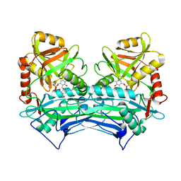
Crystal structure of SARS-CoV-2 Main Protease after incubation with Sulfo-Calpeptin
Descriptor: 3C-like proteinase nsp5, CHLORIDE ION, Calpetin, ...
Authors:Reinke, P.Y.A, Falke, S, Lieske, J, Ewert, W, Loboda, J, Rahmani Mashhour, A, Hauser, M, Karnicar, K, Usenik, A, Lindic, N, Lach, M, Boehler, H, Beck, T, Cox, R, Chapman, H.N, Hinrichs, W, Turk, D, Guenther, S, Meents, A.
Deposit date:2022-03-02
Release date:2023-03-22
Last modified:2024-07-10
Method:X-RAY DIFFRACTION (1.72 Å)
Cite:Calpeptin is a potent cathepsin inhibitor and drug candidate for SARS-CoV-2 infections.
Commun Biol, 6, 2023
8SS4
DownloadVisualize
BU of 8ss4 by Molmil
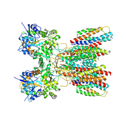
Structure of LBD-TMD of AMPA receptor GluA2 in complex with auxiliary subunits TARP gamma-5 and cornichon-2 (apo state)
Descriptor: Glutamate receptor 2, Voltage-dependent calcium channel gamma-5 subunit chimera, Protein cornichon homolog 2, ...
Authors:Yen, L.Y, Gangwar, S.P, Yelshanskaya, M.V, Sobolevsky, A.I.
Deposit date:2023-05-08
Release date:2023-09-06
Last modified:2023-11-01
Method:ELECTRON MICROSCOPY (3.3 Å)
Cite:Modulation of GluA2-gamma 5 synaptic complex desensitization, polyamine block and antiepileptic perampanel inhibition by auxiliary subunit cornichon-2.
Nat.Struct.Mol.Biol., 30, 2023
8SS8
DownloadVisualize
BU of 8ss8 by Molmil
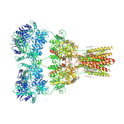
Structure of AMPA receptor GluA2 complex with auxiliary subunit TARP gamma-5 bound to competitive antagonist ZK and antiepileptic drug perampanel (closed state)
Descriptor: 1,2-DIOLEOYL-SN-GLYCERO-3-PHOSPHOCHOLINE, 2-(6'-oxo-1'-phenyl[1',6'-dihydro[2,3'-bipyridine]]-5'-yl)benzonitrile, CHOLESTEROL, ...
Authors:Gangwar, S.P, Yen, L.Y, Yelshanskaya, M.V, Sobolevsky, A.I.
Deposit date:2023-05-08
Release date:2023-09-06
Last modified:2023-10-25
Method:ELECTRON MICROSCOPY (2.81 Å)
Cite:Modulation of GluA2-gamma 5 synaptic complex desensitization, polyamine block and antiepileptic perampanel inhibition by auxiliary subunit cornichon-2.
Nat.Struct.Mol.Biol., 30, 2023
8SSA
DownloadVisualize
BU of 8ssa by Molmil
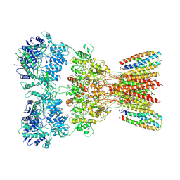
Structure of AMPA receptor GluA2 complex with auxiliary subunits TARP gamma-5 and cornichon-2 bound to glutamate and channel blocker spermidine (desensitized state)
Descriptor: 1,2-DIOLEOYL-SN-GLYCERO-3-PHOSPHOCHOLINE, GLUTAMIC ACID, Glutamate receptor 2, ...
Authors:Gangwar, S.P, Yen, L.Y, Yelshanskaya, M.V, Sobolevsky, A.I.
Deposit date:2023-05-08
Release date:2023-09-06
Last modified:2023-11-01
Method:ELECTRON MICROSCOPY (3.88 Å)
Cite:Modulation of GluA2-gamma 5 synaptic complex desensitization, polyamine block and antiepileptic perampanel inhibition by auxiliary subunit cornichon-2.
Nat.Struct.Mol.Biol., 30, 2023
8SS5
DownloadVisualize
BU of 8ss5 by Molmil
Structure of LBD-TMD of AMPA receptor GluA2 in complex with auxiliary subunit TARP gamma-5 (apo state)
Descriptor: Glutamate receptor 2, Voltage-dependent calcium channel gamma-5 subunit chimera, SODIUM ION
Authors:Yen, L.Y, Gangwar, S.P, Yelshanskaya, M.V, Sobolevsky, A.I.
Deposit date:2023-05-08
Release date:2023-09-06
Last modified:2023-10-25
Method:ELECTRON MICROSCOPY (3.56 Å)
Cite:Modulation of GluA2-gamma 5 synaptic complex desensitization, polyamine block and antiepileptic perampanel inhibition by auxiliary subunit cornichon-2.
Nat.Struct.Mol.Biol., 30, 2023

222624

数据于2024-07-17公开中

PDB statisticsPDBj update infoContact PDBjnumon