Loading
PDBj
MenuPDBj@FacebookPDBj@TwitterPDBj@YouTubewwPDB FoundationwwPDB
RCSB PDBPDBeBMRBAdv. SearchSearch help

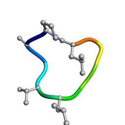
7UBC
DownloadVisualize
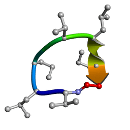
BU of 7ubc by Molmil
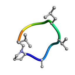
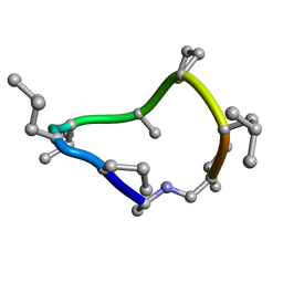
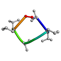
Solution NMR structure of 9-residue Rosetta-designed cyclic peptide D9.16 in d6-DMSO with cis/trans switching
Descriptor: Cyclic peptide D9.16 DPR-MAA-ALA-DVA-MLE-LEU-LEU-PRO-DLE
Authors:Ramelot, T.A, Tejero, R, Monteltione, G.T.
Deposit date:2022-03-14
Release date:2022-09-14
Last modified:2022-09-28
Method:SOLUTION NMR
Cite:Accurate de novo design of membrane-traversing macrocycles.
Cell, 185, 2022
7UBE
DownloadVisualize
BU of 7ube by Molmil
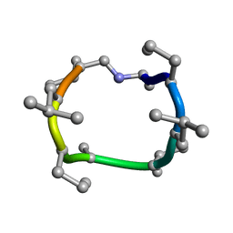
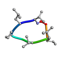
Solution NMR structure of 8-residue Rosetta-designed cyclic peptide D8.21 in d6-DMSO with cis/trans switching
Descriptor: Cyclic peptide D8.21 DVA-MLE-DPR-LEU-DVA-MLE-DPR-LEU
Authors:Ramelot, T.A, Tejero, R, Montelione, G.T.
Deposit date:2022-03-14
Release date:2022-09-14
Last modified:2022-09-28
Method:SOLUTION NMR
Cite:Accurate de novo design of membrane-traversing macrocycles.
Cell, 185, 2022
7UBG
DownloadVisualize
BU of 7ubg by Molmil
Solution NMR structure of 9-residue Rosetta-designed cyclic peptide D9.16 in CDCl3 with cis/trans switching (A-TT conformation)
Descriptor: Cyclic peptide D9.16 DPR-MAA-ALA-DVA-MLE-LEU-LEU-PRO-DLE
Authors:Ramelot, T.A, Tejero, R, Montelione, G.T.
Deposit date:2022-03-14
Release date:2022-09-14
Last modified:2022-09-28
Method:SOLUTION NMR
Cite:Accurate de novo design of membrane-traversing macrocycles.
Cell, 185, 2022
7UBH
DownloadVisualize
BU of 7ubh by Molmil
Solution NMR structure of 8-residue Rosetta-designed cyclic peptide D8.31 in CDCl3 with cis/trans switching
Descriptor: Cyclic peptide D8.31 DAL-DPR-MLU-DVA-DAL-DPR-MLU-DVA
Authors:Ramelot, T.A, Tejero, R, Montelione, G.T.
Deposit date:2022-03-14
Release date:2022-09-14
Last modified:2022-09-28
Method:SOLUTION NMR
Cite:Accurate de novo design of membrane-traversing macrocycles.
Cell, 185, 2022
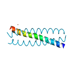
6EGO
DownloadVisualize
BU of 6ego by Molmil
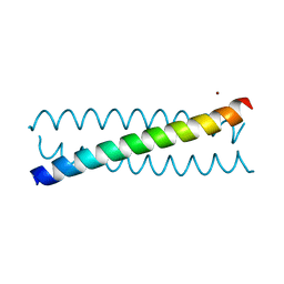
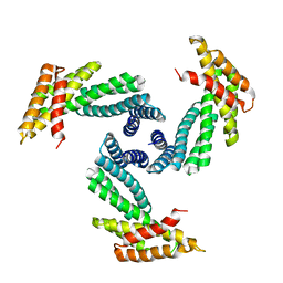
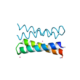
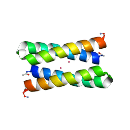
Crystal Structure of a de Novo Three-stranded Coiled Coil Peptide Containing an Ala Residue in the Second Coordination Sphere of the Hg(II)S3 Binding Site
Descriptor: Hg(II)(GRAND CoilSerL12AL16C)3-, MERCURY (II) ION, ZINC ION
Authors:Ruckthong, L, Stuckey, J.A, Pecoraro, V.L.
Deposit date:2018-08-20
Release date:2019-04-03
Last modified:2023-10-11
Method:X-RAY DIFFRACTION (1.93 Å)
Cite:How Outer Coordination Sphere Modifications Can Impact Metal Structures in Proteins: A Crystallographic Evaluation.
Chemistry, 25, 2019
6EGL
DownloadVisualize
BU of 6egl by Molmil
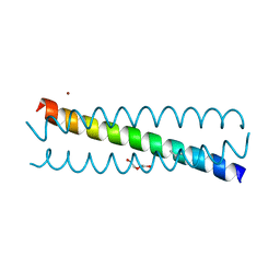
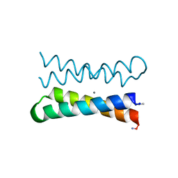
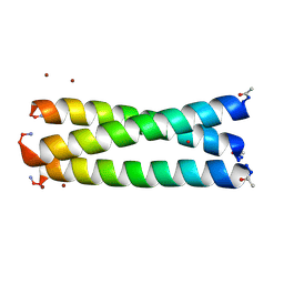
Crystal Structure of a de Novo Three-stranded Coiled Coil Peptide Containing a D-Leu in the Second Coordination Sphere of a Non-metalated Tris-thiolate Binding Site
Descriptor: Apo-(GRAND CoilSerL12(DLE)L16C)3, POLYETHYLENE GLYCOL (N=34), ZINC ION
Authors:Ruckthong, L, Stuckey, J.A, Pecoraro, V.L.
Deposit date:2018-08-20
Release date:2019-04-03
Last modified:2023-10-11
Method:X-RAY DIFFRACTION (1.4 Å)
Cite:How Outer Coordination Sphere Modifications Can Impact Metal Structures in Proteins: A Crystallographic Evaluation.
Chemistry, 25, 2019
1QCZ
DownloadVisualize
BU of 1qcz by Molmil
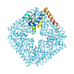
CRYSTAL STRUCTURE OF E. COLI PURE, AN UNUSUAL MUTASE THAT CATALYZES THE CONVERSION OF N5-CARBOXYAMINOIMIDAZOLE RIBONUCLEOTIDE (N5-CAIR) TO 4-CARBOXYAMINOIMIDAZOLE RIBONUCLEOTIDE (CAIR) IN THE PURINE BIOSYNTHETIC PATHWAY
Descriptor: N5-CARBOXYAMINOIMIDAZOLE RIBONUCLEOTIDE MUTASE
Authors:Ealick, S.E, Mathews, I.I.
Deposit date:1999-05-10
Release date:1999-11-10
Last modified:2018-01-31
Method:X-RAY DIFFRACTION (1.5 Å)
Cite:Crystal structure of Escherichia coli PurE, an unusual mutase in the purine biosynthetic pathway.
Structure Fold.Des., 7, 1999
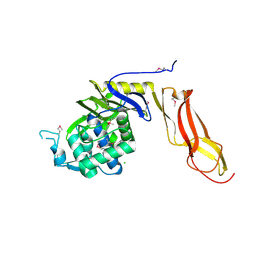
7BG1
DownloadVisualize
BU of 7bg1 by Molmil
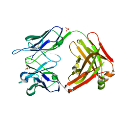
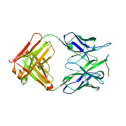
Structure of anti-FLAG M2 Fab domain remodeled based on proteomic sequencing
Descriptor: CHLORIDE ION, SULFATE ION, anti-FLAG M2 heavy chain, ...
Authors:Pronker, M.F, Snijder, J.
Deposit date:2021-01-05
Release date:2021-07-21
Last modified:2024-01-31
Method:X-RAY DIFFRACTION (1.86 Å)
Cite:Mass Spectrometry-Based De Novo Sequencing of Monoclonal Antibodies Using Multiple Proteases and a Dual Fragmentation Scheme.
J.Proteome Res., 20, 2021
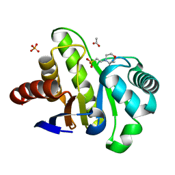
4F2V
DownloadVisualize
BU of 4f2v by Molmil
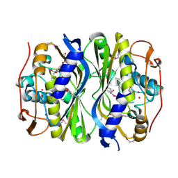
Crystal Structure of de novo designed serine hydrolase, Northeast Structural Genomics Consortium (NESG) Target OR165
Descriptor: DI(HYDROXYETHYL)ETHER, DODECYL-ALPHA-D-MALTOSIDE, De novo designed serine hydrolase
Authors:Kuzin, A, Lew, S, Seetharaman, J, Maglaqui, M, Xiao, R, Kohan, E, Rajagopalan, S, Everett, J.K, Acton, T.B, Montelione, G.T, Tong, L, Hunt, J.F, Northeast Structural Genomics Consortium (NESG)
Deposit date:2012-05-08
Release date:2012-05-30
Last modified:2023-12-06
Method:X-RAY DIFFRACTION (2.493 Å)
Cite:Design of activated serine-containing catalytic triads with atomic-level accuracy.
Nat.Chem.Biol., 10, 2014
8CTO
DownloadVisualize
BU of 8cto by Molmil
Solution NMR structure of 8-residue Rosetta-designed cyclic peptide D8.31 in d6-DMSO with cis/trans switching (B-CT conformation)
Descriptor: Cyclic peptide D8.31 DAL-DPR-MLU-DVA-DAL-DPR-MLU-DVA
Authors:Ramelot, T.A, Tejero, R, Montelione, G.T.
Deposit date:2022-05-16
Release date:2022-09-14
Last modified:2022-09-28
Method:SOLUTION NMR
Cite:Accurate de novo design of membrane-traversing macrocycles.
Cell, 185, 2022
4J19
DownloadVisualize
BU of 4j19 by Molmil
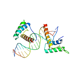
Structure of a novel telomere repeat binding protein bound to DNA
Descriptor: CHLORIDE ION, DNA (5'-D(*CP*TP*GP*TP*TP*AP*GP*GP*GP*TP*TP*AP*GP*GP*GP*TP*TP*AP*G)-3'), DNA (5'-D(*TP*CP*TP*AP*AP*CP*CP*CP*TP*AP*AP*CP*CP*CP*TP*AP*AP*CP*A)-3'), ...
Authors:Kappei, D, Butter, F, Benda, C, Scheibe, M, Draskovic, I, Stevense, M, Lopes Novo, C, Basquin, C, Krastev, D.B, Kittler, R, Jessberger, R, Londono-Vallejo, A.J, Mann, M, Buchholz, F.
Deposit date:2013-02-01
Release date:2013-05-29
Last modified:2023-09-20
Method:X-RAY DIFFRACTION (2.9 Å)
Cite:HOT1 is a mammalian direct telomere repeat-binding protein contributing to telomerase recruitment.
Embo J., 32, 2013
8CUN
DownloadVisualize
BU of 8cun by Molmil
Solution NMR structure of 8-residue Rosetta-designed cyclic peptide D8.21 in 50% d6-DMSO and 50% water with cis/trans switching (CC conformation, 50%)
Descriptor: Cyclic peptide D8.21 DVA-MLE-DPR-LEU-DVA-MLE-DPR-LEU
Authors:Ramelot, T.A, Tejero, R, Montelione, G.T.
Deposit date:2022-05-17
Release date:2022-09-14
Last modified:2022-09-28
Method:SOLUTION NMR
Cite:Accurate de novo design of membrane-traversing macrocycles.
Cell, 185, 2022
8E1E
DownloadVisualize
BU of 8e1e by Molmil
Scaffolding protein functional sites using deep learning
Descriptor: SG122_C3
Authors:Bera, A.K, Gerben, S, Baker, D.
Deposit date:2022-08-10
Release date:2023-08-16
Method:X-RAY DIFFRACTION (4.27 Å)
Cite:Design of Diverse Asymmetric Pockets in De Novo Homo-oligomeric Proteins.
Biochemistry, 62, 2023
8CWA
DownloadVisualize
BU of 8cwa by Molmil
Solution NMR structure of 8-residue Rosetta-designed cyclic peptide D8.21 in CDCl3 with cis/trans switching (TC conformation, 53%)
Descriptor: Cyclic peptide D8.21 DVA-MLE-DPR-LEU-DVA-MLE-DPR-LEU
Authors:Ramelot, T.A, Tejero, R, Montelione, G.T.
Deposit date:2022-05-18
Release date:2022-09-14
Last modified:2022-09-28
Method:SOLUTION NMR
Cite:Accurate de novo design of membrane-traversing macrocycles.
Cell, 185, 2022
1OVU
DownloadVisualize
BU of 1ovu by Molmil
CRYSTAL STRUCTURE OF FOUR-HELIX BUNDLE MODEL di-Co(II)-DF1-L13A (form I)
Descriptor: COBALT (II) ION, four-helix bundle model di-Co(II)-DF1-L13A (form I)
Authors:Di Costanzo, L, Geremia, S.
Deposit date:2003-03-27
Release date:2004-04-06
Last modified:2019-11-20
Method:X-RAY DIFFRACTION (3.1 Å)
Cite:Response of a designed metalloprotein to changes in metal ion coordination, exogenous ligands, and active site volume determined by X-ray crystallography.
J.Am.Chem.Soc., 127, 2005
1OVR
DownloadVisualize
BU of 1ovr by Molmil
CRYSTAL STRUCTURE OF FOUR-HELIX BUNDLE MODEL di-Mn(II)-DF1-L13
Descriptor: MANGANESE (II) ION, four-helix bundle model di-Mn(II)-DF1-L13
Authors:Di Costanzo, L, Geremia, S.
Deposit date:2003-03-27
Release date:2004-05-18
Last modified:2017-10-11
Method:X-RAY DIFFRACTION (2.99 Å)
Cite:Response of a designed metalloprotein to changes in metal ion coordination, exogenous ligands, and active site volume determined by X-ray crystallography.
J.Am.Chem.Soc., 127, 2005
3Q7W
DownloadVisualize
BU of 3q7w by Molmil
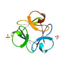
Crystal structure of Symfoil-4P/PV1: de novo designed beta-trefoil architecture with symmetric primary structure, primitive version 1
Descriptor: SULFATE ION, de novo designed beta-trefoil architecture with symmetric primary structure
Authors:Blaber, M, Lee, J.
Deposit date:2011-01-05
Release date:2012-01-11
Last modified:2024-02-21
Method:X-RAY DIFFRACTION (1.5 Å)
Cite:Simplified protein design biased for prebiotic amino acids yields a foldable, halophilic protein.
Proc.Natl.Acad.Sci.USA, 110, 2013
3Q7X
DownloadVisualize
BU of 3q7x by Molmil
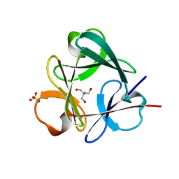
Crystal structure of Symfoil-4P/PV1: de novo designed beta-trefoil architecture with symmetric primary structure, primitive version 1
Descriptor: 2-AMINO-2-HYDROXYMETHYL-PROPANE-1,3-DIOL, SULFATE ION, de novo designed beta-trefoil architecture with symmetric primary structure
Authors:Blaber, M, Lee, J.
Deposit date:2011-01-05
Release date:2012-01-11
Last modified:2024-02-21
Method:X-RAY DIFFRACTION (1.4 Å)
Cite:Simplified protein design biased for prebiotic amino acids yields a foldable, halophilic protein.
Proc.Natl.Acad.Sci.USA, 110, 2013
1OVV
DownloadVisualize
BU of 1ovv by Molmil
CRYSTAL STRUCTURE OF FOUR-HELIX BUNDLE MODEL di-Co(II)-DF1-L13A (form II)
Descriptor: COBALT (II) ION, FOUR-HELIX BUNDLE MODEL di-Co(II)-DF1-L13A (form II)
Authors:Di Costanzo, L, Geremia, S.
Deposit date:2003-03-27
Release date:2004-04-06
Last modified:2019-11-20
Method:X-RAY DIFFRACTION (2.9 Å)
Cite:Response of a designed metalloprotein to changes in metal ion coordination, exogenous ligands, and active site volume determined by X-ray crystallography.
J.Am.Chem.Soc., 127, 2005
8P6I
DownloadVisualize
BU of 8p6i by Molmil
Crystal structure of the 139H2 Fab fragment bound to Muc1 peptide epitope
Descriptor: 139H2 HC, 139H2 LC, Mucin-1
Authors:Beugelink, J.W, Peng, W, Siborova, M, Pronker, M.F, Snijder, J, Janssen, B.J.C.
Deposit date:2023-05-26
Release date:2024-04-03
Last modified:2024-04-10
Method:X-RAY DIFFRACTION (2.5 Å)
Cite:Reverse-engineering the anti-MUC1 antibody 139H2 by mass spectrometry-based de novo sequencing.
Life Sci Alliance, 7, 2024
2JGO
DownloadVisualize
BU of 2jgo by Molmil
Structure of the arsenated de novo designed peptide Coil Ser L9C
Descriptor: ARSENIC, COIL SER L9C, ZINC ION
Authors:Touw, D.S, Nordman, C.E, Stuckey, J.A, Pecoraro, V.L.
Deposit date:2007-02-13
Release date:2007-07-10
Last modified:2023-12-13
Method:X-RAY DIFFRACTION (1.81 Å)
Cite:Identifying Important Structural Characteristics of Arsenic Resistance Proteins by Using Designed Three-Stranded Coiled Coils.
Proc.Natl.Acad.Sci.USA, 104, 2007
4G4L
DownloadVisualize
BU of 4g4l by Molmil
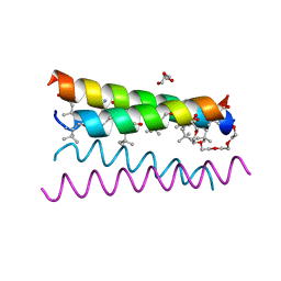
Crystal structure of the de novo designed peptide alpha4tbA6
Descriptor: ACETYL GROUP, DI(HYDROXYETHYL)ETHER, HEXAETHYLENE GLYCOL, ...
Authors:Buer, B.C, Meagher, J.L, Stuckey, J.A, Marsh, E.N.G.
Deposit date:2012-07-16
Release date:2012-10-31
Last modified:2017-11-15
Method:X-RAY DIFFRACTION (1.54 Å)
Cite:Comparison of the structures and stabilities of coiled-coil proteins containing hexafluoroleucine and t-butylalanine provides insight into the stabilizing effects of highly fluorinated amino acid side-chains.
Protein Sci., 21, 2012
4DRT
DownloadVisualize
BU of 4drt by Molmil
Three dimensional structure of de novo designed serine hydrolase OSH26, Northeast Structural Genomics Consortium (NESG) target OR89
Descriptor: CHLORIDE ION, SODIUM ION, de novo designed serine hydrolase, ...
Authors:Kuzin, A, Su, M, Rajagopalan, S, Seetharaman, J, Sahdev, S, Xiao, R, Ciccosanti, C, Baker, D, Everett, J.K, Acton, T.B, Montelione, G.T, Tong, L, Hunt, J.F, Northeast Structural Genomics Consortium (NESG)
Deposit date:2012-02-17
Release date:2012-04-18
Last modified:2023-12-06
Method:X-RAY DIFFRACTION (2.002 Å)
Cite:Design of activated serine-containing catalytic triads with atomic-level accuracy.
Nat.Chem.Biol., 10, 2014
6EGM
DownloadVisualize
BU of 6egm by Molmil
Crystal Structure of a de novo Three-stranded Coiled Coil Peptide Containing D-Leu in a d-site Position of a Tris-thiolate Binding Site
Descriptor: Apo-(GRAND CoilSerL16CL19(DLE))3, ZINC ION
Authors:Ruckthong, L, Stuckey, J.A, Pecoraro, V.L.
Deposit date:2018-08-20
Release date:2019-04-03
Last modified:2019-12-18
Method:X-RAY DIFFRACTION (1.84 Å)
Cite:How Outer Coordination Sphere Modifications Can Impact Metal Structures in Proteins: A Crystallographic Evaluation.
Chemistry, 25, 2019
4GVW
DownloadVisualize
BU of 4gvw by Molmil
Three-dimensional structure of the de novo designed serine hydrolase 2bfq_3, Northeast Structural Genomics Consortium (NESG) Target OR248
Descriptor: 2-(N-MORPHOLINO)-ETHANESULFONIC ACID, ACETIC ACID, De novo designed serine hydrolase, ...
Authors:Kuzin, A, Lew, S, Seetharaman, J, Rajagopalan, S, Everett, J.K, Acton, T.B, Montelione, G.T, Tong, L, Hunt, J.F, Northeast Structural Genomics Consortium (NESG)
Deposit date:2012-08-31
Release date:2012-09-12
Last modified:2023-09-13
Method:X-RAY DIFFRACTION (2.113 Å)
Cite:Northeast Structural Genomics Consortium Target OR248
To be Published

222926

数据于2024-07-24公开中

PDB statisticsPDBj update infoContact PDBjnumon