Loading
PDBj
MenuPDBj@FacebookPDBj@TwitterPDBj@YouTubewwPDB FoundationwwPDB
RCSB PDBPDBeBMRBAdv. SearchSearch help

6DN7
DownloadVisualize
BU of 6dn7 by Molmil
SPRY domain-containing SOCS box protein 2 complexed with WDINNN(BAL) Cyclic peptide inhibitor
Descriptor: CHLORIDE ION, SPRY domain-containing SOCS box protein 4, WDINNN(BAL) Cyclic peptide inhibitor
Authors:Law, R.H.P, Caradoc-Davies, T.T, Norton, R.S.
Deposit date:2018-06-06
Release date:2019-04-17
Last modified:2020-01-08
Method:X-RAY DIFFRACTION (1.4 Å)
Cite:A Cyclic Peptide Inhibitor of the iNOS-SPSB Protein-Protein Interaction as a Potential Anti-Infective Agent.
ACS Chem. Biol., 13, 2018
2IHS
DownloadVisualize
BU of 2ihs by Molmil
Crystal structure of the B30.2/SPRY domain of GUSTAVUS in complex with a 20-residue VASA peptide
Descriptor: 20-mer from ATP-dependent RNA helicase vasa, CG2944-PF, isoform F
Authors:Woo, J.S, Park, S.Y, Oh, B.H.
Deposit date:2006-09-27
Release date:2007-01-16
Last modified:2023-10-25
Method:X-RAY DIFFRACTION (2.2 Å)
Cite:Structural Basis for Protein Recognition by B30.2/SPRY Domains
Mol.Cell, 24, 2006
2JK9
DownloadVisualize
BU of 2jk9 by Molmil
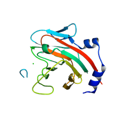
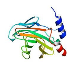
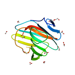
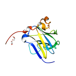
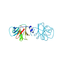
The structure of splA-ryanodine receptor domain and SOCS box containing 1 in complex with a PAR-4 peptide
Descriptor: PRKC APOPTOSIS WT1 REGULATOR PROTEIN, SPRY DOMAIN-CONTAINING SOCS BOX PROTEIN 1
Authors:Filippakopoulos, P, Bullock, A, Keates, T, Savitsky, P, Murray, J.W, von Delft, F, Arrowsmith, C.H, Edwards, A.M, Wickstroem, M, Bountra, C, Knapp, S.
Deposit date:2008-08-22
Release date:2008-09-16
Last modified:2023-12-13
Method:X-RAY DIFFRACTION (1.79 Å)
Cite:Structural Basis for Par-4 Recognition by the Spry Domain-and Socs Box-Containing Proteins Spsb1, Spsb2, and Spsb4.
J.Mol.Biol., 401, 2010
5JI7
DownloadVisualize
BU of 5ji7 by Molmil
The Crystal Structure Of IUS-SPRY Domain From RanBPM/9
Descriptor: Ran-binding protein 9
Authors:Hong, S.K, Kim, K.-H, Kim, E.E.
Deposit date:2016-04-22
Release date:2016-11-09
Last modified:2024-03-20
Method:X-RAY DIFFRACTION (1.51 Å)
Cite:Structural Basis for the Interaction between the IUS-SPRY Domain of RanBPM and DDX-4 in Germ Cell Development.
J.Mol.Biol., 428, 2016
5JI9
DownloadVisualize
BU of 5ji9 by Molmil
The Crystal Structure Of IUS-SPRY Domain From RanBPM/9
Descriptor: Ran-binding protein 9
Authors:Hong, S.K, Kim, K.-H, Kim, E.E.
Deposit date:2016-04-22
Release date:2016-11-09
Last modified:2023-11-08
Method:X-RAY DIFFRACTION (2.5 Å)
Cite:Structural Basis for the Interaction between the IUS-SPRY Domain of RanBPM and DDX-4 in Germ Cell Development.
J.Mol.Biol., 428, 2016
5JIU
DownloadVisualize
BU of 5jiu by Molmil
The crystal structure of RanBPM/9 IUS-SPRY domain in complex with DDX-4 peptide
Descriptor: CHLORIDE ION, Probable ATP-dependent RNA helicase DDX4, Ran-binding protein 9
Authors:Hong, S.K, Kim, K.-H, Kim, E.E.
Deposit date:2016-04-22
Release date:2016-11-09
Last modified:2023-11-08
Method:X-RAY DIFFRACTION (2.054 Å)
Cite:Structural Basis for the Interaction between the IUS-SPRY Domain of RanBPM and DDX-4 in Germ Cell Development.
J.Mol.Biol., 428, 2016
5JIA
DownloadVisualize
BU of 5jia by Molmil
The Crystal Structure Of IUS-SPRY Domain From RanBP10
Descriptor: IMIDAZOLE, Ran-binding protein 10
Authors:Hong, S.K, Kim, K.-H, Kim, E.E.
Deposit date:2016-04-22
Release date:2016-11-09
Last modified:2023-11-08
Method:X-RAY DIFFRACTION (1.8 Å)
Cite:Structural Basis for the Interaction between the IUS-SPRY Domain of RanBPM and DDX-4 in Germ Cell Development.
J.Mol.Biol., 428, 2016
7QRZ
DownloadVisualize
BU of 7qrz by Molmil
Crystal structure of B30.2 PRYSPRY domain of MID2
Descriptor: 1,2-ETHANEDIOL, Probable E3 ubiquitin-protein ligase MID2, SULFATE ION
Authors:Chaikuad, A, Zhubi, R, Knapp, S, Structural Genomics Consortium (SGC)
Deposit date:2022-01-12
Release date:2022-05-04
Last modified:2024-01-31
Method:X-RAY DIFFRACTION (1.57 Å)
Cite:Crystal structure of B30.2 PRYSPRY domain of MID2
To Be Published
7QS5
DownloadVisualize
BU of 7qs5 by Molmil
Crystal structure of B30.2 PRYSPRY domain of TRIM67
Descriptor: 1,2-ETHANEDIOL, Tripartite motif-containing protein 67
Authors:Chaikuad, A, Zhubi, R, Knapp, S, Structural Genomics Consortium (SGC)
Deposit date:2022-01-12
Release date:2022-05-04
Last modified:2024-01-31
Method:X-RAY DIFFRACTION (1.65 Å)
Cite:Crystal structure of B30.2 PRYSPRY domain of TRIM67
To Be Published
7QRY
DownloadVisualize
BU of 7qry by Molmil
Crystal structure of B30.2 PRYSPRY domain of MID1
Descriptor: E3 ubiquitin-protein ligase Midline-1
Authors:Chaikuad, A, Knapp, S, Structural Genomics Consortium (SGC)
Deposit date:2022-01-12
Release date:2022-05-04
Last modified:2024-01-31
Method:X-RAY DIFFRACTION (2.07 Å)
Cite:Crystal structure of B30.2 PRYSPRY domain of MID1
To Be Published
6J6L
DownloadVisualize
BU of 6j6l by Molmil
Crystal structure of mouse Ryanodine Receptor 2 SPRY1 Domain (650-844) disease mutant I784F
Descriptor: ISOPROPYL ALCOHOL, Ryanodine receptor 2
Authors:Ma, R, Lin, L, Yuchi, Z.
Deposit date:2019-01-15
Release date:2020-01-15
Last modified:2023-11-22
Method:X-RAY DIFFRACTION (1.452 Å)
Cite:A SPRY1 domain cardiac ryanodine receptor variant associated with short-coupled torsade de pointes.
Sci Rep, 11, 2021
6JKJ
DownloadVisualize
BU of 6jkj by Molmil
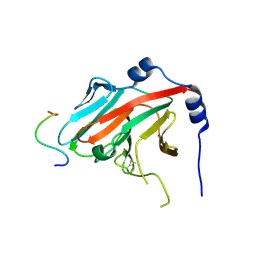
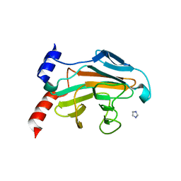
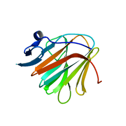
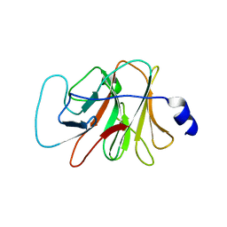
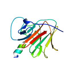
Crystal structure of human SPSB2 in the apo-state
Descriptor: SPRY domain-containing SOCS box protein 2
Authors:Luo, Y, Kuang, Z.
Deposit date:2019-03-01
Release date:2019-07-03
Last modified:2023-11-22
Method:X-RAY DIFFRACTION (1.9 Å)
Cite:Crystal structure of the SPRY domain of human SPSB2 in the apo state.
Acta Crystallogr.,Sect.F, 75, 2019
6KEY
DownloadVisualize
BU of 6key by Molmil
Structural basis for the regulation of inducible nitric oxide synthase (iNOS) by the SPRY domain-containing SOCS box protein 2 (SPSB2)
Descriptor: Nitric oxide synthase, inducible, SPRY domain-containing SOCS box protein 2
Authors:Li, K, Kuang, Z.
Deposit date:2019-07-05
Release date:2020-07-01
Last modified:2024-03-27
Method:X-RAY DIFFRACTION (1.24 Å)
Cite:Structural basis for the regulation of inducible nitric oxide synthase by the SPRY domain-containing SOCS box protein SPSB2, an E3 ubiquitin ligase.
Nitric Oxide, 113-114, 2021
6LT9
DownloadVisualize
BU of 6lt9 by Molmil
The crystal structure of diamondback moth ryanodine receptor SPRY1 domain
Descriptor: GLYCEROL, Ryanodine receptor 1, SODIUM ION
Authors:Zhou, Y, Lin, L, Yuchi, Z.
Deposit date:2020-01-22
Release date:2021-01-27
Last modified:2023-11-29
Method:X-RAY DIFFRACTION (1.801 Å)
Cite:The crystal structure of diamondback moth ryanodine receptor SPRY1 domain
To Be Published
6JWM
DownloadVisualize
BU of 6jwm by Molmil
Crystal structure of the SPRY domain of SPSB2 in complex with cR7, a potent cyclic peptide inhibitor of SPSB2-iNOS interaction
Descriptor: Nitric oxide synthase, inducible, SPRY domain-containing SOCS box protein 2
Authors:Li, K, Kuang, Z.
Deposit date:2019-04-21
Release date:2020-04-22
Last modified:2024-03-27
Method:X-RAY DIFFRACTION (1.23 Å)
Cite:Crystal structure of SPSB2 in complex with cR7, a potent cyclic peptide inhibitor of SPSB2-iNOS interaction
To Be Published
6JWN
DownloadVisualize
BU of 6jwn by Molmil
Crystal structure of the SPRY domain of SPSB2 in complex with cR9, a cyclic peptide inhibitor of SPSB-iNOS interaction
Descriptor: Nitric oxide synthase, inducible, SPRY domain-containing SOCS box protein 2
Authors:Li, K, Kuang, Z.
Deposit date:2019-04-21
Release date:2020-04-22
Last modified:2024-03-27
Method:X-RAY DIFFRACTION (1.61 Å)
Cite:Crystal structure of SPSB2 in complex with cR9, a cyclic peptide inhibitor of SPSB-iNOS interaction
To Be Published
6KIM
DownloadVisualize
BU of 6kim by Molmil
Crystal structure of diamondback moth ryanodine receptor SPRY2 domain
Descriptor: GLYCEROL, Ryanodine receptor
Authors:Zhou, Y, Lin, L, Yuchi, Z.
Deposit date:2019-07-19
Release date:2020-07-22
Last modified:2023-11-22
Method:X-RAY DIFFRACTION (2.057 Å)
Cite:Crystal structure of diamondback moth ryanodine receptor SPRY2 domain
To Be Published
3KB5
DownloadVisualize
BU of 3kb5 by Molmil
PRY-SPRY domain of human TRIM72
Descriptor: Tripartite motif-containing protein 72
Authors:Park, E.Y, Kwon, O.-B, Jeong, B.-C, Song, H.K.
Deposit date:2009-10-20
Release date:2009-12-22
Last modified:2024-03-20
Method:X-RAY DIFFRACTION (1.5 Å)
Cite:Crystal structure of PRY-SPRY domain of human TRIM72
Proteins, 2009
8HJT
DownloadVisualize
BU of 8hjt by Molmil
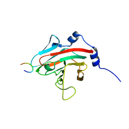
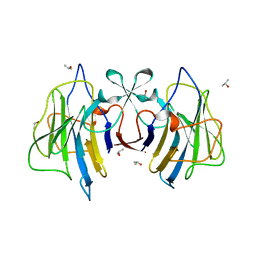
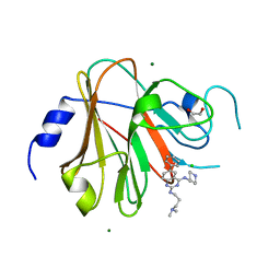
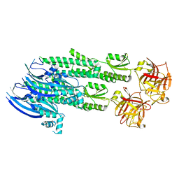
Crystal Structure of Intracellular B30.2 Domain of VpBTN3 and VpBTN2 in Complex with HMBPP
Descriptor: (2E)-4-hydroxy-3-methylbut-2-en-1-yl trihydrogen diphosphate, 4-(2-HYDROXYETHYL)-1-PIPERAZINE ETHANESULFONIC ACID, Butyrophylin 3, ...
Authors:Yang, Y.Y, Shen, P.P, Li, X, Yi, S.M, Zhang, M.T, Huang, J.-W, Chen, C.-C, Guo, R.-T.
Deposit date:2022-11-23
Release date:2023-09-13
Last modified:2023-10-18
Method:X-RAY DIFFRACTION (2.91 Å)
Cite:Phosphoantigens glue butyrophilin 3A1 and 2A1 to activate V gamma 9V delta 2 T cells.
Nature, 621, 2023
4UWE
DownloadVisualize
BU of 4uwe by Molmil
Structure of the ryanodine receptor at resolution of 8.5 A in partially open state
Descriptor: RYANODINE RECEPTOR 1
Authors:Efremov, R.G, Leitner, A, Aebersold, R, Raunser, S.
Deposit date:2014-08-11
Release date:2014-12-10
Last modified:2024-05-08
Method:ELECTRON MICROSCOPY (8.5 Å)
Cite:Architecture and Conformational Switch Mechanism of the Ryanodine Receptor.
Nature, 517, 2015
8PD6
DownloadVisualize
BU of 8pd6 by Molmil
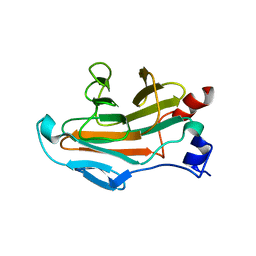
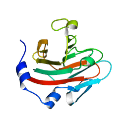
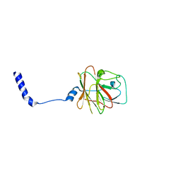
Crystal structure of the TRIM58 PRY-SPRY domain in complex with TRIM-473
Descriptor: 1,2-ETHANEDIOL, CHLORIDE ION, E3 ubiquitin-protein ligase TRIM58, ...
Authors:Renatus, M, Hoegenauer, K, Schroeder, M.
Deposit date:2023-06-11
Release date:2024-01-31
Last modified:2024-02-07
Method:X-RAY DIFFRACTION (1.3 Å)
Cite:Discovery of Ligands for TRIM58, a Novel Tissue-Selective E3 Ligase.
Acs Med.Chem.Lett., 14, 2023
8PD4
DownloadVisualize
BU of 8pd4 by Molmil
Crystal structure of TRIM58 PRY-SPRY domain
Descriptor: E3 ubiquitin-protein ligase TRIM58
Authors:Renatus, M, Schroeder, M.
Deposit date:2023-06-11
Release date:2024-01-31
Last modified:2024-02-07
Method:X-RAY DIFFRACTION (2.714 Å)
Cite:Discovery of Ligands for TRIM58, a Novel Tissue-Selective E3 Ligase.
Acs Med.Chem.Lett., 14, 2023
4V1P
DownloadVisualize
BU of 4v1p by Molmil
BTN3 Structure
Descriptor: BUTYROPHILIN SUBFAMILY 3 MEMBER A1
Authors:James, L.C.
Deposit date:2014-09-30
Release date:2015-07-22
Last modified:2024-05-08
Method:X-RAY DIFFRACTION (2.04 Å)
Cite:Activation of Human Gammadelta T Cells by Cytosolic Interactions of Btn3A1 with Soluble Phosphoantigens and the Cytoskeletal Adaptor Periplakin.
J.Immunol., 194, 2015
4WVM
DownloadVisualize
BU of 4wvm by Molmil
Stonustoxin structure
Descriptor: Stonustoxin subunit alpha, Stonustoxin subunit beta
Authors:Ellisdon, A.M, Panjikar, S, Whisstock, J.C, McGowan, S.
Deposit date:2014-11-06
Release date:2015-12-02
Last modified:2023-12-27
Method:X-RAY DIFFRACTION (3.1 Å)
Cite:Stonefish toxin defines an ancient branch of the perforin-like superfamily.
Proc.Natl.Acad.Sci.USA, 112, 2015
7OVX
DownloadVisualize
BU of 7ovx by Molmil
E3 RING ligase binding domain
Descriptor: E3 ubiquitin-protein ligase TRIM7, Peptide G
Authors:James, L.C.
Deposit date:2021-06-15
Release date:2022-06-22
Last modified:2024-01-31
Method:X-RAY DIFFRACTION (1.7 Å)
Cite:E3 ligase targeting domain
To Be Published

223532

건을2024-08-07부터공개중

PDB statisticsPDBj update infoContact PDBjnumon