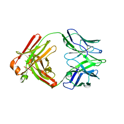
Loading
PDBj
MenuPDBj@FacebookPDBj@TwitterPDBj@YouTubewwPDB FoundationwwPDB
RCSB PDBPDBeBMRBAdv. SearchSearch help

3LW0
DownloadVisualize
BU of 3lw0 by Molmil
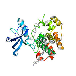
IGF-1RK in complex with ligand MSC1609119A-1
Descriptor: 3-cyano-N-{1-[4-(5-cyano-1H-indol-3-yl)butyl]piperidin-4-yl}-1H-indole-7-carboxamide, GLYCEROL, Insulin-like growth factor 1 receptor
Authors:Graedler, U, Heinrich, T, Boettcher, H, Blaukat, A, Shutes, A, Askew, B.
Deposit date:2010-02-23
Release date:2010-09-29
Last modified:2023-11-01
Method:X-RAY DIFFRACTION (1.79 Å)
Cite:Allosteric IGF-1R Inhibitors.
Acs Med.Chem.Lett., 1, 2010
3LL3
DownloadVisualize
BU of 3ll3 by Molmil
The crystal structure of ligand bound xylulose kinase from Lactobacillus acidophilus
Descriptor: 1-DEOXY-D-XYLULOSE-5-PHOSPHATE, ADENOSINE-5'-DIPHOSPHATE, ADENOSINE-5'-TRIPHOSPHATE, ...
Authors:Zhang, Z, Burley, S.K, Swaminathan, S, New York SGX Research Center for Structural Genomics (NYSGXRC)
Deposit date:2010-01-28
Release date:2010-03-23
Last modified:2023-11-22
Method:X-RAY DIFFRACTION (2.002 Å)
Cite:The crystal structure of xylulose kinase from Lactobacillus acidophilus
To be Published
3LT8
DownloadVisualize
BU of 3lt8 by Molmil
A non-biological ATP binding protein with a single point mutation (D65V), that contributes to optimized folding and ligand binding, crystallized in the presence of 100 mM ATP.
Descriptor: ADENOSINE-5'-DIPHOSPHATE, ATP BINDING PROTEIN-D65V, CHLORIDE ION, ...
Authors:Simmons, C.R, Magee, C.L, Allen, J.P, Chaput, J.C.
Deposit date:2010-02-15
Release date:2010-09-22
Last modified:2023-09-06
Method:X-RAY DIFFRACTION (2.55 Å)
Cite:Three-dimensional structures reveal multiple ADP/ATP binding modes for a synthetic class of artificial proteins.
Biochemistry, 49, 2010
3LT9
DownloadVisualize
BU of 3lt9 by Molmil
A non-biological ATP binding protein with a single point mutation (D65V), that contributes to optimized folding and ligand binding
Descriptor: ADENOSINE-5'-DIPHOSPHATE, ATP BINDING PROTEIN-D65V, CHLORIDE ION, ...
Authors:Simmons, C.R, Magee, C.L, Allen, J.P, Chaput, J.C.
Deposit date:2010-02-15
Release date:2010-09-22
Last modified:2023-09-06
Method:X-RAY DIFFRACTION (2.55 Å)
Cite:Three-dimensional structures reveal multiple ADP/ATP binding modes for a synthetic class of artificial proteins.
Biochemistry, 49, 2010
4DT2
DownloadVisualize
BU of 4dt2 by Molmil
Crystal structure of red kidney bean purple acid phosphatase in complex with Maybridge fragment CC27209
Descriptor: (2,2-dimethyl-2,3-dihydro-1-benzofuran-7-yl)methanol, 1,2-ETHANEDIOL, 2-acetamido-2-deoxy-beta-D-glucopyranose, ...
Authors:Feder, D, Hussein, W.M, Clayton, D.J, Kan, M, Schenk, G, McGeary, R.P, Guddat, L.W.
Deposit date:2012-02-20
Release date:2012-09-19
Last modified:2020-07-29
Method:X-RAY DIFFRACTION (2.7 Å)
Cite:Identification of purple acid phosphatase inhibitors by fragment-based screening: promising new leads for osteoporosis therapeutics.
Chem.Biol.Drug Des., 80, 2012
6VG3
DownloadVisualize
BU of 6vg3 by Molmil
Structure of unliganded, inactive PTK7 kinase domain
Descriptor: Inactive tyrosine-protein kinase 7
Authors:Malhotra, K, Stayrook, S.E, Sheetz, J, Lemmon, M.A.
Deposit date:2020-01-07
Release date:2020-05-27
Last modified:2023-10-11
Method:X-RAY DIFFRACTION (1.95 Å)
Cite:Structural insights into receptor tyrosine kinase pseudokinase function
Mol.Cell, 2020
4YVC
DownloadVisualize
BU of 4yvc by Molmil
ROCK 1 bound to thiazole inhibitor
Descriptor: 2-fluoro-N-[4-(pyridin-4-yl)-1,3-thiazol-2-yl]benzamide, Rho-associated protein kinase 1
Authors:Jacobs, M.D.
Deposit date:2015-03-19
Release date:2015-06-10
Last modified:2023-09-27
Method:X-RAY DIFFRACTION (3.2 Å)
Cite:Design, Synthesis, and Structure-Activity Relationships of Pyridine-Based Rho Kinase (ROCK) Inhibitors.
J.Med.Chem., 58, 2015
3LTA
DownloadVisualize
BU of 3lta by Molmil
Crystal structure of a non-biological ATP binding protein with a TYR-PHE mutation within the ligand binding domain
Descriptor: ADENOSINE-5'-TRIPHOSPHATE, ATP BINDING PROTEIN-DX, CHLORIDE ION, ...
Authors:Simmons, C.R, Magee, C.L, Allen, J.P, Chaput, J.C.
Deposit date:2010-02-15
Release date:2010-09-22
Last modified:2023-09-06
Method:X-RAY DIFFRACTION (2.7 Å)
Cite:Three-dimensional structures reveal multiple ADP/ATP binding modes for a synthetic class of artificial proteins.
Biochemistry, 49, 2010
5QLV
DownloadVisualize
BU of 5qlv by Molmil
Group deposition of library data - Crystal Structure of EcDsbA after initial refinement with no ligand modelled (structure D2_1)
Descriptor: Thiol:disulfide interchange protein
Authors:Ilyichova, O.V, Bentley, M.R, Doak, B.C, Scanlon, M.J.
Deposit date:2019-01-27
Release date:2020-02-05
Last modified:2020-07-22
Method:X-RAY DIFFRACTION (2.341 Å)
Cite:Rapid Elaboration of Fragments into Leads by X-ray Crystallographic Screening of Parallel Chemical Libraries (REFiLX).
J.Med.Chem., 63, 2020
5QMA
DownloadVisualize
BU of 5qma by Molmil
Group deposition of library data - Crystal Structure of EcDsbA after initial refinement with no ligand modelled (structure E5_1)
Descriptor: Thiol:disulfide interchange protein
Authors:Ilyichova, O.V, Bentley, M.R, Doak, B.C, Scanlon, M.J.
Deposit date:2019-01-27
Release date:2020-02-05
Last modified:2020-07-22
Method:X-RAY DIFFRACTION (2.136 Å)
Cite:Rapid Elaboration of Fragments into Leads by X-ray Crystallographic Screening of Parallel Chemical Libraries (REFiLX).
J.Med.Chem., 63, 2020
5QMF
DownloadVisualize
BU of 5qmf by Molmil
Group deposition of library data - Crystal Structure of EcDsbA after initial refinement with no ligand modelled (structure F1_1)
Descriptor: Thiol:disulfide interchange protein
Authors:Ilyichova, O.V, Bentley, M.R, Doak, B.C, Scanlon, M.J.
Deposit date:2019-01-27
Release date:2020-02-05
Last modified:2020-07-22
Method:X-RAY DIFFRACTION (2.005 Å)
Cite:Rapid Elaboration of Fragments into Leads by X-ray Crystallographic Screening of Parallel Chemical Libraries (REFiLX).
J.Med.Chem., 63, 2020
5QMP
DownloadVisualize
BU of 5qmp by Molmil
Group deposition of library data - Crystal Structure of EcDsbA after initial refinement with no ligand modelled (structure F7_1)
Descriptor: Thiol:disulfide interchange protein
Authors:Ilyichova, O.V, Bentley, M.R, Doak, B.C, Scanlon, M.J.
Deposit date:2019-01-27
Release date:2020-02-05
Last modified:2020-07-22
Method:X-RAY DIFFRACTION (2.005 Å)
Cite:Rapid Elaboration of Fragments into Leads by X-ray Crystallographic Screening of Parallel Chemical Libraries (REFiLX).
J.Med.Chem., 63, 2020
5QMV
DownloadVisualize
BU of 5qmv by Molmil
Group deposition of library data - Crystal Structure of EcDsbA after initial refinement with no ligand modelled (structure G12_1)
Descriptor: Thiol:disulfide interchange protein
Authors:Ilyichova, O.V, Bentley, M.R, Doak, B.C, Scanlon, M.J.
Deposit date:2019-01-27
Release date:2020-02-05
Last modified:2020-07-22
Method:X-RAY DIFFRACTION (1.907 Å)
Cite:Rapid Elaboration of Fragments into Leads by X-ray Crystallographic Screening of Parallel Chemical Libraries (REFiLX).
J.Med.Chem., 63, 2020
5QN7
DownloadVisualize
BU of 5qn7 by Molmil
Group deposition of library data - Crystal Structure of EcDsbA after initial refinement with no ligand modelled (structure H1_1)
Descriptor: Thiol:disulfide interchange protein
Authors:Ilyichova, O.V, Bentley, M.R, Doak, B.C, Scanlon, M.J.
Deposit date:2019-01-27
Release date:2020-02-05
Last modified:2020-07-22
Method:X-RAY DIFFRACTION (2.136 Å)
Cite:Rapid Elaboration of Fragments into Leads by X-ray Crystallographic Screening of Parallel Chemical Libraries (REFiLX).
J.Med.Chem., 63, 2020
5QNC
DownloadVisualize
BU of 5qnc by Molmil
Group deposition of library data - Crystal Structure of EcDsbA after initial refinement with no ligand modelled (structure H6_1)
Descriptor: Thiol:disulfide interchange protein
Authors:Ilyichova, O.V, Bentley, M.R, Doak, B.C, Scanlon, M.J.
Deposit date:2019-01-27
Release date:2020-02-05
Last modified:2020-07-22
Method:X-RAY DIFFRACTION (2.14 Å)
Cite:Rapid Elaboration of Fragments into Leads by X-ray Crystallographic Screening of Parallel Chemical Libraries (REFiLX).
J.Med.Chem., 63, 2020
5IIE
DownloadVisualize
BU of 5iie by Molmil
STRUCTURE OF THE UNLIGANDED ANTI-HIV ANTIBODY DH501 THAT BINDS GP120 V3 GLYCAN AND THE BASE OF V3
Descriptor: antibody DH501 FabH chain, antibody DH501 light chain
Authors:Nicely, N.I, Haynes, B.F.
Deposit date:2016-03-01
Release date:2017-03-01
Last modified:2023-09-27
Method:X-RAY DIFFRACTION (2.8 Å)
Cite:Vaccine Elicitation of High Mannose-Dependent Neutralizing Antibodies against the V3-Glycan Broadly Neutralizing Epitope in Nonhuman Primates.
Cell Rep, 18, 2017
5QMI
DownloadVisualize
BU of 5qmi by Molmil
Group deposition of library data - Crystal Structure of EcDsbA after initial refinement with no ligand modelled (structure F12_2)
Descriptor: Thiol:disulfide interchange protein
Authors:Ilyichova, O.V, Bentley, M.R, Doak, B.C, Scanlon, M.J.
Deposit date:2019-01-27
Release date:2020-02-05
Last modified:2020-07-22
Method:X-RAY DIFFRACTION (1.671 Å)
Cite:Rapid Elaboration of Fragments into Leads by X-ray Crystallographic Screening of Parallel Chemical Libraries (REFiLX).
J.Med.Chem., 63, 2020
5QN0
DownloadVisualize
BU of 5qn0 by Molmil
Group deposition of library data - Crystal Structure of EcDsbA after initial refinement with no ligand modelled (structure G6_2)
Descriptor: Thiol:disulfide interchange protein
Authors:Ilyichova, O.V, Bentley, M.R, Doak, B.C, Scanlon, M.J.
Deposit date:2019-01-27
Release date:2020-02-05
Last modified:2020-07-22
Method:X-RAY DIFFRACTION (2.007 Å)
Cite:Rapid Elaboration of Fragments into Leads by X-ray Crystallographic Screening of Parallel Chemical Libraries (REFiLX).
J.Med.Chem., 63, 2020
5QKF
DownloadVisualize
BU of 5qkf by Molmil
Group deposition of library data - Crystal Structure of EcDsbA after initial refinement with no ligand modelled (structure A12_1)
Descriptor: Thiol:disulfide interchange protein
Authors:Ilyichova, O.V, Bentley, M.R, Doak, B.C, Scanlon, M.J.
Deposit date:2019-01-27
Release date:2020-02-05
Last modified:2020-07-22
Method:X-RAY DIFFRACTION (2.141 Å)
Cite:Rapid Elaboration of Fragments into Leads by X-ray Crystallographic Screening of Parallel Chemical Libraries (REFiLX).
J.Med.Chem., 63, 2020
5QKY
DownloadVisualize
BU of 5qky by Molmil
Group deposition of library data - Crystal Structure of EcDsbA after initial refinement with no ligand modelled (structure B12_1)
Descriptor: Thiol:disulfide interchange protein
Authors:Ilyichova, O.V, Bentley, M.R, Doak, B.C, Scanlon, M.J.
Deposit date:2019-01-27
Release date:2020-02-05
Last modified:2020-07-22
Method:X-RAY DIFFRACTION (2.416 Å)
Cite:Rapid Elaboration of Fragments into Leads by X-ray Crystallographic Screening of Parallel Chemical Libraries (REFiLX).
J.Med.Chem., 63, 2020
5QLA
DownloadVisualize
BU of 5qla by Molmil
Group deposition of library data - Crystal Structure of EcDsbA after initial refinement with no ligand modelled (structure C1_1)
Descriptor: Thiol:disulfide interchange protein
Authors:Ilyichova, O.V, Bentley, M.R, Doak, B.C, Scanlon, M.J.
Deposit date:2019-01-27
Release date:2020-02-05
Last modified:2020-07-22
Method:X-RAY DIFFRACTION (1.864 Å)
Cite:Rapid Elaboration of Fragments into Leads by X-ray Crystallographic Screening of Parallel Chemical Libraries (REFiLX).
J.Med.Chem., 63, 2020
5QLU
DownloadVisualize
BU of 5qlu by Molmil
Group deposition of library data - Crystal Structure of EcDsbA after initial refinement with no ligand modelled (structure D2_2)
Descriptor: Thiol:disulfide interchange protein
Authors:Ilyichova, O.V, Bentley, M.R, Doak, B.C, Scanlon, M.J.
Deposit date:2019-01-27
Release date:2020-02-05
Last modified:2020-07-22
Method:X-RAY DIFFRACTION (2.197 Å)
Cite:Rapid Elaboration of Fragments into Leads by X-ray Crystallographic Screening of Parallel Chemical Libraries (REFiLX).
J.Med.Chem., 63, 2020
5QMB
DownloadVisualize
BU of 5qmb by Molmil
Group deposition of library data - Crystal Structure of EcDsbA after initial refinement with no ligand modelled (structure E7_1)
Descriptor: Thiol:disulfide interchange protein
Authors:Ilyichova, O.V, Bentley, M.R, Doak, B.C, Scanlon, M.J.
Deposit date:2019-01-27
Release date:2020-02-05
Last modified:2020-07-22
Method:X-RAY DIFFRACTION (1.897 Å)
Cite:Rapid Elaboration of Fragments into Leads by X-ray Crystallographic Screening of Parallel Chemical Libraries (REFiLX).
J.Med.Chem., 63, 2020
5QMR
DownloadVisualize
BU of 5qmr by Molmil
Group deposition of library data - Crystal Structure of EcDsbA after initial refinement with no ligand modelled (structure F9_1)
Descriptor: Thiol:disulfide interchange protein
Authors:Ilyichova, O.V, Bentley, M.R, Doak, B.C, Scanlon, M.J.
Deposit date:2019-01-27
Release date:2020-02-05
Last modified:2020-07-22
Method:X-RAY DIFFRACTION (2.004 Å)
Cite:Rapid Elaboration of Fragments into Leads by X-ray Crystallographic Screening of Parallel Chemical Libraries (REFiLX).
J.Med.Chem., 63, 2020
5QN6
DownloadVisualize
BU of 5qn6 by Molmil
Group deposition of library data - Crystal Structure of EcDsbA after initial refinement with no ligand modelled (structure G9_1)
Descriptor: Thiol:disulfide interchange protein
Authors:Ilyichova, O.V, Bentley, M.R, Doak, B.C, Scanlon, M.J.
Deposit date:2019-01-27
Release date:2020-02-05
Last modified:2020-07-22
Method:X-RAY DIFFRACTION (1.897 Å)
Cite:Rapid Elaboration of Fragments into Leads by X-ray Crystallographic Screening of Parallel Chemical Libraries (REFiLX).
J.Med.Chem., 63, 2020

224931

건을2024-09-11부터공개중

PDB statisticsPDBj update infoContact PDBjnumon