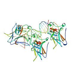
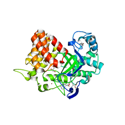
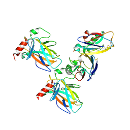
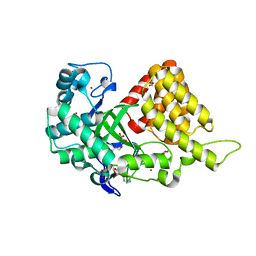
1TUP
 
 | | TUMOR SUPPRESSOR P53 COMPLEXED WITH DNA | | 分子名称: | DNA (5'-D(*AP*TP*AP*AP*TP*TP*GP*GP*GP*CP*AP*AP*GP*TP*CP*TP*A P*GP*GP*AP*A)-3'), DNA (5'-D(*TP*TP*TP*CP*CP*TP*AP*GP*AP*CP*TP*TP*GP*CP*CP*CP*A P*AP*TP*TP*A)-3'), PROTEIN (P53 TUMOR SUPPRESSOR ), ... | | 著者 | Cho, Y, Gorina, S, Jeffrey, P.D, Pavletich, N.P. | | 登録日 | 1995-07-11 | | 公開日 | 1995-07-11 | | 最終更新日 | 2024-02-14 | | 実験手法 | X-RAY DIFFRACTION (2.2 Å) | | 主引用文献 | Crystal structure of a p53 tumor suppressor-DNA complex: understanding tumorigenic mutations.
Science, 265, 1994
|
|
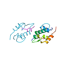
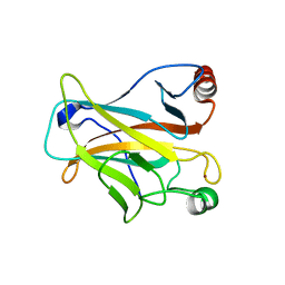
1TSR
 | | P53 CORE DOMAIN IN COMPLEX WITH DNA | | 分子名称: | DNA (5'-D(*AP*TP*AP*AP*TP*TP*GP*GP*GP*CP*AP*AP*GP*TP*CP*TP*A P*GP*GP*AP*A)-3'), DNA (5'-D(*TP*TP*TP*CP*CP*TP*AP*GP*AP*CP*TP*TP*GP*CP*CP*CP*A P*AP*TP*TP*A)-3'), PROTEIN (P53 TUMOR SUPPRESSOR), ... | | 著者 | Cho, Y, Gorina, S, Jeffrey, P, Pavletich, N. | | 登録日 | 1995-07-28 | | 公開日 | 1996-01-29 | | 最終更新日 | 2024-02-14 | | 実験手法 | X-RAY DIFFRACTION (2.2 Å) | | 主引用文献 | Crystal structure of a p53 tumor suppressor-DNA complex: understanding tumorigenic mutations.
Science, 265, 1994
|
|
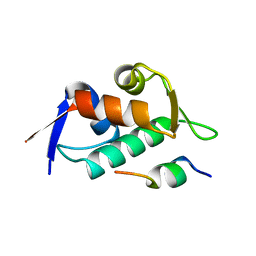
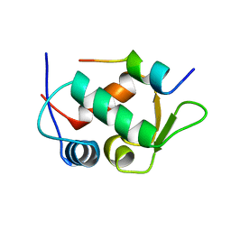
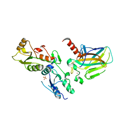
1YCS
 | | P53-53BP2 COMPLEX | | 分子名称: | 53BP2, P53, ZINC ION | | 著者 | Gorina, S, Pavletich, N.P. | | 登録日 | 1996-09-30 | | 公開日 | 1997-11-19 | | 最終更新日 | 2024-02-14 | | 実験手法 | X-RAY DIFFRACTION (2.2 Å) | | 主引用文献 | Structure of the p53 tumor suppressor bound to the ankyrin and SH3 domains of 53BP2.
Science, 274, 1996
|
|
1C26
 | |
3DAC
 | |
3DAB
 | |
3TG5
 | | Structure of SMYD2 in complex with p53 and SAH | | 分子名称: | Cellular tumor antigen p53, GLYCEROL, N-lysine methyltransferase SMYD2, ... | | 著者 | Zhao, K, Wang, L. | | 登録日 | 2011-08-17 | | 公開日 | 2011-08-31 | | 最終更新日 | 2023-11-01 | | 実験手法 | X-RAY DIFFRACTION (2.3 Å) | | 主引用文献 | Structure of human SMYD2 reveals the basis of p53 tumor suppressor methylation
J.Biol.Chem., 2011
|
|
1YCQ
 | |
1YCR
 | |
2H1L
 | |
7YGI
 | | Crystal structure of p53 DBD domain in complex with azurin | | 分子名称: | Azurin, Cellular tumor antigen p53, PHOSPHATE ION, ... | | 著者 | Jiang, W.X, Zuo, J.Q, Hu, J.J, Chen, X.Q, Ma, L.X, Liu, Z, Xing, Q. | | 登録日 | 2022-07-11 | | 公開日 | 2023-02-08 | | 実験手法 | X-RAY DIFFRACTION (2.1 Å) | | 主引用文献 | Structural basis of bacterial effector protein azurin targeting tumor suppressor p53 and inhibiting its ubiquitination.
Commun Biol, 6, 2023
|
|
2OCJ
 | |
1GZH
 | |
1UOL
 | |
2BIP
 | |
2BIQ
 | |
2BIN
 | |
2BIO
 | |
2BIM
 | |
5XZC
 | |
3TG4
 | | Structure of SMYD2 in complex with SAM | | 分子名称: | GLYCEROL, N-lysine methyltransferase SMYD2, S-ADENOSYLMETHIONINE, ... | | 著者 | Zhao, K, Wang, L. | | 登録日 | 2011-08-17 | | 公開日 | 2011-08-31 | | 最終更新日 | 2023-11-01 | | 実験手法 | X-RAY DIFFRACTION (2 Å) | | 主引用文献 | Structure of human SMYD2 reveals the basis of p53 tumor suppressor methylation
J.Biol.Chem., 2011
|
|
2M86
 | | Solution structure of Hdm2 with engineered cyclotide | | 分子名称: | E3 ubiquitin-protein ligase Mdm2, MCo-PMI | | 著者 | Majumder, S, Ji, Y, Millard, M, Borra, R, Bi, T, Elnagar, A.Y, Neamati, N, Camarero, J.A. | | 登録日 | 2013-05-07 | | 公開日 | 2013-07-31 | | 最終更新日 | 2023-06-14 | | 実験手法 | SOLUTION NMR | | 主引用文献 | In Vivo Activation of the p53 Tumor Suppressor Pathway by an Engineered Cyclotide.
J.Am.Chem.Soc., 135, 2013
|
|
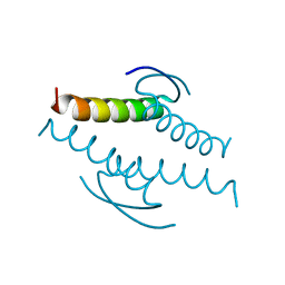
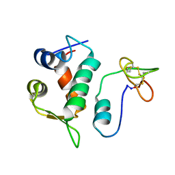
1A1U
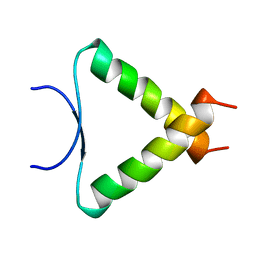 | | SOLUTION STRUCTURE DETERMINATION OF A P53 MUTANT DIMERIZATION DOMAIN, NMR, MINIMIZED AVERAGE STRUCTURE | | 分子名称: | P53 | | 著者 | Mccoy, M.A, Stavridi, E.S, Waterman, J.L.F, Wieczorek, A, Opella, S.J, Halezonetis, T.D. | | 登録日 | 1997-12-16 | | 公開日 | 1998-04-08 | | 最終更新日 | 2024-05-22 | | 実験手法 | SOLUTION NMR | | 主引用文献 | Hydrophobic side-chain size is a determinant of the three-dimensional structure of the p53 oligomerization domain.
EMBO J., 16, 1997
|
|
1KZY
 | | Crystal Structure of the 53bp1 BRCT Region Complexed to Tumor Suppressor P53 | | 分子名称: | CELLULAR TUMOR ANTIGEN P53, TUMOR SUPPRESSOR P53-BINDING PROTEIN 1, ZINC ION | | 著者 | Joo, W.S, Jeffrey, P.D, Cantor, S.B, Finnin, M.S, Livingston, D.M, Pavletich, N.P. | | 登録日 | 2002-02-08 | | 公開日 | 2002-03-20 | | 最終更新日 | 2011-07-13 | | 実験手法 | X-RAY DIFFRACTION (2.5 Å) | | 主引用文献 | Structure of the 53BP1 BRCT region bound to p53 and its comparison to the Brca1 BRCT structure.
Genes Dev., 16, 2002
|
|
1HU8
 | | CRYSTAL STRUCTURE OF THE MOUSE P53 CORE DNA-BINDING DOMAIN AT 2.7A RESOLUTION | | 分子名称: | CELLULAR TUMOR ANTIGEN P53, ZINC ION | | 著者 | Zhao, K, Chai, X, Johnston, K, Clements, A, Marmorstein, R. | | 登録日 | 2001-01-04 | | 公開日 | 2001-07-04 | | 最終更新日 | 2023-08-09 | | 実験手法 | X-RAY DIFFRACTION (2.7 Å) | | 主引用文献 | Crystal structure of the mouse p53 core DNA-binding domain at 2.7 A resolution.
J.Biol.Chem., 276, 2001
|
|


