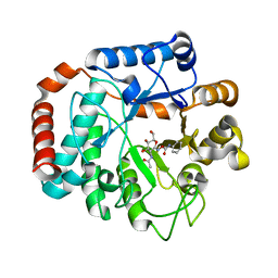
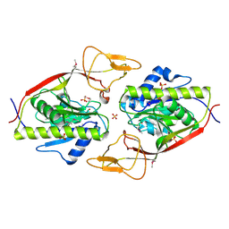
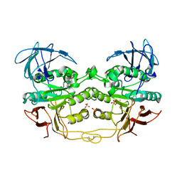
2OYT
 
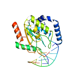 | | Crystal Structure of UNG2/DNA(TM) | | 分子名称: | DNA strand1, DNA strand2, Uracil-DNA glycosylase | | 著者 | Bianchet, M.A, Krosky, D.J, Stivers, J.T, Amzel, L.M. | | 登録日 | 2007-02-22 | | 公開日 | 2007-10-30 | | 最終更新日 | 2023-08-30 | | 実験手法 | X-RAY DIFFRACTION (2 Å) | | 主引用文献 | Enzymatic capture of an extrahelical thymine in the search for uracil in DNA.
Nature, 449, 2007
|
|
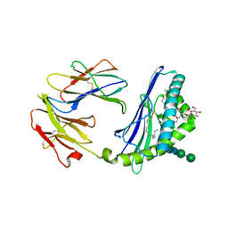
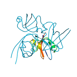
2OZO
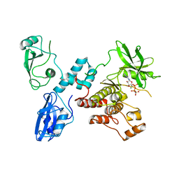 | | Autoinhibited intact human ZAP-70 | | 分子名称: | MAGNESIUM ION, PHOSPHOAMINOPHOSPHONIC ACID-ADENYLATE ESTER, Tyrosine-protein kinase ZAP-70 | | 著者 | Deindl, S, Kadlecek, T.A, Brdicka, T, Cao, X, Weiss, A, Kuriyan, J. | | 登録日 | 2007-02-26 | | 公開日 | 2007-05-22 | | 最終更新日 | 2024-02-21 | | 実験手法 | X-RAY DIFFRACTION (2.6 Å) | | 主引用文献 | Structural Basis for the Inhibition of Tyrosine Kinase Activity of ZAP-70.
Cell(Cambridge,Mass.), 129, 2007
|
|
2P6E
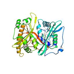 | |
2P2F
 | | Acetyl-CoA Synthetase, wild-type with acetate, AMP, and CoA bound | | 分子名称: | ACETATE ION, ADENOSINE MONOPHOSPHATE, Acetyl-coenzyme A synthetase, ... | | 著者 | Reger, A.S, Gulick, A.M. | | 登録日 | 2007-03-07 | | 公開日 | 2007-05-29 | | 最終更新日 | 2024-03-13 | | 実験手法 | X-RAY DIFFRACTION (2.58 Å) | | 主引用文献 | Biochemical and Crystallographic Analysis of Substrate Binding and Conformational Changes in Acetyl-CoA Synthetase.
Biochemistry, 46, 2007
|
|
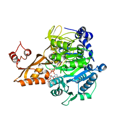
2P5L
 | | Crystal structure of a dimer of N-terminal domains of AhrC in complex with an 18bp DNA operator site | | 分子名称: | Arginine repressor, DNA (5'-D(*DCP*DAP*DTP*DGP*DAP*DAP*DTP*DAP*DAP*DAP*DAP*DAP*DTP*DTP*DCP*DAP*DAP*DG)-3'), DNA (5'-D(*DCP*DTP*DTP*DGP*DAP*DAP*DTP*DTP*DTP*DTP*DTP*DAP*DTP*DTP*DCP*DAP*DTP*DG)-3'), ... | | 著者 | Garnett, J.A, Marincs, F, Baumberg, S, Stockley, P.G, Phillips, S.E.V. | | 登録日 | 2007-03-15 | | 公開日 | 2008-03-11 | | 最終更新日 | 2023-08-30 | | 実験手法 | X-RAY DIFFRACTION (2.85 Å) | | 主引用文献 | Structure and function of the arginine repressor-operator complex from Bacillus subtilis.
J.Mol.Biol., 379, 2008
|
|
2PD8
 | |
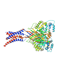
2OTG
 | | Rigor-like structures of muscle myosins reveal key mechanical elements in the transduction pathways of this allosteric motor | | 分子名称: | ADENOSINE-5'-DIPHOSPHATE, CALCIUM ION, MAGNESIUM ION, ... | | 著者 | Yang, Y, Gourinath, S, Kovacs, M, Nyitray, L, Reutzel, R, Himmel, D.M, O'Neall-Hennessey, E, Reshetnikova, L, Szent-Gyorgyi, A.G, Brown, J.H, Cohen, C. | | 登録日 | 2007-02-08 | | 公開日 | 2007-05-29 | | 最終更新日 | 2023-08-30 | | 実験手法 | X-RAY DIFFRACTION (3.12 Å) | | 主引用文献 | Rigor-like Structures from Muscle Myosins Reveal Key Mechanical Elements in the Transduction Pathways of This Allosteric Motor.
Structure, 15, 2007
|
|
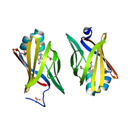
2ONT
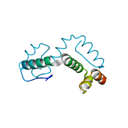 | | A swapped dimer of the HIV-1 capsid C-terminal domain | | 分子名称: | Capsid protein p24 | | 著者 | Ivanov, D, Tsodikov, O.V, Kasanov, J, Ellenberger, T, Wagner, G, Collins, T. | | 登録日 | 2007-01-24 | | 公開日 | 2007-02-20 | | 最終更新日 | 2023-08-30 | | 実験手法 | X-RAY DIFFRACTION (2.4 Å) | | 主引用文献 | Domain-swapped dimerization of the HIV-1 capsid C-terminal domain
Proc.Natl.Acad.Sci.Usa, 104, 2007
|
|
2OXN
 | |
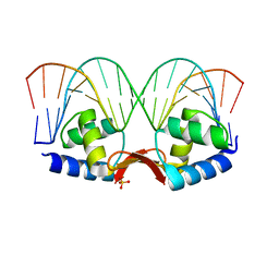
2Q81
 | | Crystal Structure of the Miz-1 BTB/POZ domain | | 分子名称: | Miz-1 protein, TETRAETHYLENE GLYCOL | | 著者 | Stead, M.A, Trinh, C.H, Garnett, J.A, Carr, S.B, Edwards, T.A, Wright, S.C. | | 登録日 | 2007-06-08 | | 公開日 | 2007-11-06 | | 最終更新日 | 2023-08-30 | | 実験手法 | X-RAY DIFFRACTION (2.1 Å) | | 主引用文献 | A Beta-Sheet Interaction Interface Directs the Tetramerisation of the Miz-1 POZ Domain
J.Mol.Biol., 373, 2007
|
|
2Q4C
 | | Ensemble refinement of the protein crystal structure of annexin from Arabidopsis thaliana gene At1g35720 | | 分子名称: | Annexin D1 | | 著者 | Levin, E.J, Kondrashov, D.A, Wesenberg, G.E, Phillips Jr, G.N, Center for Eukaryotic Structural Genomics (CESG) | | 登録日 | 2007-05-31 | | 公開日 | 2007-06-19 | | 最終更新日 | 2023-08-30 | | 実験手法 | X-RAY DIFFRACTION (2.508 Å) | | 主引用文献 | Ensemble refinement of protein crystal structures: validation and application.
Structure, 15, 2007
|
|
2Q4O
 | | Ensemble refinement of the crystal structure of a lysine decarboxylase-like protein from Arabidopsis thaliana gene At2g37210 | | 分子名称: | MAGNESIUM ION, SULFATE ION, Uncharacterized protein At2g37210/T2N18.3 | | 著者 | Levin, E.J, Kondrashov, D.A, Wesenberg, G.E, Phillips Jr, G.N, Center for Eukaryotic Structural Genomics (CESG) | | 登録日 | 2007-05-31 | | 公開日 | 2007-06-19 | | 最終更新日 | 2023-11-15 | | 実験手法 | X-RAY DIFFRACTION (1.95 Å) | | 主引用文献 | Ensemble refinement of protein crystal structures: validation and application.
Structure, 15, 2007
|
|
2Q4Z
 | | Ensemble refinement of the protein crystal structure of an aspartoacylase from Rattus norvegicus | | 分子名称: | Aspartoacylase, SULFATE ION, ZINC ION | | 著者 | Levin, E.J, Kondrashov, D.A, Wesenberg, G.E, Phillips Jr, G.N, Center for Eukaryotic Structural Genomics (CESG) | | 登録日 | 2007-05-31 | | 公開日 | 2007-06-19 | | 最終更新日 | 2023-11-15 | | 実験手法 | X-RAY DIFFRACTION (1.8 Å) | | 主引用文献 | Ensemble refinement of protein crystal structures: validation and application.
Structure, 15, 2007
|
|
1S31
 | | Crystal Structure Analysis of the human Tub protein (isoform a) spanning residues 289 through 561 | | 分子名称: | TRIETHYLENE GLYCOL, tubby isoform a | | 著者 | Boutboul, S, Carroll, K.J, Basdevant, A, Gomez, C, Nandrot, E, Clement, K, Shapiro, L, Abitbol, M. | | 登録日 | 2004-01-12 | | 公開日 | 2005-01-25 | | 最終更新日 | 2023-08-23 | | 実験手法 | X-RAY DIFFRACTION (2.704 Å) | | 主引用文献 | A novel human obesity and sensory deficit syndrome resulting from a mutation in the TUB gene
To be Published
|
|
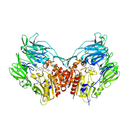
2Q7Y
 | | Structure of the endogenous iNKT cell ligand iGb3 bound to mCD1d | | 分子名称: | 2-acetamido-2-deoxy-beta-D-glucopyranose, 2-acetamido-2-deoxy-beta-D-glucopyranose-(1-4)-2-acetamido-2-deoxy-beta-D-glucopyranose, Beta-2-microglobulin, ... | | 著者 | Zajonc, D.M, Wilson, I.A, Teyton, L. | | 登録日 | 2007-06-07 | | 公開日 | 2008-04-01 | | 最終更新日 | 2023-08-30 | | 実験手法 | X-RAY DIFFRACTION (1.95 Å) | | 主引用文献 | Crystal Structures of Mouse CD1d-iGb3 Complex and its Cognate Valpha14 T Cell Receptor Suggest a Model for Dual Recognition of Foreign and Self Glycolipids.
J.Mol.Biol., 377, 2008
|
|
2QBO
 | | Crystal structure of the P450cam G248V mutant in the cyanide bound state | | 分子名称: | CAMPHOR, CYANIDE ION, Cytochrome P450-cam, ... | | 著者 | von Koenig, K, Makris, T.M, Sligar, S.D, Schlichting, I. | | 登録日 | 2007-06-18 | | 公開日 | 2007-12-25 | | 最終更新日 | 2023-08-30 | | 実験手法 | X-RAY DIFFRACTION (1.9 Å) | | 主引用文献 | Alteration of P450 Distal Pocket Solvent Leads to Impaired Proton Delivery and Changes in Heme Geometry.
Biochemistry, 46, 2007
|
|
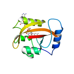
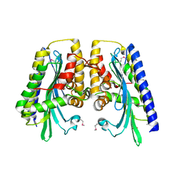
2QDV
 | | Structure of the Cu(II) form of the M51A mutant of amicyanin | | 分子名称: | Amicyanin, COPPER (II) ION, PHOSPHATE ION | | 著者 | Carrell, C.J, Ma, J.K, Wang, Y, Davidson, V.L, Mathews, F.S. | | 登録日 | 2007-06-21 | | 公開日 | 2007-12-11 | | 最終更新日 | 2021-10-20 | | 実験手法 | X-RAY DIFFRACTION (0.89 Å) | | 主引用文献 | A single methionine residue dictates the kinetic mechanism of interprotein electron transfer from methylamine dehydrogenase to amicyanin.
Biochemistry, 46, 2007
|
|
2QFQ
 | | E. coli EPSP synthase Pro101Leu liganded with S3P | | 分子名称: | 3-phosphoshikimate 1-carboxyvinyltransferase, FORMIC ACID, SHIKIMATE-3-PHOSPHATE | | 著者 | Schonbrunn, E, Healy-Fried, M.L. | | 登録日 | 2007-06-27 | | 公開日 | 2007-10-02 | | 最終更新日 | 2023-08-30 | | 実験手法 | X-RAY DIFFRACTION (1.5 Å) | | 主引用文献 | Structural basis of glyphosate tolerance resulting from mutations of Pro101 in Escherichia coli 5-enolpyruvylshikimate-3-phosphate synthase.
J.Biol.Chem., 282, 2007
|
|
2QFR
 | | Crystal structure of red kidney bean purple acid phosphatase with bound sulfate | | 分子名称: | 2-acetamido-2-deoxy-alpha-D-glucopyranose, 2-acetamido-2-deoxy-beta-D-glucopyranose, FE (III) ION, ... | | 著者 | Guddat, L.W, Schenk, G, Gahan, L.R, Elliot, T.W, Leung, E. | | 登録日 | 2007-06-27 | | 公開日 | 2008-10-14 | | 最終更新日 | 2023-08-30 | | 実験手法 | X-RAY DIFFRACTION (2.4 Å) | | 主引用文献 | Crystal structures of a purple acid phosphatase, representing different steps of this enzyme's catalytic cycle.
Bmc Struct.Biol., 8, 2008
|
|
2QKT
 | |
2QM9
 | | Troglitazone Bound to Fatty Acid Binding Protein 4 | | 分子名称: | (5R)-5-(4-{[(2R)-6-HYDROXY-2,5,7,8-TETRAMETHYL-3,4-DIHYDRO-2H-CHROMEN-2-YL]METHOXY}BENZYL)-1,3-THIAZOLIDINE-2,4-DIONE, Fatty acid-binding protein, adipocyte, ... | | 著者 | Gillilan, R.E, Ayers, S.D, Noy, N. | | 登録日 | 2007-07-14 | | 公開日 | 2007-10-09 | | 最終更新日 | 2024-02-21 | | 実験手法 | X-RAY DIFFRACTION (2.31 Å) | | 主引用文献 | Structural Basis for Activation of Fatty Acid-binding Protein 4
J.Mol.Biol., 372, 2007
|
|
2QT8
 | |
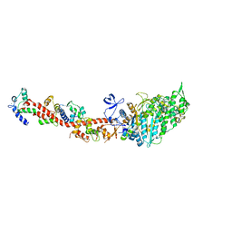
2QTS
 | | Structure of an acid-sensing ion channel 1 at 1.9 A resolution and low pH | | 分子名称: | 2-acetamido-2-deoxy-beta-D-glucopyranose, Acid-sensing ion channel, CHLORIDE ION, ... | | 著者 | Jasti, J, Furukawa, H, Gonzales, E.B, Gouaux, E. | | 登録日 | 2007-08-02 | | 公開日 | 2007-09-25 | | 最終更新日 | 2020-07-29 | | 実験手法 | X-RAY DIFFRACTION (1.9 Å) | | 主引用文献 | Structure of acid-sensing ion channel 1 at 1.9A resolution and low pH
Nature, 449, 2007
|
|
2QOE
 | | Human Dipeptidyl Peptidase IV in complex with a Triazolopiperazine-based beta amino acid Inhibitor | | 分子名称: | (2R)-4-[(8R)-8-METHYL-2-(TRIFLUOROMETHYL)-5,6-DIHYDRO[1,2,4]TRIAZOLO[1,5-A]PYRAZIN-7(8H)-YL]-4-OXO-1-(2,4,5-TRIFLUOROPHENYL)BUTAN-2-AMINE, 2-acetamido-2-deoxy-alpha-D-glucopyranose-(1-4)-2-acetamido-2-deoxy-beta-D-glucopyranose, 2-acetamido-2-deoxy-beta-D-glucopyranose, ... | | 著者 | Scapin, G. | | 登録日 | 2007-07-20 | | 公開日 | 2007-11-06 | | 最終更新日 | 2021-10-20 | | 実験手法 | X-RAY DIFFRACTION (2.3 Å) | | 主引用文献 | Design, synthesis, and biological evaluation of triazolopiperazine-based beta-amino amides as potent, orally active dipeptidyl peptidase IV (DPP-4) inhibitors.
Bioorg.Med.Chem.Lett., 17, 2007
|
|
2QVF
 | | mouse E-cadherin domains 1,2 | | 分子名称: | CALCIUM ION, Epithelial-cadherin (E-cadherin) (Uvomorulin) (Cadherin-1) (ARC-1) (CD324 antigen) | | 著者 | Carroll, K.J. | | 登録日 | 2007-08-08 | | 公開日 | 2008-08-12 | | 最終更新日 | 2023-08-30 | | 実験手法 | X-RAY DIFFRACTION (2.4 Å) | | 主引用文献 | E-cadherin domains 1,2
To be Published
|
|


