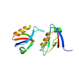
8BP5
 
 | |
4JSZ
 
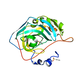 | | Benzenesulfonamide bound to hCAII H94C | | 分子名称: | Carbonic anhydrase 2, ZINC ION, benzenesulfonamide | | 著者 | Martin, D.P, Hann, Z.S, Cohen, S.M. | | 登録日 | 2013-03-22 | | 公開日 | 2013-06-19 | | 最終更新日 | 2023-09-20 | | 実験手法 | X-RAY DIFFRACTION (1.9 Å) | | 主引用文献 | Metalloprotein-Inhibitor Binding: Human Carbonic Anhydrase II as a Model for Probing Metal-Ligand Interactions in a Metalloprotein Active Site.
Inorg.Chem., 52, 2013
|
|
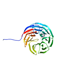
7AXQ
 
 | |
5C1T
 
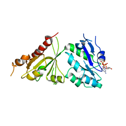 | | Crystal structure of the GTP-bound wild type EhRabX3 from Entamoeba histolytica | | 分子名称: | GUANOSINE-5'-TRIPHOSPHATE, MAGNESIUM ION, Small GTPase EhRabX3 | | 著者 | Srivastava, V.K, Chandra, M, Datta, S. | | 登録日 | 2015-06-15 | | 公開日 | 2016-04-27 | | 最終更新日 | 2024-03-20 | | 実験手法 | X-RAY DIFFRACTION (2.801 Å) | | 主引用文献 | Crystal Structure Analysis of Wild Type and Fast Hydrolyzing Mutant of EhRabX3, a Tandem Ras Superfamily GTPase from Entamoeba histolytica.
J.Mol.Biol., 428, 2016
|
|
3TKC
 
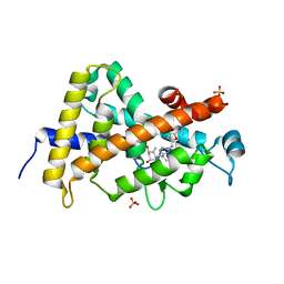 | | Design, Synthesis, Evaluation and Structure of Vitamin D Analogues with Furan Side Chains | | 分子名称: | (1S,3R,5Z,7E,14beta,17alpha,20S)-20-[5-(1-hydroxy-1-methylethyl)furan-2-yl]-9,10-secopregna-5,7,10-triene-1,3-diol, SULFATE ION, Vitamin D3 receptor | | 著者 | Huet, T, Moras, D, Rochel, N. | | 登録日 | 2011-08-26 | | 公開日 | 2012-03-07 | | 最終更新日 | 2023-09-13 | | 実験手法 | X-RAY DIFFRACTION (1.75 Å) | | 主引用文献 | Design, synthesis, evaluation, and structure of vitamin D analogues with furan side chains.
Chemistry, 18, 2012
|
|
8BM2
 
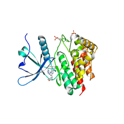 | | Crystal structure of JAK2 JH1 in complex with gandotinib | | 分子名称: | 3-[(4-chloranyl-2-fluoranyl-phenyl)methyl]-2-methyl-~{N}-(5-methyl-1~{H}-pyrazol-3-yl)-8-(morpholin-4-ylmethyl)imidazo[1,2-b]pyridazin-6-amine, Tyrosine-protein kinase JAK2 | | 著者 | Miao, Y, Haikarainen, T. | | 登録日 | 2022-11-10 | | 公開日 | 2023-11-22 | | 最終更新日 | 2024-11-06 | | 実験手法 | X-RAY DIFFRACTION (1.5 Å) | | 主引用文献 | Functional and Structural Characterization of Clinical-Stage Janus Kinase 2 Inhibitors Identifies Determinants for Drug Selectivity.
J.Med.Chem., 67, 2024
|
|
8BN2
 
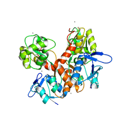 | | Crystal structure of the ligand-binding domain (LBD) of human iGluR Delta-1 (GluD1) in complex with D-Serine | | 分子名称: | 1,2-ETHANEDIOL, 2-acetamido-2-deoxy-beta-D-glucopyranose, CALCIUM ION, ... | | 著者 | Heroven, C, Malinauskas, T, Aricescu, A.R. | | 登録日 | 2022-11-11 | | 公開日 | 2023-11-22 | | 最終更新日 | 2024-10-23 | | 実験手法 | X-RAY DIFFRACTION (1.63 Å) | | 主引用文献 | GluD1 binds GABA and controls inhibitory plasticity.
Science, 382, 2023
|
|
7AXX
 
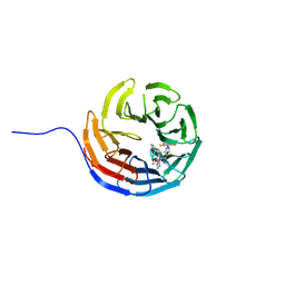 | |
3P3Z
 
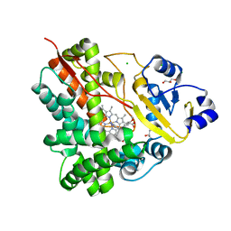 | | Crystal Structure of the Cytochrome P450 Monooxygenase AurH from Streptomyces Thioluteus in Complex with Ancymidol | | 分子名称: | (S)-cyclopropyl(4-methoxyphenyl)pyrimidin-5-ylmethanol, CHLORIDE ION, Cytochrome P450, ... | | 著者 | Zocher, G, Richter, M.E.A, Mueller, U, Hertweck, C. | | 登録日 | 2010-10-05 | | 公開日 | 2011-02-16 | | 最終更新日 | 2024-02-21 | | 実験手法 | X-RAY DIFFRACTION (2.3 Å) | | 主引用文献 | Structural fine-tuning of a multifunctional cytochrome p450 monooxygenase.
J.Am.Chem.Soc., 133, 2011
|
|
6YZU
 
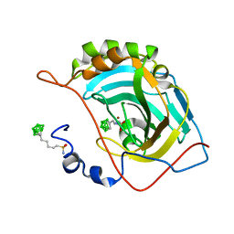 | | Carborane nido-pentyl-sulfonamide in complex with CA II | | 分子名称: | Carbonic anhydrase 2, Carborane nido-pentyl-sulfonamide, ZINC ION | | 著者 | Kugler, M, Brynda, J, Pospisilova, K, Rezacova, P. | | 登録日 | 2020-05-07 | | 公開日 | 2020-10-28 | | 最終更新日 | 2024-01-24 | | 実験手法 | X-RAY DIFFRACTION (1 Å) | | 主引用文献 | The structural basis for the selectivity of sulfonamido dicarbaboranes toward cancer-associated carbonic anhydrase IX.
J Enzyme Inhib Med Chem, 35, 2020
|
|
1FEP
 
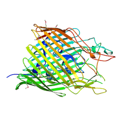 | | FERRIC ENTEROBACTIN RECEPTOR | | 分子名称: | FERRIC ENTEROBACTIN RECEPTOR | | 著者 | Buchanan, S.K, Smith, B.S, Ventatramani, L, Xia, D, Esser, L, Palnitkar, M, Chakraborty, R, Van Der Helm, D, Deisenhofer, J. | | 登録日 | 1998-11-24 | | 公開日 | 1999-01-13 | | 最終更新日 | 2024-11-06 | | 実験手法 | X-RAY DIFFRACTION (2.4 Å) | | 主引用文献 | Crystal structure of the outer membrane active transporter FepA from Escherichia coli.
Nat.Struct.Biol., 6, 1999
|
|
8BP4
 
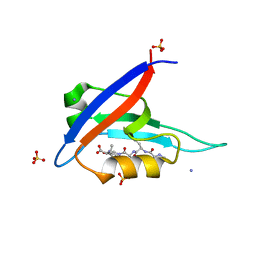 | |
7AXP
 
 | |
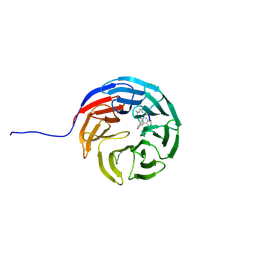
7AXS
 
 | |
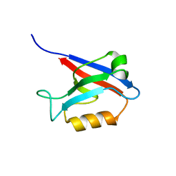
8BOJ
 
 | |
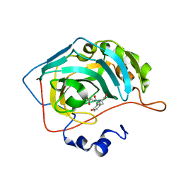
3P4V
 
 | |
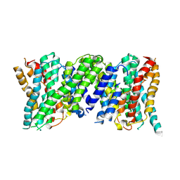
6Z3Y
 
 | |
3DLA
 
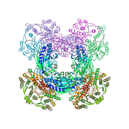 | | X-ray crystal structure of glutamine-dependent NAD+ synthetase from Mycobacterium tuberculosis bound to NaAD+ and DON | | 分子名称: | 5-OXO-L-NORLEUCINE, GLYCEROL, Glutamine-dependent NAD(+) synthetase, ... | | 著者 | LaRonde-LeBlanc, N.A, Resto, M, Gerratana, B. | | 登録日 | 2008-06-26 | | 公開日 | 2009-03-10 | | 最終更新日 | 2019-10-23 | | 実験手法 | X-RAY DIFFRACTION (2.35 Å) | | 主引用文献 | Regulation of active site coupling in glutamine-dependent NAD(+) synthetase.
Nat.Struct.Mol.Biol., 16, 2009
|
|
6N3N
 
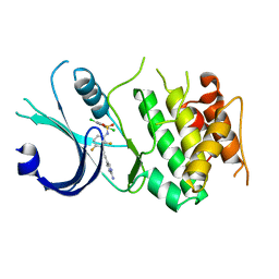 | | Identification of novel, potent and selective GCN2 inhibitors as first-in-class anti-tumor agents | | 分子名称: | N-{3-[(2-aminopyrimidin-5-yl)ethynyl]-2,4-difluorophenyl}-2,5-dichloro-3-(hydroxymethyl)benzene-1-sulfonamide, eIF-2-alpha kinase GCN2,eIF-2-alpha kinase GCN2 | | 著者 | Hoffman, I.D, Fujimoto, J, Kurasawa, O, Takagi, T, Klein, M.G, Kefala, G, Ding, S.C, Cary, D.R, Mizojiri, R. | | 登録日 | 2018-11-15 | | 公開日 | 2019-10-09 | | 最終更新日 | 2024-03-13 | | 実験手法 | X-RAY DIFFRACTION (3.01 Å) | | 主引用文献 | Identification of Novel, Potent, and Orally Available GCN2 Inhibitors with Type I Half Binding Mode.
Acs Med.Chem.Lett., 10, 2019
|
|
5C41
 
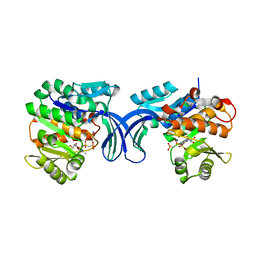 | | Crystal structure of human ribokinase in complex with AMPPCP in P21 spacegroup and with 4 protomers | | 分子名称: | PHOSPHATE ION, PHOSPHOMETHYLPHOSPHONIC ACID ADENYLATE ESTER, Ribokinase, ... | | 著者 | Park, J, Chakrabarti, J, Singh, B, Gupta, R.S, Junop, M.S. | | 登録日 | 2015-06-17 | | 公開日 | 2016-06-15 | | 最終更新日 | 2023-09-27 | | 実験手法 | X-RAY DIFFRACTION (1.95 Å) | | 主引用文献 | Crystal structure of human ribokinase in complex with AMPPCP in P21 spacegroup and with 4 protomers
To Be Published
|
|
8BN5
 
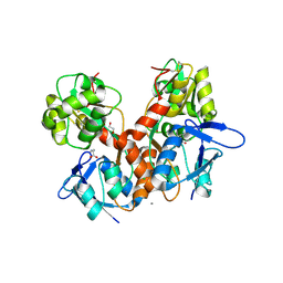 | | Crystal structure of the ligand-binding domain (LBD) of human iGluR Delta-1 (GluD1) in complex with GABA | | 分子名称: | 1,2-ETHANEDIOL, 2-acetamido-2-deoxy-beta-D-glucopyranose, CALCIUM ION, ... | | 著者 | Heroven, C, Malinauskas, T, Aricescu, A.R. | | 登録日 | 2022-11-12 | | 公開日 | 2023-11-22 | | 最終更新日 | 2024-10-23 | | 実験手法 | X-RAY DIFFRACTION (1.9 Å) | | 主引用文献 | GluD1 binds GABA and controls inhibitory plasticity.
Science, 382, 2023
|
|
8BLJ
 
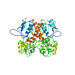 | | Crystal structure of the ligand-binding domain (LBD) of human iGluR Delta-1 (GluD1), apo state | | 分子名称: | 1,2-ETHANEDIOL, 2-acetamido-2-deoxy-beta-D-glucopyranose, CALCIUM ION, ... | | 著者 | Heroven, C, Malinauskas, T, Aricescu, A.R. | | 登録日 | 2022-11-09 | | 公開日 | 2023-11-22 | | 最終更新日 | 2024-10-16 | | 実験手法 | X-RAY DIFFRACTION (2.18 Å) | | 主引用文献 | GluD1 binds GABA and controls inhibitory plasticity.
Science, 382, 2023
|
|
8BPM
 
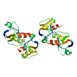 | |
8ET7
 
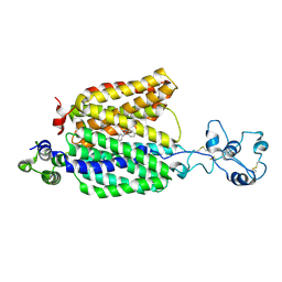 | | Cryo-EM structure of the organic cation transporter 1 in complex with diphenhydramine | | 分子名称: | 2-acetamido-2-deoxy-beta-D-glucopyranose-(1-4)-2-acetamido-2-deoxy-beta-D-glucopyranose, N-[2-(BENZHYDRYLOXY)ETHYL]-N,N-DIMETHYLAMINE, OCT1 | | 著者 | Suo, Y, Wright, N.J, Lee, S.-Y. | | 登録日 | 2022-10-16 | | 公開日 | 2023-05-31 | | 最終更新日 | 2024-10-23 | | 実験手法 | ELECTRON MICROSCOPY (3.77 Å) | | 主引用文献 | Molecular basis of polyspecific drug and xenobiotic recognition by OCT1 and OCT2.
Nat.Struct.Mol.Biol., 30, 2023
|
|
8BRZ
 
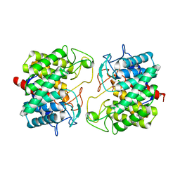 | | Room-temperature structure of Pedobacter heparinus N-acetylglucosamine 2-epimerase at 52 MPa helium gas pressure in a sapphire capillary | | 分子名称: | CHLORIDE ION, N-acylglucosamine 2-epimerase, PHOSPHATE ION | | 著者 | Lieske, J, Saouane, S, Assmann, M, Zaun, H, Kuballa, J, Meents, A. | | 登録日 | 2022-11-24 | | 公開日 | 2023-12-13 | | 実験手法 | X-RAY DIFFRACTION (1.7 Å) | | 主引用文献 | High-pressure macromolecular crystallography to explore the conformational space of proteins
To Be Published
|
|
