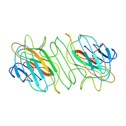
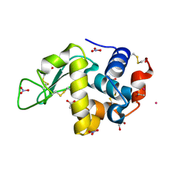
8VW4
 
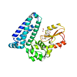 | | Crystal structure of Cbl-b TKB bound to compound 26 | | 分子名称: | (7-methoxy-2-{2-[(1S,3S,4S)-3-(3-methoxy-2-methyl-5-nitrophenyl)-1-methyl-5-oxo-1,5-dihydroimidazo[1,5-a]pyridin-2(3H)-yl]-2-oxoethoxy}quinolin-8-yl)acetic acid, DI(HYDROXYETHYL)ETHER, E3 ubiquitin-protein ligase CBL-B, ... | | 著者 | Yu, C, Murray, J, Hsu, P.L. | | 登録日 | 2024-01-31 | | 公開日 | 2024-07-03 | | 実験手法 | X-RAY DIFFRACTION (2.4 Å) | | 主引用文献 | Optimization of a Novel DEL Hit That Binds in the Cbl-b SH2 Domain and Blocks Substrate Binding.
Acs Med.Chem.Lett., 15, 2024
|
|
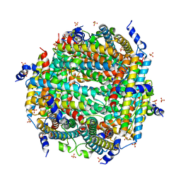
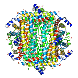
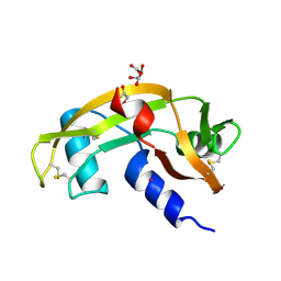
8YT4
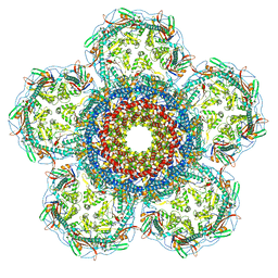
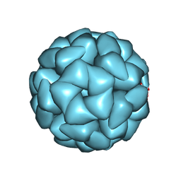 | | Structure of Aquifex aeolicus Lumazine Synthase by Cryo-Electron Microscopy to 1.42 Angstrom Resolution | | 分子名称: | 6,7-dimethyl-8-ribityllumazine synthase, PHOSPHATE ION | | 著者 | Savva, C.G, Sobhy, M.A, De Biasio, A, Hamdan, S.M. | | 登録日 | 2024-03-24 | | 公開日 | 2024-04-10 | | 最終更新日 | 2024-07-24 | | 実験手法 | ELECTRON MICROSCOPY (1.42 Å) | | 主引用文献 | Structure of Aquifex aeolicus lumazine synthase by cryo-electron microscopy to 1.42 angstrom resolution.
Iucrj, 2024
|
|
9BJL
 | |
8X3R
 | |
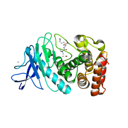
8OW3
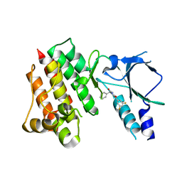 | | Crystal structure of wild-type c-MET bound by compound 2 | | 分子名称: | 5-[3,5-bis(fluoranyl)phenyl]-1-[(1S)-1-phenylethyl]pyrimidine-2,4-dione, Hepatocyte growth factor receptor | | 著者 | Collie, G.W. | | 登録日 | 2023-04-26 | | 公開日 | 2023-07-05 | | 最終更新日 | 2024-06-19 | | 実験手法 | X-RAY DIFFRACTION (2.27 Å) | | 主引用文献 | Discovery and Optimization of the First ATP Competitive Type-III c-MET Inhibitor.
J.Med.Chem., 66, 2023
|
|
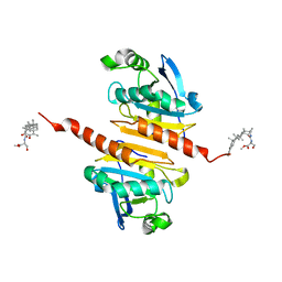
8W1E
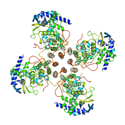
 | | Crystal Structure of DPS-like protein PA4880 from Pseudomonas aeruginosa (dodecamer) | | 分子名称: | DPS-LIKE PROTEIN, FE (II) ION, SULFATE ION | | 著者 | Lovell, S, Liu, L, Seibold, S, Battaile, K.P, Rivera, M. | | 登録日 | 2024-02-15 | | 公開日 | 2024-05-29 | | 最終更新日 | 2024-06-19 | | 実験手法 | X-RAY DIFFRACTION (2.9 Å) | | 主引用文献 | Pseudomonas aeruginosa gene PA4880 encodes a Dps-like protein with a Dps fold, bacterioferritin-type ferroxidase centers, and endonuclease activity.
Front Mol Biosci, 11, 2024
|
|
8P5O
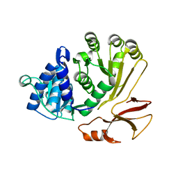 | | Proline activating adenylation domain of gramicidin S synthetase 2 - GrsB1-Acore | | 分子名称: | Gramicidin S synthase 2 | | 著者 | Stephan, P, Basquin, J, Caputi, L, O'Connor, S.E, Kries, H. | | 登録日 | 2023-05-24 | | 公開日 | 2023-07-05 | | 最終更新日 | 2023-08-30 | | 実験手法 | X-RAY DIFFRACTION (2.6 Å) | | 主引用文献 | Directed Evolution of Piperazic Acid Incorporation by a Nonribosomal Peptide Synthetase.
Angew.Chem.Int.Ed.Engl., 62, 2023
|
|
8YXK
 | |
9B8P
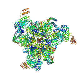 | | Synaptic Vesicle V-ATPase with synaptophysin and SidK, State 3, V1 | | 分子名称: | ADENOSINE-5'-DIPHOSPHATE, ATPase H+-transporting V1 subunit D, H(+)-transporting two-sector ATPase, ... | | 著者 | Coupland, E.M, Rubinstein, J.L. | | 登録日 | 2024-03-31 | | 公開日 | 2024-07-03 | | 最終更新日 | 2024-07-24 | | 実験手法 | ELECTRON MICROSCOPY (3.2 Å) | | 主引用文献 | High-resolution electron cryomicroscopy of V-ATPase in native synaptic vesicles.
Science, 385, 2024
|
|
8XQD
 | |
8W1F
 | | Crystal Structure of DPS-like protein PA4880 from Pseudomonas aeruginosa (dodecamer, Mg bound) | | 分子名称: | DPS-LIKE PROTEIN, FE (II) ION, MAGNESIUM ION, ... | | 著者 | Lovell, S, Liu, L, Seibold, S, Battaile, K.P, Rivera, M. | | 登録日 | 2024-02-15 | | 公開日 | 2024-05-29 | | 最終更新日 | 2024-06-19 | | 実験手法 | X-RAY DIFFRACTION (3 Å) | | 主引用文献 | Pseudomonas aeruginosa gene PA4880 encodes a Dps-like protein with a Dps fold, bacterioferritin-type ferroxidase centers, and endonuclease activity.
Front Mol Biosci, 11, 2024
|
|
9B33
 | |
9BKN
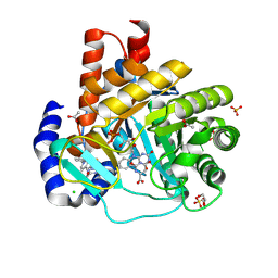 | | DHODH in complex with Ligand 16 | | 分子名称: | (2P,6P)-6-[4-ethyl-3-(hydroxymethyl)-5-oxo-4,5-dihydro-1H-1,2,4-triazol-1-yl]-7-fluoro-2-(2-methylphenyl)-4-(propan-2-yl)isoquinolin-1(2H)-one, ACETATE ION, CHLORIDE ION, ... | | 著者 | Shaffer, P.L. | | 登録日 | 2024-04-29 | | 公開日 | 2024-07-03 | | 最終更新日 | 2024-07-24 | | 実験手法 | X-RAY DIFFRACTION (1.3 Å) | | 主引用文献 | Discovery of JNJ-74856665: A Novel Isoquinolinone DHODH Inhibitor for the Treatment of AML.
J.Med.Chem., 67, 2024
|
|
8OWI
 | |
9BBL
 | |
8OV7
 | | Crystal structure of D1228V c-MET bound by compound 10 | | 分子名称: | 5-[3,5-bis(fluoranyl)phenyl]-1-[(1S)-1-[3-(1H-imidazol-5-yl)phenyl]ethyl]pyrimidine-2,4-dione, Hepatocyte growth factor receptor | | 著者 | Collie, G.W. | | 登録日 | 2023-04-25 | | 公開日 | 2023-07-05 | | 最終更新日 | 2024-06-19 | | 実験手法 | X-RAY DIFFRACTION (1.95 Å) | | 主引用文献 | Discovery and Optimization of the First ATP Competitive Type-III c-MET Inhibitor.
J.Med.Chem., 66, 2023
|
|
9BKO
 | | DHODH in complex with Ligand 26 | | 分子名称: | (2P,6P)-6-[4-ethyl-3-(hydroxymethyl)-5-oxo-4,5-dihydro-1H-1,2,4-triazol-1-yl]-7-fluoro-2-(2-methylphenyl)-4-[(2R)-1,1,1-trifluoropropan-2-yl]isoquinolin-1(2H)-one, ACETATE ION, CHLORIDE ION, ... | | 著者 | Shaffer, P.L. | | 登録日 | 2024-04-29 | | 公開日 | 2024-07-03 | | 最終更新日 | 2024-07-24 | | 実験手法 | X-RAY DIFFRACTION (1.44 Å) | | 主引用文献 | Discovery of JNJ-74856665: A Novel Isoquinolinone DHODH Inhibitor for the Treatment of AML.
J.Med.Chem., 67, 2024
|
|
8XOU
 | |
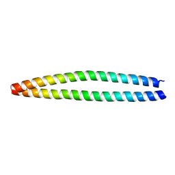
8XH6
 | | Structure of EBV LMP1 dimer | | 分子名称: | Latent membrane protein 1 | | 著者 | Gao, P, Huang, J.F. | | 登録日 | 2023-12-17 | | 公開日 | 2024-06-26 | | 最終更新日 | 2024-07-31 | | 実験手法 | ELECTRON MICROSCOPY (3.52 Å) | | 主引用文献 | Assembly and activation of EBV latent membrane protein 1.
Cell, 2024
|
|
9EO2
 | |
8OO3
 | |
8ZM4
 | |
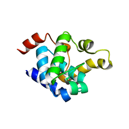
8X73
 | | Crystal structure of Peroxiredoxin I in complex with compound 19-069 | | 分子名称: | Peroxiredoxin-1, methyl (2~{S})-2-[[(2~{R},4~{a}~{S},6~{a}~{R},6~{a}~{S},14~{a}~{S},14~{b}~{R})-2,4~{a},6~{a},6~{a},9,14~{a}-hexamethyl-10-oxidanyl-11-oxidanylidene-1,3,4,5,6,13,14,14~{b}-octahydropicen-2-yl]carbamoylamino]-3-oxidanyl-propanoate | | 著者 | Zhang, H, Luo, C. | | 登録日 | 2023-11-22 | | 公開日 | 2024-06-19 | | 実験手法 | X-RAY DIFFRACTION (1.61 Å) | | 主引用文献 | Discovery of a Novel Orally Bioavailable FLT3-PROTAC Degrader for Efficient Treatment of Acute Myeloid Leukemia and Overcoming Resistance of FLT3 Inhibitors.
J.Med.Chem., 67, 2024
|
|
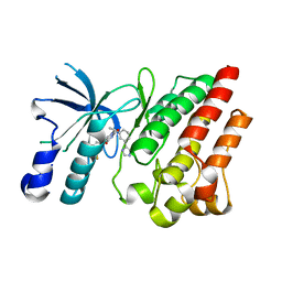
8XPG
 | | The Crystal Structure of polo box domain of Plk4 from Biortus. | | 分子名称: | SULFATE ION, Serine/threonine-protein kinase PLK4 | | 著者 | Wang, F, Cheng, W, Lv, Z, Meng, Q, Zhang, B. | | 登録日 | 2024-01-03 | | 公開日 | 2024-03-06 | | 実験手法 | X-RAY DIFFRACTION (2.7 Å) | | 主引用文献 | The Crystal Structure of polo box domain of Plk4 from Biortus.
To Be Published
|
|
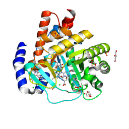
8XPX
 | | The Crystal Structure of PARP12 from Biortus. | | 分子名称: | 1,2-ETHANEDIOL, ACETATE ION, DI(HYDROXYETHYL)ETHER, ... | | 著者 | Wang, F, Cheng, W, Lv, Z, Qi, J, Shen, Z. | | 登録日 | 2024-01-04 | | 公開日 | 2024-03-06 | | 実験手法 | X-RAY DIFFRACTION (1.75 Å) | | 主引用文献 | The Crystal Structure of PARP12 from Biortus.
To Be Published
|
|


