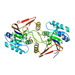
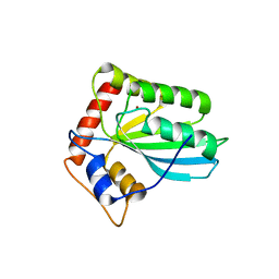
1A21
 
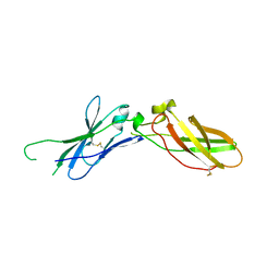 | | TISSUE FACTOR (TF) FROM RABBIT | | 分子名称: | TISSUE FACTOR | | 著者 | Muller, Y.A, De Vos, A.M. | | 登録日 | 1998-01-14 | | 公開日 | 1998-05-27 | | 最終更新日 | 2023-08-02 | | 実験手法 | X-RAY DIFFRACTION (2.35 Å) | | 主引用文献 | Hinge bending within the cytokine receptor superfamily revealed by the 2.4 A crystal structure of the extracellular domain of rabbit tissue factor.
Protein Sci., 7, 1998
|
|
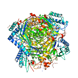
5MLM
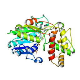 | | Plantago Major multifunctional oxidoreductase V150M mutant in complex with progesterone and NADP+ | | 分子名称: | NADP NICOTINAMIDE-ADENINE-DINUCLEOTIDE PHOSPHATE, PROGESTERONE, Progesterone 5-beta-reductase | | 著者 | Fellows, R, Russo, C.M, Lee, S.G, Jez, J.M, Chisholm, J.D, Zubieta, C, Nanao, M. | | 登録日 | 2016-12-07 | | 公開日 | 2018-08-01 | | 最終更新日 | 2024-01-17 | | 実験手法 | X-RAY DIFFRACTION (2.563 Å) | | 主引用文献 | A multisubstrate reductase from Plantago major: structure-function in the short chain reductase superfamily.
Sci Rep, 8, 2018
|
|
2V3K
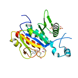 | | The yeast ribosome synthesis factor Emg1 alpha beta knot fold methyltransferase | | 分子名称: | ESSENTIAL FOR MITOTIC GROWTH 1, S-ADENOSYLMETHIONINE, SULFATE ION | | 著者 | Leulliot, N, Bohnsack, M.T, Graille, M, Tollervey, D, VanTilbeurgh, H. | | 登録日 | 2007-06-18 | | 公開日 | 2007-07-10 | | 最終更新日 | 2023-12-13 | | 実験手法 | X-RAY DIFFRACTION (2 Å) | | 主引用文献 | The Yeast Ribosome Synthesis Factor Emg1 is a Novel Member of the Superfamily of Alpha/Beta Knot Fold Methyltransferases.
Nucleic Acids Res., 36, 2008
|
|
8AEP
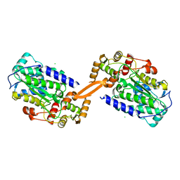 | | Reductase domain of the carboxylate reductase of Neurospora crassa | | 分子名称: | Acetyl-CoA synthetase-like protein, CHLORIDE ION, SULFATE ION | | 著者 | Daniel, B, Schrufer, A, Marlene, L, Sagmeister, T, Pavkov-Keller, T. | | 登録日 | 2022-07-13 | | 公開日 | 2023-03-08 | | 最終更新日 | 2024-02-07 | | 実験手法 | X-RAY DIFFRACTION (2.3 Å) | | 主引用文献 | Structure of the Reductase Domain of a Fungal Carboxylic Acid Reductase and Its Substrate Scope in Thioester and Aldehyde Reduction.
Acs Catalysis, 12, 2022
|
|
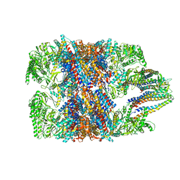
6D90
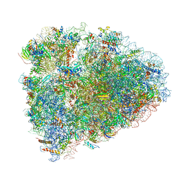 | | Mammalian 80S ribosome with a double translocated CrPV-IRES, P-site tRNA and eRF1. | | 分子名称: | 18S rRNA, 28S rRNA, 5.8S rRNA, ... | | 著者 | Pisareva, V.P, Pisarev, A.V, Fernandez, I.S. | | 登録日 | 2018-04-27 | | 公開日 | 2018-06-06 | | 最終更新日 | 2019-12-18 | | 実験手法 | ELECTRON MICROSCOPY (3.2 Å) | | 主引用文献 | Dual tRNA mimicry in the Cricket Paralysis Virus IRES uncovers an unexpected similarity with the Hepatitis C Virus IRES.
Elife, 7, 2018
|
|
6NRA
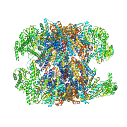 | | hTRiC-hPFD Class1 (No PFD) | | 分子名称: | T-complex protein 1 subunit alpha, T-complex protein 1 subunit beta, T-complex protein 1 subunit delta, ... | | 著者 | Gestaut, D.R, Roh, S.H, Ma, B, Pintilie, G, Joachimiak, L.A, Leitner, A, Walzthoeni, T, Aebersold, R, Chiu, W, Frydman, J. | | 登録日 | 2019-01-23 | | 公開日 | 2019-06-19 | | 最終更新日 | 2024-03-20 | | 実験手法 | ELECTRON MICROSCOPY (7.7 Å) | | 主引用文献 | The Chaperonin TRiC/CCT Associates with Prefoldin through a Conserved Electrostatic Interface Essential for Cellular Proteostasis.
Cell, 177, 2019
|
|
7UGR
 | | Crystal structure of hyperfolder YFP | | 分子名称: | 1,2-ETHANEDIOL, DI(HYDROXYETHYL)ETHER, Hyperfolder yellow fluorescent protein, ... | | 著者 | Campbell, B.C, Liu, C.F, Petsko, G.A. | | 登録日 | 2022-03-25 | | 公開日 | 2022-10-26 | | 最終更新日 | 2023-11-15 | | 実験手法 | X-RAY DIFFRACTION (1.74 Å) | | 主引用文献 | Chemically stable fluorescent proteins for advanced microscopy.
Nat.Methods, 19, 2022
|
|
6NRC
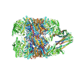 | | hTRiC-hPFD Class3 | | 分子名称: | Prefoldin subunit 1, Prefoldin subunit 2, Prefoldin subunit 3, ... | | 著者 | Gestaut, D, Roh, S.H, Ma, B, Pintilie, G, Joachimiak, L.A, Leitner, A, Walzthoeni, T, Aebersold, R, Chiu, W, Frydman, J. | | 登録日 | 2019-01-23 | | 公開日 | 2019-06-19 | | 最終更新日 | 2024-03-20 | | 実験手法 | ELECTRON MICROSCOPY (8.3 Å) | | 主引用文献 | The Chaperonin TRiC/CCT Associates with Prefoldin through a Conserved Electrostatic Interface Essential for Cellular Proteostasis.
Cell, 177, 2019
|
|
6NR9
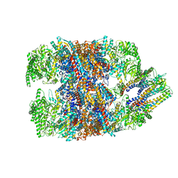 | | hTRiC-hPFD Class5 | | 分子名称: | Prefoldin subunit 1, Prefoldin subunit 2, Prefoldin subunit 3, ... | | 著者 | Gestaut, D, Roh, S.H, Ma, B, Pintilie, G, Joachimiak, L.A, Leitner, A, Walzthoeni, T, Aebersold, R, Chiu, W, Frydman, J. | | 登録日 | 2019-01-23 | | 公開日 | 2019-06-19 | | 最終更新日 | 2024-03-20 | | 実験手法 | ELECTRON MICROSCOPY (8.5 Å) | | 主引用文献 | The Chaperonin TRiC/CCT Associates with Prefoldin through a Conserved Electrostatic Interface Essential for Cellular Proteostasis.
Cell, 177, 2019
|
|
6NRD
 | | hTRiC-hPFD Class4 | | 分子名称: | Prefoldin subunit 1, Prefoldin subunit 2, Prefoldin subunit 3, ... | | 著者 | Gestaut, D.R, Roh, S.H, Ma, B, Pintilie, G, Joachimiak, L.A, Leitner, A, Walzthoeni, T, Aebersold, R, Chiu, W, Frydman, J. | | 登録日 | 2019-01-23 | | 公開日 | 2019-06-19 | | 最終更新日 | 2024-03-20 | | 実験手法 | ELECTRON MICROSCOPY (8.2 Å) | | 主引用文献 | The Chaperonin TRiC/CCT Associates with Prefoldin through a Conserved Electrostatic Interface Essential for Cellular Proteostasis.
Cell, 177, 2019
|
|
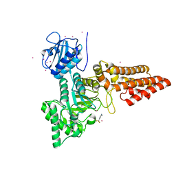
6JTZ
 | | Crystal Structure of hRecQ1_D2-Zn-WH containing mutation on beta-hairpin | | 分子名称: | ATP-dependent DNA helicase Q1, ZINC ION | | 著者 | Das, T, Mukhopadhyay, S, Bose, M, Das, A.K, Ganguly, A. | | 登録日 | 2019-04-12 | | 公開日 | 2020-04-15 | | 最終更新日 | 2023-11-22 | | 実験手法 | X-RAY DIFFRACTION (2.797 Å) | | 主引用文献 | Residues at the interface between zinc binding and winged helix domains of human RECQ1 play a significant role in DNA strand annealing activity.
Nucleic Acids Res., 2021
|
|
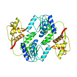
5G3P
 | | Bacillus cereus formamidase (BceAmiF) acetylated at the active site. | | 分子名称: | 1,2-ETHANEDIOL, ACETATE ION, DI(HYDROXYETHYL)ETHER, ... | | 著者 | Gavira, J.A, Conejero-Muriel, M, Martinez-Rodriguez, S. | | 登録日 | 2016-04-29 | | 公開日 | 2017-04-12 | | 最終更新日 | 2024-01-10 | | 実験手法 | X-RAY DIFFRACTION (1.78 Å) | | 主引用文献 | A novel cysteine carbamoyl-switch is responsible for the inhibition of formamidase, a nitrilase superfamily member.
Arch.Biochem.Biophys., 662, 2019
|
|
5OXD
 | |
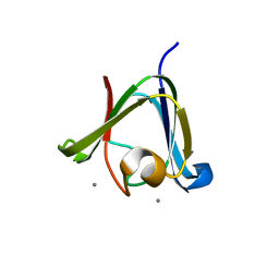
6A4R
 | | Crystal structure of aspartate bound peptidase E from Salmonella enterica | | 分子名称: | ASPARTIC ACID, Peptidase E | | 著者 | Yadav, P, Chandravanshi, K, Goyal, V.D, Singh, R, Kumar, A, Gokhale, S.M, Makde, R.D. | | 登録日 | 2018-06-20 | | 公開日 | 2018-10-24 | | 最終更新日 | 2023-11-22 | | 実験手法 | X-RAY DIFFRACTION (1.828 Å) | | 主引用文献 | Structure of Asp-bound peptidase E from Salmonella enterica: Active site at dimer interface illuminates Asp recognition.
FEBS Lett., 592, 2018
|
|
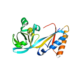
6I1M
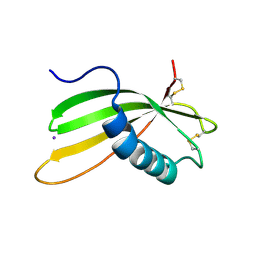 | | Secreted type 1 cystatin from Fasciola hepatica | | 分子名称: | Cystatin, IODIDE ION | | 著者 | Busa, M, Rezacova, P, Pachl, P, Stefanic, S, Mares, M. | | 登録日 | 2018-10-29 | | 公開日 | 2020-08-26 | | 最終更新日 | 2023-03-29 | | 実験手法 | X-RAY DIFFRACTION (1.6 Å) | | 主引用文献 | An evolutionary molecular adaptation of an unusual stefin from the liver fluke Fasciola hepatica redefines the cystatin superfamily.
J.Biol.Chem., 299, 2023
|
|
3HZ2
 | |
3HZB
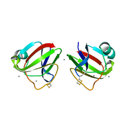 | |
6A4S
 | | Crystal structure of peptidase E with ordered active site loop from Salmonella enterica | | 分子名称: | Peptidase E | | 著者 | Yadav, P, Chandravanshi, K, Goyal, V.D, Singh, R, Kumar, A, Gokhale, S.M, Makde, R.D. | | 登録日 | 2018-06-20 | | 公開日 | 2018-10-31 | | 最終更新日 | 2023-11-22 | | 実験手法 | X-RAY DIFFRACTION (1.9 Å) | | 主引用文献 | Structure of Asp-bound peptidase E from Salmonella enterica: Active site at dimer interface illuminates Asp recognition.
FEBS Lett., 592, 2018
|
|
3VM8
 | |
5LIL
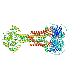 | |
5LJ8
 | | Structure of the E. coli MacB periplasmic domain (P21) | | 分子名称: | Macrolide export ATP-binding/permease protein MacB | | 著者 | Crow, A. | | 登録日 | 2016-07-18 | | 公開日 | 2017-11-15 | | 最終更新日 | 2024-01-10 | | 実験手法 | X-RAY DIFFRACTION (1.95 Å) | | 主引用文献 | Structure and mechanotransmission mechanism of the MacB ABC transporter superfamily.
Proc. Natl. Acad. Sci. U.S.A., 114, 2017
|
|
7CYH
 | |
5LJ6
 | |
5LJ9
 | | Structure of the E. coli MacB ABC domain (C2221) | | 分子名称: | Macrolide export ATP-binding/permease protein MacB | | 著者 | Crow, A. | | 登録日 | 2016-07-18 | | 公開日 | 2017-11-15 | | 最終更新日 | 2024-01-10 | | 実験手法 | X-RAY DIFFRACTION (2.3 Å) | | 主引用文献 | Structure and mechanotransmission mechanism of the MacB ABC transporter superfamily.
Proc. Natl. Acad. Sci. U.S.A., 114, 2017
|
|
8URB
 | |


