2HF5
 
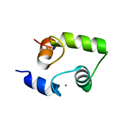 | | The structure and function of a novel two-site calcium-binding fragment of calmodulin | | 分子名称: | CALCIUM ION, Calmodulin | | 著者 | Lakowski, T.M, Lee, G.M, Reid, R.E, McIntosh, L.P. | | 登録日 | 2006-06-23 | | 公開日 | 2007-05-01 | | 最終更新日 | 2024-05-29 | | 実験手法 | SOLUTION NMR | | 主引用文献 | Calcium-induced folding of a fragment of calmodulin composed of EF-hands 2 and 3
Protein Sci., 16, 2007
|
|
2H6D
 
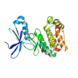 | | Protein Kinase Domain of the Human 5'-AMP-activated protein kinase catalytic subunit alpha-2 (AMPK alpha-2 chain) | | 分子名称: | 5'-AMP-activated protein kinase catalytic subunit alpha-2 | | 著者 | Littler, D.R, Walker, J.R, Wybenga-Groot, L, Newman, E.M, Butler-Cole, C, Mackenzie, F, Finerty, P.J, Weigelt, J, Sundstrom, M, Arrowsmith, C.H, Edwards, A.M, Bochkarev, A, Dhe-Paganon, S, Structural Genomics Consortium (SGC) | | 登録日 | 2006-05-31 | | 公開日 | 2006-06-27 | | 最終更新日 | 2023-08-30 | | 実験手法 | X-RAY DIFFRACTION (1.85 Å) | | 主引用文献 | A conserved mechanism of autoinhibition for the AMPK kinase domain: ATP-binding site and catalytic loop refolding as a means of regulation.
Acta Crystallogr.,Sect.F, 66, 2010
|
|
4F3A
 
 | |
4QGA
 
 | |
4QGH
 
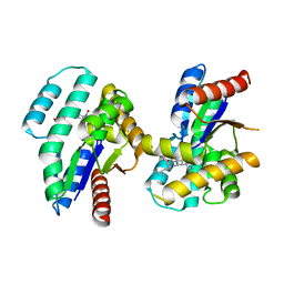 | | S.aureus TMK in complex with potent inhibitor compound 47 | | 分子名称: | 2-(3-chlorophenoxy)-3-fluoro-4-{(1S)-3-methyl-1-[(3S)-3-(5-methyl-2,4-dioxo-3,4-dihydropyrimidin-1(2H)-yl)piperidin-1-yl]butyl}benzoic acid, Thymidylate kinase | | 著者 | Olivier, N.B. | | 登録日 | 2014-05-22 | | 公開日 | 2014-06-11 | | 最終更新日 | 2024-02-28 | | 実験手法 | X-RAY DIFFRACTION (1.78 Å) | | 主引用文献 | Antibacterial inhibitors of gram-positive thymidylate kinase: structure-activity relationships and chiral preference of a new hydrophobic binding region.
J.Med.Chem., 57, 2014
|
|
2FV7
 
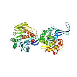 | | Crystal structure of human ribokinase | | 分子名称: | ADENOSINE-5'-DIPHOSPHATE, MAGNESIUM ION, Ribokinase, ... | | 著者 | Rabeh, W.M, Tempel, W, Nedyalkova, L, Arrowsmith, C, Edwards, A, Sundstrom, M, Weigelt, J, Bochkarev, A, Park, H, Structural Genomics Consortium (SGC) | | 登録日 | 2006-01-30 | | 公開日 | 2006-02-28 | | 最終更新日 | 2023-08-30 | | 実験手法 | X-RAY DIFFRACTION (2.1 Å) | | 主引用文献 | Crystal structure of human ribokinase
to be published
|
|
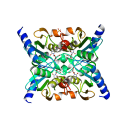
2FR5
 
 | |
2K49
 
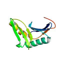 | | Solution NMR structure of UPF0339 protein SO3888 from Shewanella oneidensis. Northeast Structural Genomics Consortium target SoR190 | | 分子名称: | UPF0339 protein SO_3888 | | 著者 | Tang, Y, Wang, D, Nwosu, C, Maglaqui, M, Xiao, R, Liu, J, Baran, M.C, Swapna, G.V.T, Acton, T.B, Rost, B, Montelione, G.T, Northeast Structural Genomics Consortium (NESG) | | 登録日 | 2008-05-31 | | 公開日 | 2008-07-08 | | 最終更新日 | 2024-05-08 | | 実験手法 | SOLUTION NMR | | 主引用文献 | Solution NMR structure of UPF0339 protein SO3888 from Shewanella oneidensis.
To be Published
|
|
4QGF
 
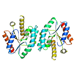 | |
6WB6
 
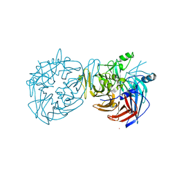
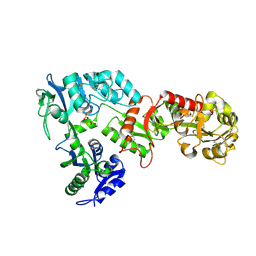 | | 2.05 A resolution structure of transferrin 1 from Manduca sexta | | 分子名称: | 2-acetamido-2-deoxy-beta-D-glucopyranose-(1-4)-2-acetamido-2-deoxy-beta-D-glucopyranose, CARBONATE ION, FE (III) ION, ... | | 著者 | Lovell, S, Kashipathy, M.M, Battaile, K.P, Weber, J.J, Gorman, M.J. | | 登録日 | 2020-03-26 | | 公開日 | 2020-11-25 | | 最終更新日 | 2024-11-13 | | 実験手法 | X-RAY DIFFRACTION (2.05 Å) | | 主引用文献 | Structural insight into the novel iron-coordination and domain interactions of transferrin-1 from a model insect, Manduca sexta.
Protein Sci., 30, 2021
|
|
2K1F
 
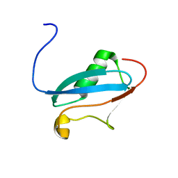 | | SUMO-3 from Drosophila melanogaster (dsmt3) | | 分子名称: | CG4494-PA | | 著者 | Kumar, D, Misra, J.R, Misra, A.K, Chugh, J, Sharma, S, Hosur, R.V. | | 登録日 | 2008-03-03 | | 公開日 | 2009-03-10 | | 最終更新日 | 2024-05-01 | | 実験手法 | SOLUTION NMR | | 主引用文献 | NMR-derived solution structure of SUMO from Drosophila melanogaster (dSmt3).
Proteins, 75, 2009
|
|
7F4V
 
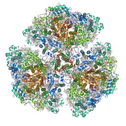 | | Cryo-EM structure of a primordial cyanobacterial photosystem I | | 分子名称: | 1,2-DIPALMITOYL-PHOSPHATIDYL-GLYCEROLE, 1,2-DISTEAROYL-MONOGALACTOSYL-DIGLYCERIDE, BETA-CAROTENE, ... | | 著者 | Kato, K, Hamaguchi, T, Nagao, R, Kawakami, K, Yonekura, K, Shen, J.R. | | 登録日 | 2021-06-21 | | 公開日 | 2022-04-06 | | 最終更新日 | 2024-10-16 | | 実験手法 | ELECTRON MICROSCOPY (2.04 Å) | | 主引用文献 | Structural basis for the absence of low-energy chlorophylls responsible for photoprotection from a primitive cyanobacterial PSI
Biorxiv, 2022
|
|
3SYZ
 
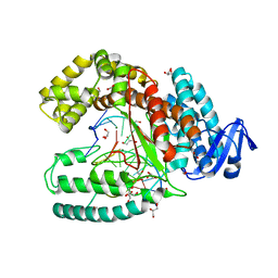 | | Crystal structure of the large fragment of DNA polymerase I from Thermus Aquaticus in an open binary complex with dNaM as templating nucleobase | | 分子名称: | (5'-D(*AP*AP*AP*(BMN)P*GP*GP*CP*GP*CP*CP*GP*TP*GP*GP*TP*C)-3'), (5'-D(*GP*AP*CP*CP*AP*CP*GP*GP*CP*GP*CP*(DOC))-3'), DNA polymerase I, ... | | 著者 | Betz, K, Diederichs, K, Marx, A. | | 登録日 | 2011-07-18 | | 公開日 | 2012-06-06 | | 最終更新日 | 2023-09-13 | | 実験手法 | X-RAY DIFFRACTION (1.952 Å) | | 主引用文献 | KlenTaq polymerase replicates unnatural base pairs by inducing a Watson-Crick geometry.
Nat.Chem.Biol., 8, 2012
|
|
2K3X
 
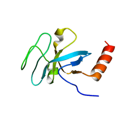 | | Solution structure of EAF3 chromo barrel domain | | 分子名称: | Chromatin modification-related protein EAF3 | | 著者 | Mer, G, Xu, C. | | 登録日 | 2008-05-19 | | 公開日 | 2008-09-16 | | 最終更新日 | 2024-05-29 | | 実験手法 | SOLUTION NMR | | 主引用文献 | Structural Basis for the Recognition of Methylated Histone H3K36 by the Eaf3 Subunit of Histone Deacetylase Complex Rpd3S.
Structure, 16, 2008
|
|
6W9V
 
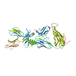 | |
4DXT
 
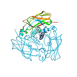 | | Human SUN2 (AA 522-717) | | 分子名称: | POTASSIUM ION, SUN domain-containing protein 2 | | 著者 | Sosa, B, Schwartz, T.U. | | 登録日 | 2012-02-28 | | 公開日 | 2012-06-06 | | 最終更新日 | 2024-11-06 | | 実験手法 | X-RAY DIFFRACTION (2.22 Å) | | 主引用文献 | LINC Complexes Form by Binding of Three KASH Peptides to Domain Interfaces of Trimeric SUN Proteins.
Cell(Cambridge,Mass.), 149, 2012
|
|
2JXY
 
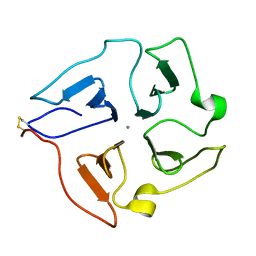 | | Solution structure of the hemopexin-like domain of MMP12 | | 分子名称: | CALCIUM ION, Macrophage metalloelastase | | 著者 | Bertini, I, Calderone, V, Fragai, M, Jaiswal, R, Luchinat, C, Melikian, M. | | 登録日 | 2007-12-01 | | 公開日 | 2008-05-27 | | 最終更新日 | 2024-11-13 | | 実験手法 | SOLUTION NMR | | 主引用文献 | Evidence of reciprocal reorientation of the catalytic and hemopexin-like domains of full-length MMP-12
J.Am.Chem.Soc., 130, 2008
|
|
4DYX
 
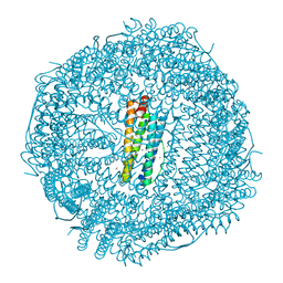 | |
2K0J
 
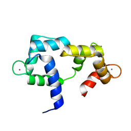 | | Solution structure of CaM complexed to DRP1p | | 分子名称: | CALCIUM ION, LANTHANUM (III) ION, calmodulin | | 著者 | Bertini, I, Luchinat, C, Parigi, G, Yuan, J, Structural Proteomics in Europe (SPINE) | | 登録日 | 2008-02-04 | | 公開日 | 2009-03-10 | | 最終更新日 | 2024-05-29 | | 実験手法 | SOLUTION NMR | | 主引用文献 | Accurate solution structures of proteins from X-ray data and a minimal set of NMR data: calmodulin-peptide complexes as examples.
J.Am.Chem.Soc., 131, 2009
|
|
4TXL
 
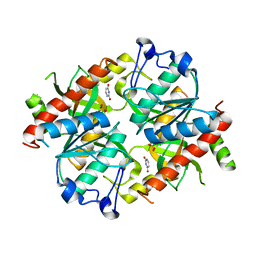 | | Crystal structure of uridine phosphorylase from Schistosoma mansoni in complex with uracil | | 分子名称: | SULFATE ION, URACIL, Uridine phosphorylase | | 著者 | Marinho, A, Torini, J, Romanello, L, Cassago, A, DeMarco, R, Brandao-Neto, J, Pereira, H.M. | | 登録日 | 2014-07-03 | | 公開日 | 2015-07-15 | | 最終更新日 | 2023-09-27 | | 実験手法 | X-RAY DIFFRACTION (1.92 Å) | | 主引用文献 | Analysis of two Schistosoma mansoni uridine phosphorylases isoforms suggests the emergence of a protein with a non-canonical function.
Biochimie, 125, 2016
|
|
3X0R
 
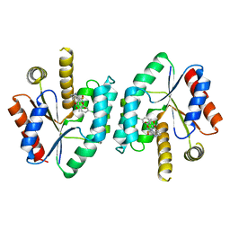
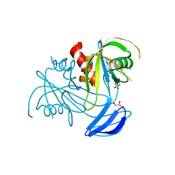 | | DP ribose pyrophosphatase from Thermus thermophilus HB8 in E'-state at reaction time of 30 min | | 分子名称: | ADP-ribose pyrophosphatase, GLYCEROL, MANGANESE (II) ION, ... | | 著者 | Furuike, Y, Akita, Y, Miyahara, I, Kamiya, N. | | 登録日 | 2014-10-17 | | 公開日 | 2016-04-27 | | 最終更新日 | 2023-11-08 | | 実験手法 | X-RAY DIFFRACTION (1.15 Å) | | 主引用文献 | ADP-Ribose Pyrophosphatase Reaction in Crystalline State Conducted by Consecutive Binding of Two Manganese(II) Ions as Cofactors
Biochemistry, 55, 2016
|
|
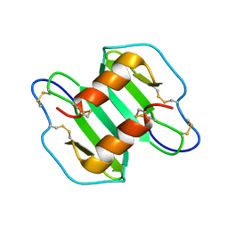
2K01
 
 | |
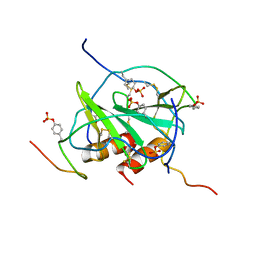
2K05
 
 | |
2JA8
 
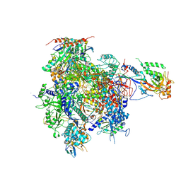 | | CPD lesion containing RNA Polymerase II elongation complex D | | 分子名称: | 5'-D(*AP*GP*CP*TP*CP*AP*AP*GP*TP*AP *CP*TP*TP*TP*TP*TTP*CP*BRUP*GP*GP*TP*CP*AP*TP*T)-3', 5'-D(*TP*AP*AP*GP*TP*AP*CP*TP*TP*GP *AP*GP*CP*T)-3', 5'-R(*UP*UP*CP*GP*AP*CP*CP*AP*GP*AP*UP)-3', ... | | 著者 | Brueckner, F, Hennecke, U, Carell, T, Cramer, P. | | 登録日 | 2006-11-23 | | 公開日 | 2007-02-20 | | 最終更新日 | 2024-11-13 | | 実験手法 | X-RAY DIFFRACTION (3.8 Å) | | 主引用文献 | Cpd Damage Recognition by Transcribing RNA Polymerase II.
Science, 315, 2007
|
|
4EQR
 
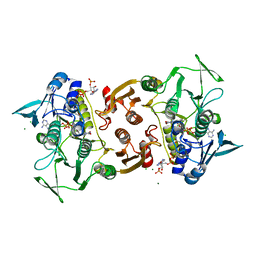 | | Crystal structure of the Y361F mutant of Staphylococcus aureus CoADR | | 分子名称: | CHLORIDE ION, COENZYME A, Coenzyme A disulfide reductase, ... | | 著者 | Wallace, B.D, Edwards, J.S, Wallen, J.R, Claiborne, A, Redinbo, M.R. | | 登録日 | 2012-04-19 | | 公開日 | 2012-10-17 | | 最終更新日 | 2024-10-16 | | 実験手法 | X-RAY DIFFRACTION (1.8 Å) | | 主引用文献 | Turnover-Dependent Covalent Inactivation of Staphylococcus aureus Coenzyme A-Disulfide Reductase by Coenzyme A-Mimetics: Mechanistic and Structural Insights.
Biochemistry, 51, 2012
|
|
