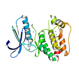
6O4Y
 
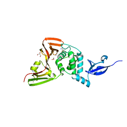
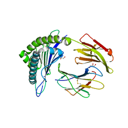 | | Structure of HLA-A2:01 with peptide MM91 | | 分子名称: | Beta-2-microglobulin, GLYCEROL, MHC class I antigen, ... | | 著者 | Ying, G, Bitra, A, Zajonc, D.M. | | 登録日 | 2019-03-01 | | 公開日 | 2020-01-15 | | 最終更新日 | 2023-10-11 | | 実験手法 | X-RAY DIFFRACTION (1.58 Å) | | 主引用文献 | Anin silico-in vitroPipeline Identifying an HLA-A*02:01+KRAS G12V+Spliced Epitope Candidate for a Broad Tumor-Immune Response in Cancer Patients.
Front Immunol, 10, 2019
|
|
6O72
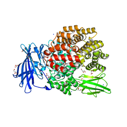
 | | Structure of the TRPM8 cold receptor by single particle electron cryo-microscopy, TC-I 2014-bound state | | 分子名称: | (1R)-2-{[(S)-(2-aminoethoxy)(hydroxy)phosphoryl]oxy}-1-[(heptanoyloxy)methyl]ethyl octadecanoate, 3-{7-(trifluoromethyl)-5-[2-(trifluoromethyl)phenyl]-1H-benzimidazol-2-yl}-1-oxa-2-azaspiro[4.5]dec-2-ene, CHOLESTEROL HEMISUCCINATE, ... | | 著者 | Diver, M.M, Cheng, Y, Julius, D. | | 登録日 | 2019-03-07 | | 公開日 | 2019-09-18 | | 最終更新日 | 2024-03-20 | | 実験手法 | ELECTRON MICROSCOPY (3 Å) | | 主引用文献 | Structural insights into TRPM8 inhibition and desensitization.
Science, 365, 2019
|
|
5Y3E
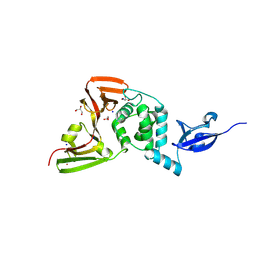 | |
6OOR
 | | Structure of 1B1 bound to mouse CD1d | | 分子名称: | Antibody 1B1 Heavy chain, Antibody 1B1 Light chain, Antigen-presenting glycoprotein CD1d1, ... | | 著者 | Ying, G, Zajonc, D.M. | | 登録日 | 2019-04-23 | | 公開日 | 2019-07-17 | | 最終更新日 | 2023-10-11 | | 実験手法 | X-RAY DIFFRACTION (2.45 Å) | | 主引用文献 | Structural basis of NKT cell inhibition using the T-cell receptor-blocking anti-CD1d antibody 1B1.
J.Biol.Chem., 294, 2019
|
|
6O5E
 | | Crystal structure of the Vitronectin hemopexin-like domain | | 分子名称: | CHLORIDE ION, IMIDAZOLE, NITRATE ION, ... | | 著者 | Lechtenberg, B.C, Shin, K, Marassi, F.M. | | 登録日 | 2019-03-01 | | 公開日 | 2019-09-18 | | 最終更新日 | 2023-10-11 | | 実験手法 | X-RAY DIFFRACTION (1.9 Å) | | 主引用文献 | Structure of human Vitronectin C-terminal domain and interaction withYersinia pestisouter membrane protein Ail.
Sci Adv, 5, 2019
|
|
5Y7D
 | | Crystal structure of human Endothelial-overexpressed LPS associated factor 1 | | 分子名称: | CHLORIDE ION, GLYCEROL, Protein CXorf40A, ... | | 著者 | Park, S.H, Kim, M.J, Park, J.S, Kim, H.J, Han, B.W. | | 登録日 | 2017-08-17 | | 公開日 | 2018-08-22 | | 最終更新日 | 2024-03-27 | | 実験手法 | X-RAY DIFFRACTION (1.71 Å) | | 主引用文献 | Crystal Structure of Human EOLA1 Implies Its Possibility of RNA Binding.
Molecules, 24, 2019
|
|
6AMC
 | |
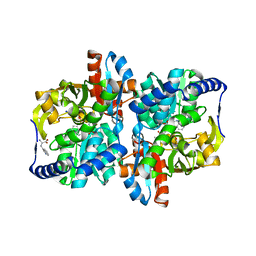
2IH1
 | | Ion selectivity in a semi-synthetic K+ channel locked in the conductive conformation | | 分子名称: | (1S)-2-HYDROXY-1-[(NONANOYLOXY)METHYL]ETHYL MYRISTATE, FAB Heavy Chain, FAB Light Chain, ... | | 著者 | Valiyaveetil, F.I, Leonetti, M, Muir, T.W, MacKinnon, R. | | 登録日 | 2006-09-25 | | 公開日 | 2006-11-21 | | 最終更新日 | 2021-10-20 | | 実験手法 | X-RAY DIFFRACTION (2.4 Å) | | 主引用文献 | Ion Selectivity in a Semisynthetic K+ Channel Locked in the Conductive Conformation.
Science, 314, 2006
|
|
5N84
 | | TTK kinase domain in complex with Mps-BAY2b | | 分子名称: | 2-(2-METHOXYETHOXY)ETHANOL, Dual specificity protein kinase TTK, SODIUM ION, ... | | 著者 | Uitdehaag, J, Willemsen-Seegers, N, de Man, J, Buijsman, R.C, Zaman, G.J.R. | | 登録日 | 2017-02-22 | | 公開日 | 2017-05-31 | | 最終更新日 | 2024-01-17 | | 実験手法 | X-RAY DIFFRACTION (2.3 Å) | | 主引用文献 | Target Residence Time-Guided Optimization on TTK Kinase Results in Inhibitors with Potent Anti-Proliferative Activity.
J. Mol. Biol., 429, 2017
|
|
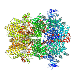
6J8M
 | | Low-dose structure of bovine heart cytochrome c oxidase in the fully oxidized state determined using 30 keV X-ray | | 分子名称: | (1R)-2-{[{[(2S)-2,3-DIHYDROXYPROPYL]OXY}(HYDROXY)PHOSPHORYL]OXY}-1-[(PALMITOYLOXY)METHYL]ETHYL (11E)-OCTADEC-11-ENOATE, (1S)-2-{[(2-AMINOETHOXY)(HYDROXY)PHOSPHORYL]OXY}-1-[(STEAROYLOXY)METHYL]ETHYL (5E,8E,11E,14E)-ICOSA-5,8,11,14-TETRAENOATE, (7R,17E,20E)-4-HYDROXY-N,N,N-TRIMETHYL-9-OXO-7-[(PALMITOYLOXY)METHYL]-3,5,8-TRIOXA-4-PHOSPHAHEXACOSA-17,20-DIEN-1-AMINIUM 4-OXIDE, ... | | 著者 | Ueno, G, Shimada, A, Yamashita, E, Hasegawa, K, Kumasaka, T, Shinzawa-Itoh, K, Yoshikawa, S, Tsukihara, T, Yamamoto, M. | | 登録日 | 2019-01-20 | | 公開日 | 2019-06-26 | | 最終更新日 | 2023-11-22 | | 実験手法 | X-RAY DIFFRACTION (1.9 Å) | | 主引用文献 | Low-dose X-ray structure analysis of cytochrome c oxidase utilizing high-energy X-rays.
J.Synchrotron Radiat., 26, 2019
|
|
6AM8
 | |
6AWW
 | | Structure of PR 10 Allergen Ara h 8.01 in complex with 3-Hydroxy-2-naphthoic acid | | 分子名称: | 3-hydroxynaphthalene-2-carboxylic acid, Ara h 8 allergen, SODIUM ION | | 著者 | Offermann, L.R, Yarbrough, J, McBride, J, Hurlburt, B.K, Maleki, S.J, Pote, S.S, Chruszcz, M. | | 登録日 | 2017-09-06 | | 公開日 | 2018-09-12 | | 最終更新日 | 2023-10-04 | | 実験手法 | X-RAY DIFFRACTION (2.3 Å) | | 主引用文献 | Structure of PR-10 Allergen Ara h 8.01.
To Be Published
|
|
4QJE
 | | 1.85 Angstrom resolution crystal structure of apo betaine aldehyde dehydrogenase (betB) G234S mutant from Staphylococcus aureus (IDP00699) with BME-free sulfinic acid form of Cys289 | | 分子名称: | 2-AMINO-2-HYDROXYMETHYL-PROPANE-1,3-DIOL, 2-[3-(2-HYDROXY-1,1-DIHYDROXYMETHYL-ETHYLAMINO)-PROPYLAMINO]-2-HYDROXYMETHYL-PROPANE-1,3-DIOL, Betaine aldehyde dehydrogenase, ... | | 著者 | Halavaty, A.S, Minasov, G, Chen, C, Joo, J.C, Yakunin, A.F, Anderson, W.F, Center for Structural Genomics of Infectious Diseases (CSGID) | | 登録日 | 2014-06-03 | | 公開日 | 2014-06-18 | | 最終更新日 | 2023-09-20 | | 実験手法 | X-RAY DIFFRACTION (1.85 Å) | | 主引用文献 | Structural and functional analysis of betaine aldehyde dehydrogenase from Staphylococcus aureus.
Acta Crystallogr.,Sect.D, 71, 2015
|
|
6LZH
 | | Crystal structure of Alpha/beta hydrolase GrgF from Penicillium sp. sh18 | | 分子名称: | GrgF, SODIUM ION | | 著者 | Wang, H, Yu, J, Wang, W.G, Matsuda, Y, Yao, M. | | 登録日 | 2020-02-19 | | 公開日 | 2020-06-24 | | 最終更新日 | 2023-11-29 | | 実験手法 | X-RAY DIFFRACTION (1.9 Å) | | 主引用文献 | Molecular Basis for the Biosynthesis of an Unusual Chain-Fused Polyketide, Gregatin A.
J.Am.Chem.Soc., 142, 2020
|
|
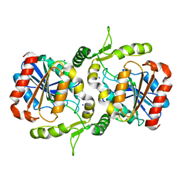
2IH3
 | | Ion selectivity in a semi-synthetic K+ channel locked in the conductive conformation | | 分子名称: | (1S)-2-HYDROXY-1-[(NONANOYLOXY)METHYL]ETHYL MYRISTATE, FAB Heavy Chain, FAB Light Chain, ... | | 著者 | Valiyaveetil, F.I, Leonetti, M, Muir, T.W, MacKinnon, R. | | 登録日 | 2006-09-25 | | 公開日 | 2006-11-21 | | 最終更新日 | 2021-10-20 | | 実験手法 | X-RAY DIFFRACTION (1.72 Å) | | 主引用文献 | Ion Selectivity in a Semisynthetic K+ Channel Locked in the Conductive Conformation.
Science, 314, 2006
|
|
6MFM
 | |
6M38
 | | X-ray structure of a Drosophila dopamine transporter with subsiteB mutations (D121G/S426M) in S-duloxetine bound form | | 分子名称: | (3S)-N-methyl-3-(naphthalen-1-yloxy)-3-(thiophen-2-yl)propan-1-amine, Antibody fragment 9D5 heavy chain, Antibody fragment 9D5 light chain, ... | | 著者 | Shabareesh, P, Mallela, A.K, Joseph, D, Penmatsa, A. | | 登録日 | 2020-03-02 | | 公開日 | 2021-02-17 | | 最終更新日 | 2023-11-29 | | 実験手法 | X-RAY DIFFRACTION (3.001 Å) | | 主引用文献 | Structural basis of norepinephrine recognition and transport inhibition in neurotransmitter transporters.
Nat Commun, 12, 2021
|
|
6M0Z
 | | X-ray structure of Drosophila dopamine transporter with NET-like mutations (D121G/S426M/F471L) in L-norepinephrine bound form | | 分子名称: | Antibody fragment (Fab) 9D5 Light chain, Antibody fragment (Fab) 9D5 heavy chain, CHLORIDE ION, ... | | 著者 | Shabareesh, P, Mallela, A.K, Joseph, D, Penmatsa, A. | | 登録日 | 2020-02-24 | | 公開日 | 2021-02-17 | | 最終更新日 | 2023-11-29 | | 実験手法 | X-RAY DIFFRACTION (2.88 Å) | | 主引用文献 | Structural basis of norepinephrine recognition and transport inhibition in neurotransmitter transporters.
Nat Commun, 12, 2021
|
|
5MUR
 | | X-ray structure of the F14'A mutant of GLIC in complex with propofol | | 分子名称: | 2,6-BIS(1-METHYLETHYL)PHENOL, ACETATE ION, CHLORIDE ION, ... | | 著者 | Sauguet, L, Fourati, Z, Delarue, M. | | 登録日 | 2017-01-13 | | 公開日 | 2018-02-14 | | 最終更新日 | 2024-01-17 | | 実験手法 | X-RAY DIFFRACTION (3.1 Å) | | 主引用文献 | Structural Basis for a Bimodal Allosteric Mechanism of General Anesthetic Modulation in Pentameric Ligand-Gated Ion Channels.
Cell Rep, 23, 2018
|
|
5Y3Q
 | |
5MFT
 | | The crystal structure of E. coli Aminopeptidase N in complex with 7-amino-1-bromo-4-phenyl-5,7,8,9-tetrahydrobenzocyclohepten-6-one | | 分子名称: | Aminopeptidase N, CHLORIDE ION, DIMETHYL SULFOXIDE, ... | | 著者 | Peng, G, Olieric, V, McEwen, A.G, Schmitt, C, Albrecht, S, Cavarelli, J, Tarnus, C. | | 登録日 | 2016-11-18 | | 公開日 | 2017-04-19 | | 最終更新日 | 2024-01-17 | | 実験手法 | X-RAY DIFFRACTION (1.59 Å) | | 主引用文献 | Insight into the remarkable affinity and selectivity of the aminobenzosuberone scaffold for the M1 aminopeptidases family based on structure analysis.
Proteins, 85, 2017
|
|
5NEQ
 | | The structure of the G. violaceus guanidine II riboswitch P1 stem-loop with aminoguanidine | | 分子名称: | AMINOGUANIDINE, RNA (5'-R(*GP*GP*UP*GP*GP*GP*GP*AP*CP*GP*AP*CP*CP*CP*CP*AP*(CBV)P*C)-3'), SODIUM ION, ... | | 著者 | Huang, L, Wang, J, Lilley, D.M.J. | | 登録日 | 2017-03-11 | | 公開日 | 2017-05-31 | | 最終更新日 | 2024-01-17 | | 実験手法 | X-RAY DIFFRACTION (1.69 Å) | | 主引用文献 | The Structure of the Guanidine-II Riboswitch.
Cell Chem Biol, 24, 2017
|
|
5WZE
 | | The structure of Pseudomonas aeruginosa aminopeptidase PepP | | 分子名称: | 1,2-ETHANEDIOL, ALANINE, Aminopeptidase P, ... | | 著者 | Bao, R, Peng, C.T, Liu, L, He, L.H, Li, C.C, Li, T, Shen, Y.L, Zhu, Y.B, Song, Y.J. | | 登録日 | 2017-01-17 | | 公開日 | 2018-01-17 | | 最終更新日 | 2023-11-22 | | 実験手法 | X-RAY DIFFRACTION (1.783 Å) | | 主引用文献 | Structure-Function Relationship of Aminopeptidase P from Pseudomonas aeruginosa.
Front Microbiol, 8, 2017
|
|
6BFK
 | | Caspase-3 Mutant- T245A | | 分子名称: | AC-ASP-GLU-VAL-ASP-CMK, Caspase-3, SODIUM ION | | 著者 | Thomas, M.E, Grinshpon, R, Swartz, P.D, Clark, A.C. | | 登録日 | 2017-10-26 | | 公開日 | 2018-02-21 | | 最終更新日 | 2018-04-25 | | 実験手法 | X-RAY DIFFRACTION (1.753 Å) | | 主引用文献 | Modifications to a common phosphorylation network provide individualized control in caspases.
J. Biol. Chem., 293, 2018
|
|
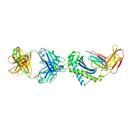
6ML1
 | | Structure of the USP15 deubiquitinase domain in complex with an affinity-matured inhibitory Ubv | | 分子名称: | 1,2-ETHANEDIOL, 2-(N-MORPHOLINO)-ETHANESULFONIC ACID, CALCIUM ION, ... | | 著者 | Singer, A.U, Teyra, J, Boehmelt, G, Lenter, M, Sicheri, F, Sidhu, S.S. | | 登録日 | 2018-09-26 | | 公開日 | 2019-01-23 | | 最終更新日 | 2023-10-25 | | 実験手法 | X-RAY DIFFRACTION (1.9 Å) | | 主引用文献 | Structural and Functional Characterization of Ubiquitin Variant Inhibitors of USP15.
Structure, 27, 2019
|
|


