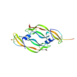
1W5W
 
 | | HIV-1 protease in complex with fluoro substituted diol-based C2- symmetric inhibitor | | 分子名称: | (2R,3R,4R,5R)-2,5-BIS[(2,4-DIFLUOROBENZYL)OXY]-3,4-DIHYDROXY-N,N'-BIS[(1R,2S)-2-HYDROXY-2,3-DIHYDRO-1H-INDEN-1-YL]HEXAN EDIAMIDE, POL POLYPROTEIN | | 著者 | Lindberg, J, Pyring, D, Loewgren, S, Rosenquist, A, Zuccarello, G, Kvarnstroem, I, Zhang, H, Vrang, L, Claesson, B, Hallberg, A, Samuelsson, B, Unge, T. | | 登録日 | 2004-08-10 | | 公開日 | 2004-12-22 | | 最終更新日 | 2024-05-08 | | 実験手法 | X-RAY DIFFRACTION (1.8 Å) | | 主引用文献 | Symmetric Fluoro-Substituted Diol-Based HIV Protease Inhibitors. Ortho-Fluorinated and Meta-Fluorinated P1/P1'-Benzyloxy Side Groups Significantly Improve the Antiviral Activity and Preserve Binding Efficacy
Eur.J.Biochem., 271, 2004
|
|
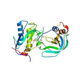
1M7E
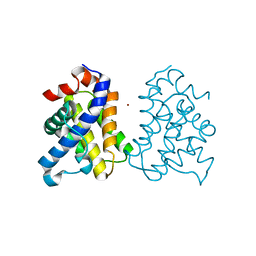
 | | Crystal structure of the phosphotyrosine binding domain(PTB) of mouse Disabled 2(Dab2):implications for Reeling signaling | | 分子名称: | Disabled homolog 2, NGYENPTYK peptide | | 著者 | Yun, M, Keshvara, L, Park, C.-G, Zhang, Y.-M, Dickerson, J.B, Zheng, J, Rock, C.O, Curran, T, Park, H.-W. | | 登録日 | 2002-07-19 | | 公開日 | 2003-08-05 | | 最終更新日 | 2024-05-22 | | 実験手法 | X-RAY DIFFRACTION (2.45 Å) | | 主引用文献 | Crystal structures of the Dab homology domains of mouse disabled 1 and 2
J.Biol.Chem., 278, 2003
|
|
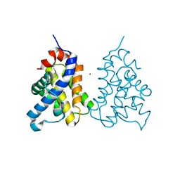
1MG5
 | | Crystal structure of Drosophila melanogaster alcohol dehydrogenase complexed with NADH and acetate at 1.6 A | | 分子名称: | 1,4-DIHYDRONICOTINAMIDE ADENINE DINUCLEOTIDE, ACETATE ION, alcohol dehydrogenase | | 著者 | Benach, J, Atrian, S, Gonzalez-Duarte, R, Ladenstein, R. | | 登録日 | 2002-08-14 | | 公開日 | 2003-10-14 | | 最終更新日 | 2024-02-14 | | 実験手法 | X-RAY DIFFRACTION (1.63 Å) | | 主引用文献 | Drosophila alcohol dehydrogenase: acetate-enzyme interactions and novel insights into the effects of electrostatics on catalysis
J.Mol.Biol., 345, 2005
|
|
1W0T
 | | hTRF1 DNA-binding domain in complex with telomeric DNA. | | 分子名称: | 5'-D(*CP*TP*GP*TP*TP*AP*GP*GP*GP*TP *TP*AP*GP*GP*GP*TP*TP*AP*G)-3', 5'-D(*TP*CP*TP*AP*AP*CP*CP*CP*TP*AP *AP*CP*CP*CP*TP*AP*AP*CP*A)-3', TELOMERIC REPEAT BINDING FACTOR 1 | | 著者 | Court, R.I, Chapman, L.M, Fairall, L, Rhodes, D. | | 登録日 | 2004-06-11 | | 公開日 | 2004-12-22 | | 最終更新日 | 2024-05-08 | | 実験手法 | X-RAY DIFFRACTION (2 Å) | | 主引用文献 | How the Human Telomeric Proteins Trf1 and Trf2 Recognize Telomeric DNA: A View from High-Resolution Crystal Structures
Embo Rep., 6, 2005
|
|
1VPP
 | | COMPLEX BETWEEN VEGF AND A RECEPTOR BLOCKING PEPTIDE | | 分子名称: | PROTEIN (PEPTIDE V108), PROTEIN (VASCULAR ENDOTHELIAL GROWTH FACTOR) | | 著者 | Wiesmann, C, Christinger, H.W, Cochran, A.G, Cunningham, B.C, Fairbrother, W.J, Keenan, C.J, Meng, G, de Vos, A.M. | | 登録日 | 1998-10-09 | | 公開日 | 1999-02-23 | | 最終更新日 | 2023-08-23 | | 実験手法 | X-RAY DIFFRACTION (1.9 Å) | | 主引用文献 | Crystal structure of the complex between VEGF and a receptor-blocking peptide.
Biochemistry, 37, 1998
|
|
4HOH
 | | RIBONUCLEASE T1 (THR93ALA MUTANT) COMPLEXED WITH 2'GMP | | 分子名称: | CALCIUM ION, GUANOSINE-2'-MONOPHOSPHATE, PROTEIN (RIBONUCLEASE T1) | | 著者 | Langhorst, U, Loris, R, Denisov, V.P, Doumen, J, Roose, P, Maes, D, Halle, B, Steyaert, J. | | 登録日 | 1998-09-14 | | 公開日 | 1998-09-23 | | 最終更新日 | 2023-09-20 | | 実験手法 | X-RAY DIFFRACTION (2.05 Å) | | 主引用文献 | Dissection of the structural and functional role of a conserved hydration site in RNase T1.
Protein Sci., 8, 1999
|
|
2J0T
 | | Crystal Structure of the Catalytic Domain of MMP-1 in Complex with the Inhibitory Domain of TIMP-1 | | 分子名称: | CALCIUM ION, INTERSTITIAL COLLAGENASE, METALLOPROTEINASE INHIBITOR 1, ... | | 著者 | Iyer, S, Wei, S, Brew, K, Acharya, K.R. | | 登録日 | 2006-08-04 | | 公開日 | 2006-10-18 | | 最終更新日 | 2023-12-13 | | 実験手法 | X-RAY DIFFRACTION (2.54 Å) | | 主引用文献 | Crystal Structure of the Catalytic Domain of Matrix Metalloproteinase-1 in Complex with the Inhibitory Domain of Tissue Inhibitor of Metalloproteinase-1.
J.Biol.Chem., 282, 2007
|
|
2IMS
 | | The X-ray Structure of a Bak Homodimer Reveals an Inhibitory Zinc Binding Site | | 分子名称: | Apoptosis regulator BAK, ZINC ION | | 著者 | Moldoveanu, T, Liu, Q, Tocilj, A, Watson, M, Shore, G.C, Gehring, K.B. | | 登録日 | 2006-10-04 | | 公開日 | 2006-12-26 | | 最終更新日 | 2017-10-18 | | 実験手法 | X-RAY DIFFRACTION (1.48 Å) | | 主引用文献 | The X-Ray Structure of a BAK Homodimer Reveals an Inhibitory Zinc Binding Site
Mol.Cell, 24, 2006
|
|
2IUH
 | | Crystal structure of the PI3-kinase p85 N-terminal SH2 domain in complex with c-Kit phosphotyrosyl peptide | | 分子名称: | C-KIT PHOSPHOTYROSYL PEPTIDE, PHOSPHATIDYLINOSITOL 3-KINASE REGULATORY ALPHA SUBUNIT | | 著者 | Nolte, R.T, Eck, M.J, Schlessinger, J, Shoelson, S.E, Harrison, S.C. | | 登録日 | 2006-06-03 | | 公開日 | 2006-06-06 | | 最終更新日 | 2021-04-28 | | 実験手法 | X-RAY DIFFRACTION (2 Å) | | 主引用文献 | Crystal Structure of the Pi 3-Kinase P85 Amino-Terminal Sh2 Domain and its Phosphopeptide Complexes
Nat.Struct.Biol., 3, 1996
|
|
4JD5
 | | Crystal Structure of Benzoylformate Decarboxylase Mutant L403E | | 分子名称: | 2-{3-[(4-AMINO-2-METHYLPYRIMIDIN-5-YL)METHYL]-4-METHYL-2-OXO-2,3-DIHYDRO-1,3-THIAZOL-5-YL}ETHYL TRIHYDROGEN DIPHOSPHATE, Benzoylformate decarboxylase, CALCIUM ION, ... | | 著者 | Novak, W.R.P, Andrews, F.H, Tom, A.R, Gunderman, P.R, McLeish, M.J. | | 登録日 | 2013-02-23 | | 公開日 | 2013-05-22 | | 最終更新日 | 2024-02-28 | | 実験手法 | X-RAY DIFFRACTION (1.33 Å) | | 主引用文献 | A bulky hydrophobic residue is not required to maintain the v-conformation of enzyme-bound thiamin diphosphate.
Biochemistry, 52, 2013
|
|
2IMT
 | | The X-ray Structure of a Bak Homodimer Reveals an Inhibitory Zinc Binding Site | | 分子名称: | Apoptosis regulator BAK, ZINC ION | | 著者 | Moldoveanu, T, Liu, Q, Tocilj, A, Watson, M, Shore, G.C, Gehring, K.B. | | 登録日 | 2006-10-04 | | 公開日 | 2007-01-23 | | 最終更新日 | 2023-08-30 | | 実験手法 | X-RAY DIFFRACTION (1.49 Å) | | 主引用文献 | The X-ray structure of a BAK homodimer reveals an inhibitory zinc binding site.
Mol.Cell, 24, 2006
|
|
2IUG
 | | Crystal structure of the PI3-kinase p85 N-terminal SH2 domain | | 分子名称: | PHOSPHATIDYLINOSITOL 3-KINASE REGULATORY ALPHA SUBUNIT | | 著者 | Nolte, R.T, Eck, M.J, Schlessinger, J, Shoelson, S.E, Harrison, S.C. | | 登録日 | 2006-06-03 | | 公開日 | 2006-06-06 | | 最終更新日 | 2024-05-08 | | 実験手法 | X-RAY DIFFRACTION (1.89 Å) | | 主引用文献 | Crystal Structure of the Pi 3-Kinase P85 Amino-Terminal Sh2 Domain and its Phosphopeptide Complexes
Nat.Struct.Biol., 3, 1996
|
|
2IUI
 | | Crystal structure of the PI3-kinase p85 N-terminal SH2 domain in complex with PDGFR phosphotyrosyl peptide | | 分子名称: | Phosphatidylinositol 3-kinase regulatory subunit alpha, Platelet-derived growth factor receptor beta | | 著者 | Nolte, R.T, Eck, M.J, Schlessinger, J, Shoelson, S.E, Harrison, S.C. | | 登録日 | 2006-06-03 | | 公開日 | 2006-06-06 | | 最終更新日 | 2021-04-28 | | 実験手法 | X-RAY DIFFRACTION (2.4 Å) | | 主引用文献 | Crystal Structure of the Pi 3-Kinase P85 Amino- Terminal Sh2 Domain and its Phosphopeptide Complexes
Nat.Struct.Biol., 3, 1996
|
|
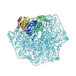
8EM7
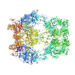 | | Cryo-EM structure of LRP2 at pH 5.2 | | 分子名称: | 2-acetamido-2-deoxy-beta-D-galactopyranose, 2-acetamido-2-deoxy-beta-D-glucopyranose, 2-acetamido-2-deoxy-beta-D-glucopyranose-(1-4)-2-acetamido-2-deoxy-beta-D-glucopyranose, ... | | 著者 | Beenken, A, Cerutti, G, Fitzpatrick, A.W, Barasch, J, Shapiro, L. | | 登録日 | 2022-09-27 | | 公開日 | 2023-02-08 | | 最終更新日 | 2023-03-08 | | 実験手法 | ELECTRON MICROSCOPY (2.97 Å) | | 主引用文献 | Structures of LRP2 reveal a molecular machine for endocytosis.
Cell, 186, 2023
|
|
8EM4
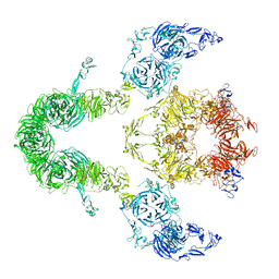 | | Cryo-EM structure of LRP2 at pH 7.5 | | 分子名称: | 2-acetamido-2-deoxy-beta-D-galactopyranose, 2-acetamido-2-deoxy-beta-D-glucopyranose, 2-acetamido-2-deoxy-beta-D-glucopyranose-(1-4)-2-acetamido-2-deoxy-beta-D-glucopyranose, ... | | 著者 | Beenken, A, Cerutti, G, Brasch, J, Fitzpatrick, A.W, Barasch, J, Shapiro, L. | | 登録日 | 2022-09-26 | | 公開日 | 2023-02-08 | | 最終更新日 | 2023-03-08 | | 実験手法 | ELECTRON MICROSCOPY (2.83 Å) | | 主引用文献 | Structures of LRP2 reveal a molecular machine for endocytosis.
Cell, 186, 2023
|
|
1WY4
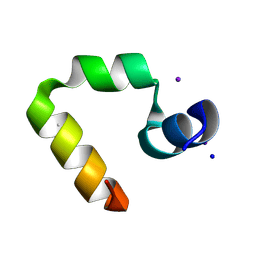 | | Chicken villin subdomain HP-35, K65(NLE), N68H, pH5.1 | | 分子名称: | IODIDE ION, SODIUM ION, Villin | | 著者 | Chiu, T.K, Kubelka, J, Herbst-Irmer, R, Eaton, W.A, Hofrichter, J, Davies, D.R. | | 登録日 | 2005-02-04 | | 公開日 | 2005-05-03 | | 最終更新日 | 2023-11-15 | | 実験手法 | X-RAY DIFFRACTION (1.55 Å) | | 主引用文献 | High-resolution x-ray crystal structures of the villin headpiece subdomain, an ultrafast folding protein.
Proc.Natl.Acad.Sci.Usa, 102, 2005
|
|
1WY3
 | | Chicken villin subdomain HP-35, K65(NLE), N68H, pH7.0 | | 分子名称: | Villin | | 著者 | Chiu, T.K, Kubelka, J, Herbst-Irmer, R, Eaton, W.A, Hofrichter, J, Davies, D.R. | | 登録日 | 2005-02-04 | | 公開日 | 2005-05-03 | | 最終更新日 | 2023-11-15 | | 実験手法 | X-RAY DIFFRACTION (0.95 Å) | | 主引用文献 | High-resolution x-ray crystal structures of the villin headpiece subdomain, an ultrafast folding protein.
Proc.Natl.Acad.Sci.Usa, 102, 2005
|
|
1X1S
 | | Crystal structure of M-Ras in complex with GppNHp | | 分子名称: | MAGNESIUM ION, PHOSPHOAMINOPHOSPHONIC ACID-GUANYLATE ESTER, Ras-related protein M-Ras | | 著者 | Ye, M, Shima, F, Muraoka, S, Liao, J, Okamoto, H, Yamamoto, M, Tamura, A, Yagi, N, Ueki, T, Kataoka, T. | | 登録日 | 2005-04-13 | | 公開日 | 2005-07-26 | | 最終更新日 | 2023-10-25 | | 実験手法 | X-RAY DIFFRACTION (2.2 Å) | | 主引用文献 | Crystal Structure of M-Ras Reveals a GTP-bound "Off" State Conformation of Ras Family Small GTPases
J.Biol.Chem., 280, 2005
|
|
1X1R
 | | Crystal structure of M-Ras in complex with GDP | | 分子名称: | GUANOSINE-5'-DIPHOSPHATE, MAGNESIUM ION, Ras-related protein M-Ras | | 著者 | Ye, M, Shima, F, Muraoka, S, Liao, J, Okamoto, H, Yamamoto, M, Tamura, A, Yagi, N, Ueki, T, Kataoka, T. | | 登録日 | 2005-04-12 | | 公開日 | 2005-07-26 | | 最終更新日 | 2023-10-25 | | 実験手法 | X-RAY DIFFRACTION (1.3 Å) | | 主引用文献 | Crystal Structure of M-Ras Reveals a GTP-bound "Off" State Conformation of Ras Family Small GTPases
J.Biol.Chem., 280, 2005
|
|
1XA5
 | | Structure of Calmodulin in complex with KAR-2, a bis-indol alkaloid | | 分子名称: | 3"-(BETA-CHLOROETHYL)-2",4"-DIOXO-3, 5"-SPIRO-OXAZOLIDINO-4-DEACETOXY-VINBLASTINE, CALCIUM ION, ... | | 著者 | Horvath, I, Harmat, V, Hlavanda, E, Naray-Szabo, G, Ovadi, J. | | 登録日 | 2004-08-25 | | 公開日 | 2004-12-21 | | 最終更新日 | 2023-10-25 | | 実験手法 | X-RAY DIFFRACTION (2.12 Å) | | 主引用文献 | The structure of the complex of calmodulin with KAR-2: a novel mode of binding explains the unique pharmacology of the drug
J.Biol.Chem., 280, 2005
|
|
1XEQ
 | | Crystal tructure of RNA binding domain of influenza B virus non-structural protein | | 分子名称: | BROMIDE ION, Nonstructural protein NS1 | | 著者 | Khan, J.A, Yin, C, Krug, R.M, Montelione, G.T, Tong, L, Northeast Structural Genomics Consortium (NESG) | | 登録日 | 2004-09-11 | | 公開日 | 2005-09-20 | | 最終更新日 | 2024-02-14 | | 実験手法 | X-RAY DIFFRACTION (2.1 Å) | | 主引用文献 | Conserved surface features form the double-stranded RNA binding site of non-structural protein 1 (NS1) from influenza A and B viruses.
J.Biol.Chem., 282, 2007
|
|
1YRI
 | | Chicken villin subdomain HP-35, N68H, pH6.4 | | 分子名称: | ACETATE ION, IODIDE ION, Villin | | 著者 | Chiu, T.K, Kubelka, J, Herbst-Irmer, R, Eaton, W.A, Hofrichter, J, Davies, D.R. | | 登録日 | 2005-02-03 | | 公開日 | 2005-05-03 | | 最終更新日 | 2023-08-23 | | 実験手法 | X-RAY DIFFRACTION (1 Å) | | 主引用文献 | High-resolution x-ray crystal structures of the villin headpiece subdomain, an ultrafast folding protein.
Proc.Natl.Acad.Sci.Usa, 102, 2005
|
|
1XM8
 | | X-RAY STRUCTURE OF GLYOXALASE II FROM ARABIDOPSIS THALIANA GENE AT2G31350 | | 分子名称: | ACETIC ACID, DI(HYDROXYETHYL)ETHER, FE (III) ION, ... | | 著者 | Wesenberg, G.E, Smith, D.W, Phillips Jr, G.N, Bitto, E, Bingman, C.A, Allard, S.T.M, Center for Eukaryotic Structural Genomics (CESG) | | 登録日 | 2004-10-01 | | 公開日 | 2004-10-12 | | 最終更新日 | 2024-02-14 | | 実験手法 | X-RAY DIFFRACTION (1.74 Å) | | 主引用文献 | Structural studies on a mitochondrial glyoxalase II.
J.Biol.Chem., 280, 2005
|
|
1XM2
 | | Crystal structure of Human PRL-1 | | 分子名称: | SULFATE ION, Tyrosine Phosphatase | | 著者 | Jeong, D.G, Kim, S.J, Kim, J.H, Son, J.H, Ryu, S.E. | | 登録日 | 2004-10-01 | | 公開日 | 2005-01-25 | | 最終更新日 | 2021-11-10 | | 実験手法 | X-RAY DIFFRACTION (2.7 Å) | | 主引用文献 | Trimeric structure of PRL-1 phosphatase reveals an active enzyme conformation and regulation mechanisms
J.Mol.Biol., 345, 2005
|
|
2A4O
 | | Dual modes of modification of Hepatitis A virus 3C protease by a serine derived beta-lactone: selective crytstallization and high resolution structure of the His102 adduct | | 分子名称: | ACETYL GROUP, N-[(BENZYLOXY)CARBONYL]-L-ALANINE, PHENYLALANINE AMIDE, ... | | 著者 | Yin, J, Bergmann, E.M, Cherney, M.M, Lall, M.S, Jain, R.P, Vederas, J.C, James, M.N.G. | | 登録日 | 2005-06-29 | | 公開日 | 2005-12-27 | | 最終更新日 | 2021-11-10 | | 実験手法 | X-RAY DIFFRACTION (1.55 Å) | | 主引用文献 | Dual Modes of Modification of Hepatitis A Virus 3C Protease by a Serine-derived beta-Lactone: Selective Crystallization and Formation of a Functional Catalytic Triad in the Active Site
J.MOL.BIOL., 354, 2005
|
|


