1P9K
 
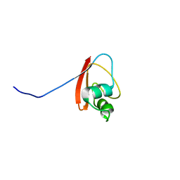 | | THE SOLUTION STRUCTURE OF YBCJ FROM E. COLI REVEALS A RECENTLY DISCOVERED ALFAL MOTIF INVOLVED IN RNA-BINDING | | 分子名称: | orf, hypothetical protein | | 著者 | Volpon, L, Lievre, C, Osborne, M.J, Gandhi, S, Iannuzzi, P, Larocque, R, Matte, A, Cygler, M, Gehring, K, Ekiel, I, Montreal-Kingston Bacterial Structural Genomics Initiative (BSGI) | | 登録日 | 2003-05-12 | | 公開日 | 2003-11-25 | | 最終更新日 | 2024-05-01 | | 実験手法 | SOLUTION NMR | | 主引用文献 | The solution structure of YbcJ from Escherichia coli reveals a recently discovered alphaL motif involved in RNA binding.
J.Bacteriol., 185, 2003
|
|
1P9L
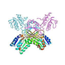 | | Structure of M. tuberculosis dihydrodipicolinate reductase in complex with NADH and 2,6 PDC | | 分子名称: | 1,4-DIHYDRONICOTINAMIDE ADENINE DINUCLEOTIDE, PYRIDINE-2,6-DICARBOXYLIC ACID, TETRAETHYLENE GLYCOL, ... | | 著者 | Cirilli, M, Zheng, R, Scapin, G, Blanchard, J.S, TB Structural Genomics Consortium (TBSGC) | | 登録日 | 2003-05-12 | | 公開日 | 2003-08-26 | | 最終更新日 | 2023-08-16 | | 実験手法 | X-RAY DIFFRACTION (2.3 Å) | | 主引用文献 | The three-dimensional structures of the Mycobacterium tuberculosis dihydrodipicolinate reductase-NADH-2,6-PDC and -NADPH-2,6-PDC complexes. Structural and mutagenic analysis of relaxed nucleotide specificity.
Biochemistry, 42, 2003
|
|
1P9M
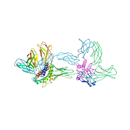 | | Crystal structure of the hexameric human IL-6/IL-6 alpha receptor/gp130 complex | | 分子名称: | Interleukin-6, Interleukin-6 receptor alpha chain, Interleukin-6 receptor beta chain | | 著者 | Boulanger, M.J, Chow, D.C, Brevnova, E.E, Garcia, K.C. | | 登録日 | 2003-05-12 | | 公開日 | 2003-07-01 | | 最終更新日 | 2023-08-16 | | 実験手法 | X-RAY DIFFRACTION (3.65 Å) | | 主引用文献 | Hexameric Structure and Assembly of the Interleukin-6/IL-6 alpha-Receptor/gp130 Complex
Science, 300, 2003
|
|
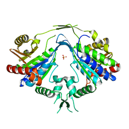
1P9N
 | | Crystal structure of Escherichia coli MobB. | | 分子名称: | Molybdopterin-guanine dinucleotide biosynthesis protein B, SULFATE ION | | 著者 | Rangarajan, S.E, Tocilj, A, Li, Y, Iannuzzi, P, Matte, A, Cygler, M, Montreal-Kingston Bacterial Structural Genomics Initiative (BSGI) | | 登録日 | 2003-05-12 | | 公開日 | 2003-05-20 | | 最終更新日 | 2018-01-31 | | 実験手法 | X-RAY DIFFRACTION (2.8 Å) | | 主引用文献 | Molecules of Escherichia coli MobB assemble into densely packed hollow cylinders in a crystal lattice with 75% solvent content.
Acta Crystallogr.,Sect.D, 59, 2003
|
|
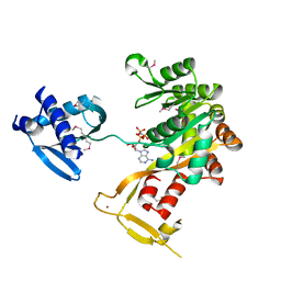
1P9O
 | | Crystal Structure of Phosphopantothenoylcysteine Synthetase | | 分子名称: | Phosphopantothenoylcysteine synthetase, SULFATE ION | | 著者 | Manoj, N, Strauss, E, Begley, T.P, Ealick, S.E. | | 登録日 | 2003-05-12 | | 公開日 | 2003-09-02 | | 最終更新日 | 2024-04-03 | | 実験手法 | X-RAY DIFFRACTION (2.3 Å) | | 主引用文献 | Structure of human phosphopantothenoylcysteine synthetase at 2.3 A resolution.
Structure, 11, 2003
|
|
1P9P
 | | The Crystal Structure of a M1G37 tRNA Methyltransferase, TrmD | | 分子名称: | S-ADENOSYL-L-HOMOCYSTEINE, tRNA (Guanine-N(1)-)-methyltransferase | | 著者 | Elkins, P.A, Watts, J.M, Zalacain, M, Van Thiel, A, Vitaszka, P.R, Redlak, M, Andraos-Selim, C, Rastinejad, F, Holmes, W.M. | | 登録日 | 2003-05-12 | | 公開日 | 2004-05-18 | | 最終更新日 | 2024-02-14 | | 実験手法 | X-RAY DIFFRACTION (2.5 Å) | | 主引用文献 | Insights into Catalysis by a Knotted TrmD tRNA Methyltransferase.
J.Mol.Biol., 333, 2003
|
|
1P9Q
 | | Structure of a hypothetical protein AF0491 from Archaeoglobus fulgidus | | 分子名称: | Hypothetical protein AF0491 | | 著者 | Savchenko, A, Evdokimova, E, Skarina, T, Arrowsmith, C.H, Edwards, A.M, Bochkarev, A. | | 登録日 | 2003-05-12 | | 公開日 | 2004-06-08 | | 最終更新日 | 2024-02-14 | | 実験手法 | X-RAY DIFFRACTION (2 Å) | | 主引用文献 | The Shwachman-Bodian-Diamond syndrome protein family is involved in RNA metabolism.
J.Biol.Chem., 280, 2005
|
|
1P9R
 | | Crystal Structure of Vibrio cholerae putative NTPase EpsE | | 分子名称: | CHLORIDE ION, General secretion pathway protein E, ZINC ION | | 著者 | Robien, M.A, Krumm, B.E, Sandkvist, M, Hol, W.G.J. | | 登録日 | 2003-05-12 | | 公開日 | 2003-10-14 | | 最終更新日 | 2011-07-13 | | 実験手法 | X-RAY DIFFRACTION (2.5 Å) | | 主引用文献 | Crystal structure of the extracellular protein secretion NTPase EpsE of Vibrio cholerae
J.Mol.Biol., 333, 2003
|
|
1P9S
 | | Coronavirus Main Proteinase (3CLpro) Structure: Basis for Design of anti-SARS Drugs | | 分子名称: | 1,4-DIETHYLENE DIOXIDE, Replicase polyprotein 1ab | | 著者 | Anand, K, Ziebuhr, J, Wadhwani, P, Mesters, J.R, Hilgenfeld, R. | | 登録日 | 2003-05-12 | | 公開日 | 2003-05-20 | | 最終更新日 | 2023-11-15 | | 実験手法 | X-RAY DIFFRACTION (2.54 Å) | | 主引用文献 | Coronavirus Main Proteinase (3CLpro) Structure: Basis for Design of anti-SARS Drugs
Science, 300, 2003
|
|
1P9U
 | | Coronavirus Main Proteinase (3CLpro) Structure: Basis for Design of anti-SARS Drugs | | 分子名称: | (4R)-2-METHYLPENTANE-2,4-DIOL, PHQ-VNSTLQ-CHLOROMETHYLKETONE INHIBITOR, SULFATE ION, ... | | 著者 | Anand, K, Ziebuhr, J, Wadhwani, P, Mesters, J.R, Hilgenfeld, R. | | 登録日 | 2003-05-12 | | 公開日 | 2003-05-20 | | 最終更新日 | 2017-10-11 | | 実験手法 | X-RAY DIFFRACTION (2.37 Å) | | 主引用文献 | Coronavirus Main Proteinase (3CLpro) Structure: Basis for Design of anti-SARS Drugs
Science, 300, 2003
|
|
1P9W
 | | Crystal Structure of Vibrio cholerae putative NTPase EpsE | | 分子名称: | General secretion pathway protein E, PHOSPHOAMINOPHOSPHONIC ACID-ADENYLATE ESTER, ZINC ION | | 著者 | Robien, M.A, Krumm, B.E, Sandkvist, M, Hol, W.G.J. | | 登録日 | 2003-05-12 | | 公開日 | 2003-10-14 | | 最終更新日 | 2011-07-13 | | 実験手法 | X-RAY DIFFRACTION (2.7 Å) | | 主引用文献 | Crystal structure of the extracellular protein secretion NTPase EpsE of Vibrio cholerae
J.Mol.Biol., 333, 2003
|
|
1P9X
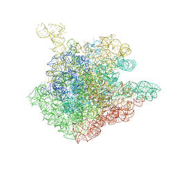 | | THE CRYSTAL STRUCTURE OF THE 50S LARGE RIBOSOMAL SUBUNIT FROM DEINOCOCCUS RADIODURANS COMPLEXED WITH TELITHROMYCIN KETOLIDE ANTIBIOTIC | | 分子名称: | 23S RIBOSOMAL RNA, TELITHROMYCIN | | 著者 | Berisio, R, Harms, J, Schluenzen, F, Zarivach, R, Hansen, H.A, Fucini, P, Yonath, A. | | 登録日 | 2003-05-13 | | 公開日 | 2003-08-05 | | 最終更新日 | 2023-08-16 | | 実験手法 | X-RAY DIFFRACTION (3.4 Å) | | 主引用文献 | Structural insight into the antibiotic action of telithromycin against resistant mutants
J.Bacteriol., 185, 2003
|
|
1P9Y
 | |
1P9Z
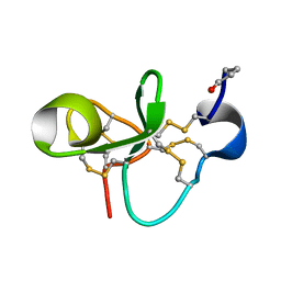 | | The Solution Structure of Antifungal Peptide Distinct With a Five-disulfide Motif from Eucommia ulmoides Oliver | | 分子名称: | Eucommia Antifungal peptide 2 | | 著者 | Huang, R.H, Xiang, Y, Tu, G.Z, Zhang, Y, Wang, D.C. | | 登録日 | 2003-05-13 | | 公開日 | 2004-05-25 | | 最終更新日 | 2019-12-25 | | 実験手法 | SOLUTION NMR | | 主引用文献 | Solution Structure of Eucommia Antifungal Peptide: A Novel Structural Model Distinct with a Five-Disulfide Motif.
Biochemistry, 43, 2004
|
|
1PA0
 | | CRYSTAL STRUCTURE OF BNSP-7, A LYS49-PHOSPHOLIPASE A2 | | 分子名称: | Myotoxic phospholipase A2-like | | 著者 | Magro, A.J, Soares, A.M, Giglio, J.R, Fontes, M.R. | | 登録日 | 2003-05-13 | | 公開日 | 2004-07-27 | | 最終更新日 | 2024-04-03 | | 実験手法 | X-RAY DIFFRACTION (2.2 Å) | | 主引用文献 | Crystal structures of BnSP-7 and BnSP-6, two Lys49-phospholipases A(2): quaternary structure and inhibition mechanism insights.
Biochem.Biophys.Res.Commun., 311, 2003
|
|
1PA1
 | | Crystal structure of the C215D mutant of protein tyrosine phosphatase 1B | | 分子名称: | CHLORIDE ION, MAGNESIUM ION, Protein-tyrosine phosphatase, ... | | 著者 | Romsicki, Y, Scapin, G, Beaulieu-Audy, V, Patel, S.B, Becker, J.W, Kennedy, B, Asante-Appiah, E. | | 登録日 | 2003-05-13 | | 公開日 | 2003-08-05 | | 最終更新日 | 2023-08-16 | | 実験手法 | X-RAY DIFFRACTION (1.6 Å) | | 主引用文献 | Functional characterization and crystal structure of the C215D mutant of protein-tyrosine phosphatase-1B
J.Biol.Chem., 278, 2003
|
|
1PA2
 | | ARABIDOPSIS THALIANA PEROXIDASE A2 | | 分子名称: | CALCIUM ION, MAGNESIUM ION, PEROXIDASE, ... | | 著者 | Henriksen, A. | | 登録日 | 1999-05-18 | | 公開日 | 2000-11-15 | | 最終更新日 | 2023-08-16 | | 実験手法 | X-RAY DIFFRACTION (1.45 Å) | | 主引用文献 | Arabidopsis ATP A2 peroxidase. Expression and high-resolution structure of a plant peroxidase with implications for lignification.
Plant Mol.Biol., 44, 2000
|
|
1PA3
 | |
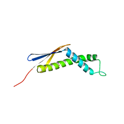
1PA4
 | | Solution structure of a putative ribosome-binding factor from Mycoplasma pneumoniae (MPN156) | | 分子名称: | Probable ribosome-binding factor A | | 著者 | Rubin, S.M, Pelton, J.G, Yokota, H, Kim, R, Wemmer, D.E, Berkeley Structural Genomics Center (BSGC) | | 登録日 | 2003-05-13 | | 公開日 | 2004-03-02 | | 最終更新日 | 2024-05-22 | | 実験手法 | SOLUTION NMR | | 主引用文献 | Solution structure of a putative ribosome binding protein from Mycoplasma pneumoniae and comparison to a distant homolog.
J.STRUCT.FUNCT.GENOM., 4, 2003
|
|
1PA6
 | |
1PA7
 | |
1PA9
 | | Yersinia Protein-Tyrosine Phosphatase complexed with pNCS (Yop51,Pasteurella X,Ptpase,Yop51delta162) (Catalytic Domain, Residues 163-468) Mutant With Cys 235 Replaced By Arg (C235r) | | 分子名称: | N,4-DIHYDROXY-N-OXO-3-(SULFOOXY)BENZENAMINIUM, Protein-tyrosine phosphatase yopH | | 著者 | Sun, J.P, Wu, L, Fedorov, A.A, Almo, S.C, Zhang, Z.Y. | | 登録日 | 2003-05-13 | | 公開日 | 2003-11-04 | | 最終更新日 | 2023-08-16 | | 実験手法 | X-RAY DIFFRACTION (2 Å) | | 主引用文献 | Crystal structure of the Yersinia protein-tyrosine phosphatase YopH complexed
with a specific small molecule inhibitor
J.BIOL.CHEM., 278, 2003
|
|
1PAA
 | | STRUCTURE OF A HISTIDINE-X4-HISTIDINE ZINC FINGER DOMAIN: INSIGHTS INTO ADR1-UAS1 PROTEIN-DNA RECOGNITION | | 分子名称: | YEAST TRANSCRIPTION FACTOR ADR1, ZINC ION | | 著者 | Bernstein, B.E, Hoffman, R.C, Horvath, S.J, Herriott, J.R, Klevit, R.E. | | 登録日 | 1994-07-15 | | 公開日 | 1994-10-15 | | 最終更新日 | 2024-05-01 | | 実験手法 | SOLUTION NMR | | 主引用文献 | Structure of a histidine-X4-histidine zinc finger domain: insights into ADR1-UAS1 protein-DNA recognition.
Biochemistry, 33, 1994
|
|
1PAD
 | |
1PAE
 | | nucleoside diphosphate kinase | | 分子名称: | Nucleoside diphosphate kinase, cytosolic, SELENIUM ATOM | | 著者 | Strub, M.-P, Hoh, F, Sanchez, J.-F, Strub, J.M, Bock, A, Aumelas, A, Dumas, C. | | 登録日 | 2003-05-14 | | 公開日 | 2003-11-11 | | 最終更新日 | 2021-10-27 | | 実験手法 | X-RAY DIFFRACTION (2.7 Å) | | 主引用文献 | Selenomethionine and Selenocysteine Double Labeling Strategy for Crystallographic Phasing
Structure, 11, 2003
|
|


