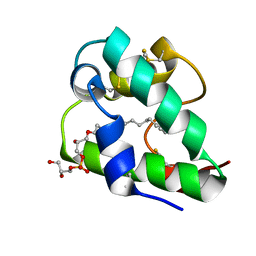
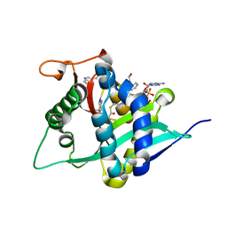
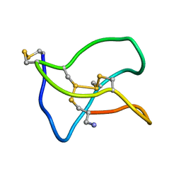
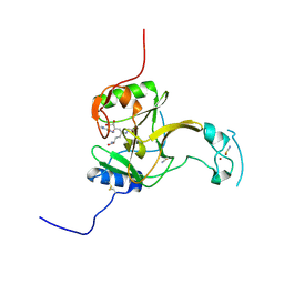
6J4B
 
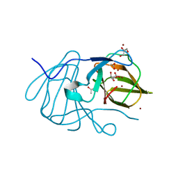 | | Crystal structure of MarH, an epimerase for biosynthesis of Maremycins in Streptomyces, under 400 mM Zinc acetate | | 分子名称: | ACETIC ACID, Cupin superfamily protein, GLYCEROL, ... | | 著者 | Hou, Y, Liu, B, Hu, K, Zhang, R. | | 登録日 | 2019-01-08 | | 公開日 | 2020-01-15 | | 最終更新日 | 2024-03-27 | | 実験手法 | X-RAY DIFFRACTION (1.58 Å) | | 主引用文献 | Structural basis of the mechanism of beta-methyl epimerization by enzyme MarH.
Org.Biomol.Chem., 17, 2019
|
|
5YVH
 | |
5LSX
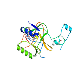 | | Structure of the Epigenetic Oncogene MMSET and inhibition by N-Alkyl Sinefungin Derivatives | | 分子名称: | Histone-lysine N-methyltransferase SETD2, ZINC ION, [(2~{R},5~{S})-1-[(2~{R},3~{S},4~{R},5~{R})-5-(6-aminopurin-9-yl)-3,4-bis(oxidanyl)oxolan-2-yl]-5-azaniumyl-6-oxidanyl-6-oxidanylidene-hexan-2-yl]-(phenylmethyl)azanium | | 著者 | Tisi, D, Pathuri, P, Heightman, T. | | 登録日 | 2016-09-05 | | 公開日 | 2016-10-05 | | 最終更新日 | 2024-05-08 | | 実験手法 | X-RAY DIFFRACTION (2.9 Å) | | 主引用文献 | Structure of the Epigenetic Oncogene MMSET and Inhibition by N-Alkyl Sinefungin Derivatives.
ACS Chem. Biol., 11, 2016
|
|
5WC7
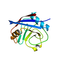 | | CypA Mutant - I97V S99T C115S | | 分子名称: | Peptidyl-prolyl cis-trans isomerase A | | 著者 | Fraser, J.S. | | 登録日 | 2017-06-29 | | 公開日 | 2018-04-18 | | 最終更新日 | 2023-10-04 | | 実験手法 | X-RAY DIFFRACTION (1.43 Å) | | 主引用文献 | Rescue of conformational dynamics in enzyme catalysis by directed evolution.
Nat Commun, 9, 2018
|
|
5LQV
 | | Spatial structure of the lentil lipid transfer protein in complex with anionic lysolipid LPPG | | 分子名称: | 1-MYRISTOYL-2-HYDROXY-SN-GLYCERO-3-[PHOSPHO-RAC-(1-GLYCEROL)], Non-specific lipid-transfer protein 2 | | 著者 | Mineev, K.S, Shenkarev, Z.O, Arseniev, A.S, Melnikova, D.N, Finkina, E.I, Ovchinnikova, T.V. | | 登録日 | 2016-08-17 | | 公開日 | 2017-06-28 | | 最終更新日 | 2019-05-08 | | 実験手法 | SOLUTION NMR | | 主引用文献 | Ligand Binding Properties of the Lentil Lipid Transfer Protein: Molecular Insight into the Possible Mechanism of Lipid Uptake.
Biochemistry, 56, 2017
|
|
5M1H
 | |
6KIQ
 | | Complex of yeast cytoplasmic dynein MTBD-High and MT with DTT | | 分子名称: | Alpha tubulin, Dynein heavy chain, cytoplasmic, ... | | 著者 | Komori, Y, Nishida, N, Shimada, I, Kikkawa, M. | | 登録日 | 2019-07-19 | | 公開日 | 2020-03-04 | | 最終更新日 | 2024-03-27 | | 実験手法 | ELECTRON MICROSCOPY (3.62 Å) | | 主引用文献 | Structural basis for two-way communication between dynein and microtubules.
Nat Commun, 11, 2020
|
|
6K7E
 | |
6K80
 | | Crystal Structure of Drosophila melanogaster Dopamine N-Acetyltransferase in Complex with CoA and Tryptophol | | 分子名称: | 2-(1H-indol-3-yl)ethanol, ACETYL COENZYME *A, Dopamine N-acetyltransferase | | 著者 | Wu, C.Y, Hu, I.C, Yang, Y.C, Cheng, H.C, Lyu, P.C. | | 登録日 | 2019-06-11 | | 公開日 | 2020-06-17 | | 最終更新日 | 2023-11-22 | | 実験手法 | X-RAY DIFFRACTION (1.28 Å) | | 主引用文献 | An essential role of acetyl coenzyme A in the catalytic cycle of insect arylalkylamine N-acetyltransferase.
Commun Biol, 3, 2020
|
|
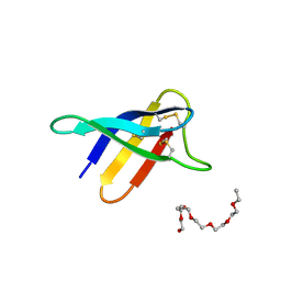
5W96
 | | Solution structure of phage derived peptide inhibitor of frizzled 7 receptor | | 分子名称: | Fz7 binding peptide | | 著者 | Nile, A.H, de Sousa e Melo, F, Mukund, S, Piskol, R, Hansen, S, Zhou, L, Zhang, Y, Fu, Y, Gogol, E.B, Komuves, L.G, Modrusan, Z, Angers, S, Franke, Y, Koth, C, Fairbrother, W.J, Wang, W, de Sauvage, F.J, Hannoush, R.N. | | 登録日 | 2017-06-22 | | 公開日 | 2018-04-18 | | 最終更新日 | 2023-06-14 | | 実験手法 | SOLUTION NMR | | 主引用文献 | A selective peptide inhibitor of Frizzled 7 receptors disrupts intestinal stem cells.
Nat. Chem. Biol., 14, 2018
|
|
6HNL
 | | Selenomethionine derivative of IdmH 96-104 loop truncation variant | | 分子名称: | Putative polyketide cyclase IdmH | | 著者 | Drulyte, I, Obajdin, J, Trinh, C, Hemsworth, G.R, Berry, A. | | 登録日 | 2018-09-16 | | 公開日 | 2019-11-06 | | 最終更新日 | 2019-11-20 | | 実験手法 | X-RAY DIFFRACTION (2.2 Å) | | 主引用文献 | Crystal structure of the putative cyclase IdmH from the indanomycin nonribosomal peptide synthase/polyketide synthase.
Iucrj, 6, 2019
|
|
5WLK
 | |
5NIP
 | |
6HAJ
 | | Crystal structure of PAF - p-sulfonatocalix[8]arene complex | | 分子名称: | 2-(2-(2-(2-(2-(2-ETHOXYETHOXY)ETHOXY)ETHOXY)ETHOXY)ETHOXY)ETHANOL, Pc24g00380 protein, sulfonato-calix[8]arene | | 著者 | Alex, J.M, Rennie, M, Engilberge, S, Batta, G, Crowley, P.B. | | 登録日 | 2018-08-07 | | 公開日 | 2019-02-13 | | 最終更新日 | 2024-01-17 | | 実験手法 | X-RAY DIFFRACTION (1.5 Å) | | 主引用文献 | Calixarene-mediated assembly of a small antifungal protein.
Iucrj, 6, 2019
|
|
6HZE
 | | BP0997, GH138 enzyme targeting pectin rhamnogalacturonan II | | 分子名称: | BPa0997, SODIUM ION, beta-D-galactopyranuronic acid | | 著者 | Basle, A, Cartmell, A, Labourel, A, Gilbert, H. | | 登録日 | 2018-10-23 | | 公開日 | 2019-03-20 | | 最終更新日 | 2020-07-29 | | 実験手法 | X-RAY DIFFRACTION (2.7 Å) | | 主引用文献 | Structural and functional analyses of glycoside hydrolase 138 enzymes targeting chain A galacturonic acid in the complex pectin rhamnogalacturonan II.
J.Biol.Chem., 294, 2019
|
|
5NPT
 | | Structure of the N-terminal domain of the yeast telomerase reverse transcriptase | | 分子名称: | Telomerase reverse transcriptase | | 著者 | Rodina, E.V, Lebedev, A.A, Hakanpaa, J, Hackenberg, C, Petrova, O.A, Zvereva, M.I, Dontsova, O.A, Lamzin, V.S. | | 登録日 | 2017-04-18 | | 公開日 | 2017-12-20 | | 最終更新日 | 2024-05-01 | | 実験手法 | X-RAY DIFFRACTION (2.4 Å) | | 主引用文献 | Structure and function of the N-terminal domain of the yeast telomerase reverse transcriptase.
Nucleic Acids Res., 46, 2018
|
|
5LSU
 | |
5ZNU
 | |
5YXI
 | | Designed protein dRafX6 | | 分子名称: | Designed protein dRafX6 | | 著者 | Liu, R. | | 登録日 | 2017-12-05 | | 公開日 | 2018-12-05 | | 最終更新日 | 2024-05-15 | | 実験手法 | SOLUTION NMR | | 主引用文献 | De novo sequence redesign of a functional Ras-binding domain globally inverted the surface charge distribution and led to extreme thermostability.
Biotechnol.Bioeng., 118, 2021
|
|
5LVF
 | | Solution structure of Rtt103 CTD-interacting domain bound to a Thr4 phosphorylated CTD peptide | | 分子名称: | PRO-SER-TYR-SER-PRO-PTH-SER-PRO-SER-TYR-SER-PRO-THR-SER-PRO-SER, Regulator of Ty1 transposition protein 103 | | 著者 | Jasnovidova, O, Kubicek, K, Krejcikova, M, Stefl, R. | | 登録日 | 2016-09-14 | | 公開日 | 2017-05-10 | | 最終更新日 | 2019-10-30 | | 実験手法 | SOLUTION NMR | | 主引用文献 | Structural insight into recognition of phosphorylated threonine-4 of RNA polymerase II C-terminal domain by Rtt103p.
EMBO Rep., 18, 2017
|
|
5LQP
 | |
5LT7
 | | Structure of the Epigenetic Oncogene MMSET and inhibition by N-Alkyl Sinefungin Derivatives | | 分子名称: | Histone-lysine N-methyltransferase SETD2, THIOCYANATE ION, ZINC ION, ... | | 著者 | Tisi, D, Pathuri, P, Heightman, T. | | 登録日 | 2016-09-06 | | 公開日 | 2016-10-05 | | 最終更新日 | 2019-10-16 | | 実験手法 | X-RAY DIFFRACTION (1.51 Å) | | 主引用文献 | Structure of the Epigenetic Oncogene MMSET and Inhibition by N-Alkyl Sinefungin Derivatives.
ACS Chem. Biol., 11, 2016
|
|
6KN4
 | |
6KML
 | | 2.09 Angstrom resolution crystal structure of tetrameric HigBA toxin-antitoxin complex from E.coli | | 分子名称: | Antitoxin HigA, mRNA interferase toxin HigB | | 著者 | Jadhav, P, Sinha, V.K, Rothweiler, U, Singh, M. | | 登録日 | 2019-07-31 | | 公開日 | 2020-11-18 | | 最終更新日 | 2023-11-22 | | 実験手法 | X-RAY DIFFRACTION (2.095 Å) | | 主引用文献 | 2.09 angstrom Resolution structure of E. coli HigBA toxin-antitoxin complex reveals an ordered DNA-binding domain and intrinsic dynamics in antitoxin.
Biochem.J., 477, 2020
|
|
6KMQ
 | | 2.3 Angstrom resolution structure of dimeric HigBA toxin-antitoxin complex from E. coli | | 分子名称: | Antitoxin HigA, mRNA interferase toxin HigB | | 著者 | Jadhav, P, Sinha, V.K, Rothweiler, U, Singh, M. | | 登録日 | 2019-07-31 | | 公開日 | 2020-11-18 | | 最終更新日 | 2023-11-22 | | 実験手法 | X-RAY DIFFRACTION (2.35 Å) | | 主引用文献 | 2.09 angstrom Resolution structure of E. coli HigBA toxin-antitoxin complex reveals an ordered DNA-binding domain and intrinsic dynamics in antitoxin.
Biochem.J., 477, 2020
|
|


