6US9
 
 | |
4I8X
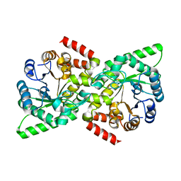
 | | Crystal structure of rabbit LDHA in complex with AP27460 | | 分子名称: | 6-phenylpyridine-3-carboxylic acid, L-lactate dehydrogenase A chain | | 著者 | Zhou, T, Stephan, Z.G, Kohlmann, A, Li, F, Commodore, L, Greenfield, M.T, Zhu, X, Dalgarno, D.C. | | 登録日 | 2012-12-04 | | 公開日 | 2013-01-23 | | 最終更新日 | 2023-09-20 | | 実験手法 | X-RAY DIFFRACTION (2.23 Å) | | 主引用文献 | Fragment growing and linking lead to novel nanomolar lactate dehydrogenase inhibitors.
J.Med.Chem., 56, 2013
|
|
3OBL
 | |
6UWR
 | |
3O00
 | | Crystal Structure of the Salmonella Type III Secretion System Tip Protein SipD-C244S | | 分子名称: | Cell invasion protein sipD, NICKEL (II) ION | | 著者 | Chatterjee, S, Zhong, D, Nordhues, B.A, Battaile, K.P, Lovell, S, DeGuzman, R.N. | | 登録日 | 2010-07-18 | | 公開日 | 2010-11-17 | | 最終更新日 | 2024-02-21 | | 実験手法 | X-RAY DIFFRACTION (1.85 Å) | | 主引用文献 | The crystal structures of the Salmonella type III secretion system tip protein SipD in complex with deoxycholate and chenodeoxycholate.
Protein Sci., 20, 2011
|
|
6VBJ
 | |
4I9N
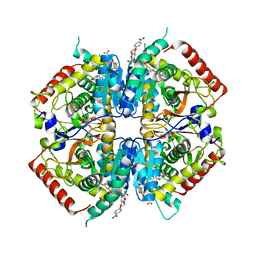 | | Crystal structure of rabbit LDHA in complex with AP28161 and AP28122 | | 分子名称: | 6-({2-[(5-chloro-4-{[(2S)-2,3-dihydroxypropyl]oxy}-2-methoxyphenyl)amino]-2-oxoethyl}sulfanyl)pyridine-3-carboxylic acid, 6-[3-(carboxymethoxy)-5-fluorophenyl]pyridine-3-carboxylic acid, L-lactate dehydrogenase A chain | | 著者 | Zhou, T, Kohlmann, A, Stephan, Z.G, Li, F, Commodore, L, Greenfield, M.T, Shakespeare, W.C, Zhu, X, Dalgarno, D.C. | | 登録日 | 2012-12-05 | | 公開日 | 2013-01-23 | | 最終更新日 | 2023-09-20 | | 実験手法 | X-RAY DIFFRACTION (2.35 Å) | | 主引用文献 | Fragment growing and linking lead to novel nanomolar lactate dehydrogenase inhibitors.
J.Med.Chem., 56, 2013
|
|
4IBG
 | | Ebola virus VP35 bound to small molecule | | 分子名称: | GLYCEROL, PHOSPHATE ION, Polymerase cofactor VP35, ... | | 著者 | Brown, C.S, Leung, D.W, Xu, W, Borek, D.M, Otwinowski, Z, Ramanan, P, Stubbs, A.J, Peterson, D.S, Binning, J.M, Amarasinghe, G.K. | | 登録日 | 2012-12-08 | | 公開日 | 2014-03-19 | | 最終更新日 | 2024-02-28 | | 実験手法 | X-RAY DIFFRACTION (1.413 Å) | | 主引用文献 | In Silico Derived Small Molecules Bind the Filovirus VP35 Protein and Inhibit Its Polymerase Cofactor Activity.
J.Mol.Biol., 426, 2014
|
|
6UWO
 | |
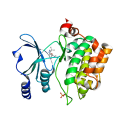
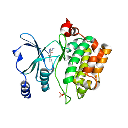
3NWV
 | | Human cytochrome c G41S | | 分子名称: | Cytochrome c, HEME C | | 著者 | Fagerlund, R.D, Wilbanks, S.M. | | 登録日 | 2010-07-11 | | 公開日 | 2011-03-09 | | 最終更新日 | 2019-10-02 | | 実験手法 | X-RAY DIFFRACTION (1.9 Å) | | 主引用文献 | The Proapoptotic G41S Mutation to Human Cytochrome c Alters the Heme Electronic Structure and Increases the Electron Self-Exchange Rate.
J.Am.Chem.Soc., 133, 2011
|
|
4IBB
 | | Ebola virus VP35 bound to small molecule | | 分子名称: | Polymerase cofactor VP35, {4-[(5R)-3-hydroxy-2-oxo-4-(thiophen-2-ylcarbonyl)-5-(2,4,5-trimethylphenyl)-2,5-dihydro-1H-pyrrol-1-yl]phenyl}acetic acid | | 著者 | Brown, C.S, Leung, D.W, Xu, W, Borek, D.M, Otwinowski, Z, Ramanan, P, Stubbs, A.J, Peterson, D.S, Binning, J.M, Amarasinghe, G.K, Center for Structural Genomics of Infectious Diseases (CSGID) | | 登録日 | 2012-12-08 | | 公開日 | 2014-02-26 | | 最終更新日 | 2024-02-28 | | 実験手法 | X-RAY DIFFRACTION (1.752 Å) | | 主引用文献 | In Silico Derived Small Molecules Bind the Filovirus VP35 Protein and Inhibit Its Polymerase Cofactor Activity.
J.Mol.Biol., 426, 2014
|
|
4IBD
 | | Ebola virus VP35 bound to small molecule | | 分子名称: | 5-[(2R)-3-benzoyl-2-(4-bromothiophen-2-yl)-4-hydroxy-5-oxo-2,5-dihydro-1H-pyrrol-1-yl]-2-methylbenzoic acid, GLYCEROL, MAGNESIUM ION, ... | | 著者 | Brown, C.S, Leung, D.W, Xu, W, Borek, D.M, Otwinowski, Z, Ramanan, P, Stubbs, A.J, Peterson, D.S, Binning, J.M, Amarasinghe, G.K. | | 登録日 | 2012-12-08 | | 公開日 | 2014-03-19 | | 最終更新日 | 2024-02-28 | | 実験手法 | X-RAY DIFFRACTION (1.84 Å) | | 主引用文献 | In Silico Derived Small Molecules Bind the Filovirus VP35 Protein and Inhibit Its Polymerase Cofactor Activity.
J.Mol.Biol., 426, 2014
|
|
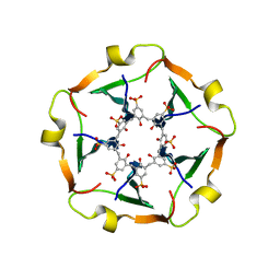
6V1N
 | | CSP1-E1A-cyc(Dap6E10) | | 分子名称: | Competence-stimulating peptide type 1 | | 著者 | Yang, Y. | | 登録日 | 2019-11-20 | | 公開日 | 2020-01-08 | | 最終更新日 | 2020-02-05 | | 実験手法 | SOLUTION NMR | | 主引用文献 | Designing cyclic competence-stimulating peptide (CSP) analogs with pan-group quorum-sensing inhibition activity inStreptococcus pneumoniae.
Proc.Natl.Acad.Sci.USA, 117, 2020
|
|
8R3C
 | |
8AHG
 | |
8AHH
 | |
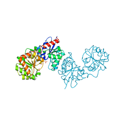
3NY2
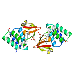
 | | Structure of the ubr-box of UBR2 ubiquitin ligase | | 分子名称: | E3 ubiquitin-protein ligase UBR2, ZINC ION | | 著者 | Matta-Camacho, E, Kozlov, G, Li, F, Gehring, K. | | 登録日 | 2010-07-14 | | 公開日 | 2010-08-11 | | 最終更新日 | 2024-02-21 | | 実験手法 | X-RAY DIFFRACTION (2.61 Å) | | 主引用文献 | Structural basis of substrate recognition and specificity in the N-end rule pathway.
Nat.Struct.Mol.Biol., 17, 2010
|
|
8AHE
 | | PAC-FragmentDEL: Photoactivated covalent capture of DNA encoded fragments for hit discovery | | 分子名称: | SULFATE ION, UDP-N-acetylglucosamine 2-epimerase, ~{N},5-dimethyl-1-phenyl-pyrazole-4-sulfonamide | | 著者 | Baker, L.M, Murray, J.B, Hubbard, R.E. | | 登録日 | 2022-07-21 | | 公開日 | 2022-09-14 | | 最終更新日 | 2024-01-31 | | 実験手法 | X-RAY DIFFRACTION (2.108 Å) | | 主引用文献 | PAC-FragmentDEL - photoactivated covalent capture of DNA-encoded fragments for hit discovery.
Rsc Med Chem, 13, 2022
|
|
8AHF
 | | PAC-FragmentDEL: Photoactivated covalent capture of DNA encoded fragments for hit discovery | | 分子名称: | (2~{R},4~{S})-4-[bis(fluoranyl)methoxy]-~{N}-methyl-1-(2~{H}-pyrazolo[4,3-b]pyridin-6-ylcarbonyl)pyrrolidine-2-carboxamide, SULFATE ION, UDP-N-acetylglucosamine 2-epimerase | | 著者 | Baker, L.M, Murray, J.B, Hubbard, R.E. | | 登録日 | 2022-07-21 | | 公開日 | 2022-09-14 | | 最終更新日 | 2024-01-31 | | 実験手法 | X-RAY DIFFRACTION (2.271 Å) | | 主引用文献 | PAC-FragmentDEL - photoactivated covalent capture of DNA-encoded fragments for hit discovery.
Rsc Med Chem, 13, 2022
|
|
8AHI
 | |
6V6M
 | |
6US8
 | |
6V6U
 | |
3NXE
 | |
3NY3
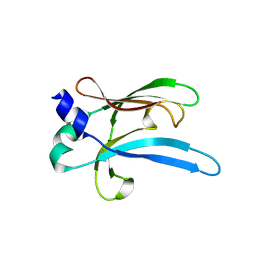
 | | Structure of the ubr-box of UBR2 in complex with N-degron | | 分子名称: | E3 ubiquitin-protein ligase UBR2, N-degron, ZINC ION | | 著者 | Matta-Camacho, E, Kozlov, G, Li, F, Gehring, K. | | 登録日 | 2010-07-14 | | 公開日 | 2010-08-11 | | 最終更新日 | 2024-02-21 | | 実験手法 | X-RAY DIFFRACTION (1.6 Å) | | 主引用文献 | Structural basis of substrate recognition and specificity in the N-end rule pathway.
Nat.Struct.Mol.Biol., 17, 2010
|
|


