3NJG
 
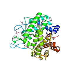
 | | K98A mutant of SO1698 protein, an aspartic peptidase from Shewanella oneidensis. | | 分子名称: | Peptidase | | 著者 | Osipiuk, J, Mulligan, R, Bargassa, M, Collart, F, Joachimiak, A, Midwest Center for Structural Genomics (MCSG) | | 登録日 | 2010-06-17 | | 公開日 | 2010-07-14 | | 最終更新日 | 2023-09-06 | | 実験手法 | X-RAY DIFFRACTION (1.92 Å) | | 主引用文献 | Characterization of member of DUF1888 protein family, self-cleaving and self-assembling endopeptidase.
J.Biol.Chem., 287, 2012
|
|
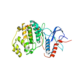
3NL6
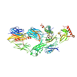
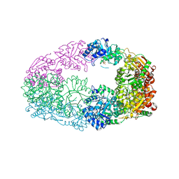 | | The Crystal Structure of Candida glabrata THI6, a Bifunctional Enzyme involved in Thiamin Biosyhthesis of Eukaryotes | | 分子名称: | MAGNESIUM ION, PHOSPHOMETHYLPHOSPHONIC ACID ADENYLATE ESTER, THIAMIN PHOSPHATE, ... | | 著者 | Paul, D, Chatterjee, A, Begley, T.P, Ealick, S.E. | | 登録日 | 2010-06-21 | | 公開日 | 2010-11-10 | | 最終更新日 | 2024-02-21 | | 実験手法 | X-RAY DIFFRACTION (2.612 Å) | | 主引用文献 | Domain Organization in Candida glabrata THI6, a Bifunctional Enzyme Required for Thiamin Biosynthesis in Eukaryotes .
Biochemistry, 49, 2010
|
|
3NJQ
 | |
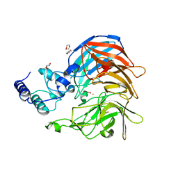
3UL5
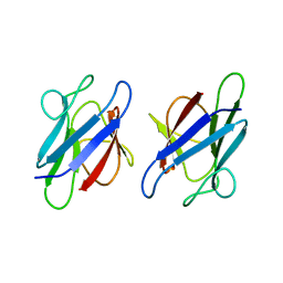
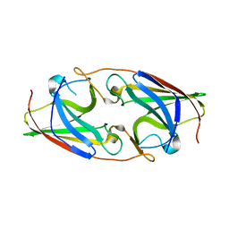
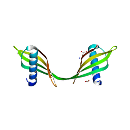 | | Saccharum officinarum canecystatin-1 in space group C2221 | | 分子名称: | Canecystatin-1, GLYCEROL, SODIUM ION | | 著者 | Valadares, N.F, Pereira, H.M, Oliveira-Silva, R, Garratt, R.C. | | 登録日 | 2011-11-10 | | 公開日 | 2012-11-28 | | 最終更新日 | 2023-09-13 | | 実験手法 | X-RAY DIFFRACTION (2.3 Å) | | 主引用文献 | X-ray crystallography and NMR studies of domain-swapped canecystatin-1.
Febs J., 280, 2013
|
|
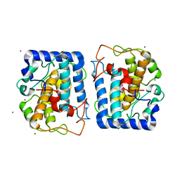
3NM7
 | | Crystal Structure of Borrelia burgdorferi Pur-alpha | | 分子名称: | 1,2-ETHANEDIOL, MAGNESIUM ION, Uncharacterized protein | | 著者 | Graebsch, A, Roche, S, Kostrewa, D, Niessing, D. | | 登録日 | 2010-06-22 | | 公開日 | 2010-10-06 | | 最終更新日 | 2023-09-06 | | 実験手法 | X-RAY DIFFRACTION (2.2 Å) | | 主引用文献 | Of Bits and Bugs - on the use of bioinformatics and a bacterial crystal structure to solve a eukaryotic repeat-protein structure.
Plos One, 5, 2010
|
|
3NMJ
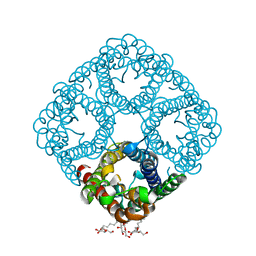
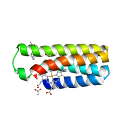
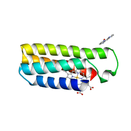 | | Crystal structure of a nickel mediated dimer for the phenanthroline-modified cytochrome cb562 variant, MBP-Phen2 | | 分子名称: | N-1,10-phenanthrolin-5-ylacetamide, NICKEL (II) ION, PROTOPORPHYRIN IX CONTAINING FE, ... | | 著者 | Radford, R.J, Tezcan, F.A. | | 登録日 | 2010-06-22 | | 公開日 | 2010-11-24 | | 最終更新日 | 2023-12-27 | | 実験手法 | X-RAY DIFFRACTION (3.1 Å) | | 主引用文献 | Porous protein frameworks with unsaturated metal centers in sterically encumbered coordination sites.
Chem.Commun.(Camb.), 47, 2011
|
|
3ULJ
 | | Crystal structure of apo Lin28B cold shock domain | | 分子名称: | ACETATE ION, GLYCEROL, Lin28b, ... | | 著者 | Mayr, F, Schuetz, A, Doege, N, Heinemann, U. | | 登録日 | 2011-11-10 | | 公開日 | 2012-08-15 | | 最終更新日 | 2024-04-03 | | 実験手法 | X-RAY DIFFRACTION (1.06 Å) | | 主引用文献 | The Lin28 cold-shock domain remodels pre-let-7 microRNA.
Nucleic Acids Res., 40, 2012
|
|
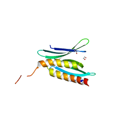
3UMZ
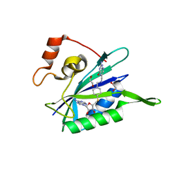
 | | Crystal Structure of the human MDC1 FHA Domain | | 分子名称: | Mediator of DNA damage checkpoint protein 1 | | 著者 | Luo, S, Ye, K. | | 登録日 | 2011-11-15 | | 公開日 | 2012-01-25 | | 最終更新日 | 2024-03-20 | | 実験手法 | X-RAY DIFFRACTION (1.65 Å) | | 主引用文献 | Structural mechanism of the phosphorylation-dependent dimerization of the MDC1 forkhead-associated domain
Nucleic Acids Res., 40, 2012
|
|
3NMB
 | |
3NMS
 | |
3NK5
 | | Crystal structure of AqpZ mutant F43W | | 分子名称: | Aquaporin Z, octyl beta-D-glucopyranoside | | 著者 | Savage, D.F, O'Connell, J.D, Stroud, R.M, Finer-Moore, J.S. | | 登録日 | 2010-06-18 | | 公開日 | 2010-08-11 | | 最終更新日 | 2024-04-03 | | 実験手法 | X-RAY DIFFRACTION (2.4 Å) | | 主引用文献 | Structural context shapes the aquaporin selectivity filter.
Proc.Natl.Acad.Sci.USA, 107, 2010
|
|
3NKR
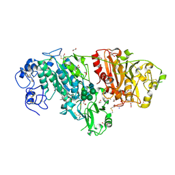 | | Crystal structure of mouse autotaxin in complex with 22:6-LPA | | 分子名称: | (2R)-2-hydroxy-3-(phosphonooxy)propyl (4Z,7E,10E,13Z,16Z,19Z)-docosa-4,7,10,13,16,19-hexaenoate, 1,2-ETHANEDIOL, 2-acetamido-2-deoxy-beta-D-glucopyranose-(1-4)-2-acetamido-2-deoxy-beta-D-glucopyranose, ... | | 著者 | Nishimasu, H, Ishitani, R, Mihara, E, Takagi, J, Aoki, J, Nureki, O. | | 登録日 | 2010-06-20 | | 公開日 | 2011-01-19 | | 最終更新日 | 2023-11-01 | | 実験手法 | X-RAY DIFFRACTION (1.704 Å) | | 主引用文献 | Crystal structure of autotaxin and insight into GPCR activation by lipid mediators
Nat.Struct.Mol.Biol., 18, 2011
|
|
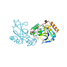
3UD5
 | | Crystal structure of E. coli HPPK in complex with bisubstrate analogue inhibitor J1A | | 分子名称: | 1,2-ETHANEDIOL, 2-amino-4-hydroxy-6-hydroxymethyldihydropteridine pyrophosphokinase, 5'-S-[1-(2-{[(2-amino-4-oxo-3,4-dihydropteridin-6-yl)methyl]amino}ethyl)piperidin-4-yl]-5'-thioadenosine | | 著者 | Shaw, G, Shi, G, Ji, X. | | 登録日 | 2011-10-27 | | 公開日 | 2012-01-04 | | 最終更新日 | 2023-09-13 | | 実験手法 | X-RAY DIFFRACTION (2 Å) | | 主引用文献 | Bisubstrate analogue inhibitors of 6-hydroxymethyl-7,8-dihydropterin pyrophosphokinase: New design with improved properties.
Bioorg.Med.Chem., 20, 2012
|
|
3NMI
 | | Crystal structure of the phenanthroline-modified cytochrome cb562 variant, MBP-Phen2 | | 分子名称: | ACETATE ION, N-1,10-phenanthrolin-5-ylacetamide, PROTOPORPHYRIN IX CONTAINING FE, ... | | 著者 | Radford, R.J, Tezcan, F.A. | | 登録日 | 2010-06-22 | | 公開日 | 2010-11-24 | | 最終更新日 | 2023-12-27 | | 実験手法 | X-RAY DIFFRACTION (2.01 Å) | | 主引用文献 | Porous protein frameworks with unsaturated metal centers in sterically encumbered coordination sites.
Chem.Commun.(Camb.), 47, 2011
|
|
3NMX
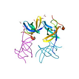
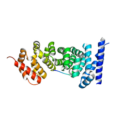 | | Crystal structure of APC complexed with Asef | | 分子名称: | APC variant protein, Rho guanine nucleotide exchange factor 4 | | 著者 | Zhang, Z, Chen, L, Gao, L, Lin, K, Wu, G. | | 登録日 | 2010-06-22 | | 公開日 | 2011-07-06 | | 最終更新日 | 2023-12-27 | | 実験手法 | X-RAY DIFFRACTION (2.3 Å) | | 主引用文献 | Structural basis for the recognition of Asef by adenomatous polyposis coli.
Cell Res., 22, 2012
|
|
3NPF
 | |
3W45
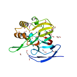
 | | Crystal structure of RsbX in complex with cobalt in space group P1 | | 分子名称: | COBALT (II) ION, Phosphoserine phosphatase RsbX | | 著者 | Makino, M, Teh, A.H, Baba, S, Shimizu, N, Yamamoto, M, Kumasaka, T. | | 登録日 | 2013-01-04 | | 公開日 | 2014-01-22 | | 最終更新日 | 2024-03-20 | | 実験手法 | X-RAY DIFFRACTION (1.7 Å) | | 主引用文献 | Structure of the RsbX phosphatase involved in the general stress response of Bacillus subtilis
Acta Crystallogr.,Sect.D, 71, 2015
|
|
3NRP
 | |
3NNB
 | |
3NO8
 | | Crystal structure of the PHR domain from human BTBD2 Protein | | 分子名称: | BTB/POZ domain-containing protein 2, GLYCEROL, SULFATE ION | | 著者 | Sampathkumar, P, Miller, S, Rutter, M, Bain, K, Gheyi, T, Atwell, S, Thompson, D.A, Emtage, J.S, Wasserman, S, Sauder, J.M, Burley, S.K, New York SGX Research Center for Structural Genomics (NYSGXRC) | | 登録日 | 2010-06-24 | | 公開日 | 2010-08-25 | | 最終更新日 | 2023-09-06 | | 実験手法 | X-RAY DIFFRACTION (2.2 Å) | | 主引用文献 | Crystal structure of the PHR domain from human BTBD2 Protein
To be Published
|
|
3R63
 | |
3NPE
 | | Structure of VP14 in complex with oxygen | | 分子名称: | 1,4-DIETHYLENE DIOXIDE, 9-cis-epoxycarotenoid dioxygenase 1, chloroplastic, ... | | 著者 | Messing, S.A, Gabelli, S.B, Amzel, L.M. | | 登録日 | 2010-06-28 | | 公開日 | 2010-11-10 | | 最終更新日 | 2023-12-27 | | 実験手法 | X-RAY DIFFRACTION (3.2 Å) | | 主引用文献 | Structural insights into maize viviparous14, a key enzyme in the biosynthesis of the phytohormone abscisic acid.
Plant Cell, 22, 2010
|
|
3NQ1
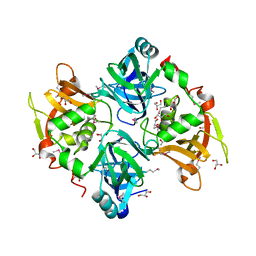
 | | Crystal Structure of Tyrosinase from Bacillus megaterium in complex with inhibitor kojic acid | | 分子名称: | 5-HYDROXY-2-(HYDROXYMETHYL)-4H-PYRAN-4-ONE, COPPER (II) ION, Tyrosinase, ... | | 著者 | Sendovski, M, Kanteev, M, Adir, N, Fishman, A. | | 登録日 | 2010-06-29 | | 公開日 | 2010-11-17 | | 最終更新日 | 2023-12-27 | | 実験手法 | X-RAY DIFFRACTION (2.3 Å) | | 主引用文献 | First structures of an active bacterial tyrosinase reveal copper plasticity.
J.Mol.Biol., 405, 2011
|
|
3NQ5
 | | Crystal Structure of Tyrosinase from Bacillus megaterium R209H mutant | | 分子名称: | COPPER (II) ION, Tyrosinase, ZINC ION | | 著者 | Sendovski, M, Kanteev, M, Adir, N, Fishman, A. | | 登録日 | 2010-06-29 | | 公開日 | 2010-11-17 | | 最終更新日 | 2023-11-01 | | 実験手法 | X-RAY DIFFRACTION (2.3 Å) | | 主引用文献 | First structures of an active bacterial tyrosinase reveal copper plasticity.
J.Mol.Biol., 405, 2011
|
|
3NVZ
 | |


