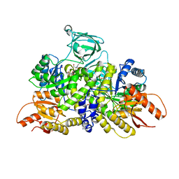
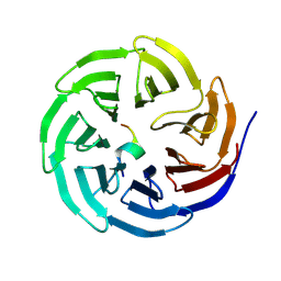
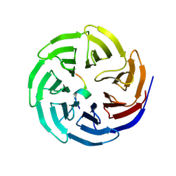
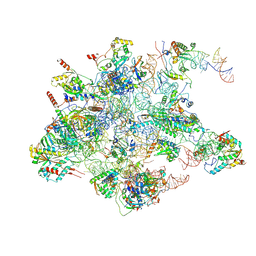
8WQB
 
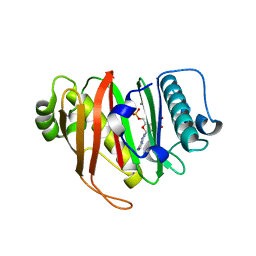
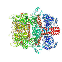
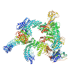 | | Cryo-EM structure of CUL2-RBX1-ELOB-ELOC-FEM1B bound with the C-degron of CCDC89 (conformation 2) | | 分子名称: | Coiled-coil domain-containing protein 89, Cullin-2, E3 ubiquitin-protein ligase RBX1, ... | | 著者 | Chen, X, Zhang, K, Xu, C. | | 登録日 | 2023-10-11 | | 公開日 | 2024-04-03 | | 最終更新日 | 2024-05-08 | | 実験手法 | ELECTRON MICROSCOPY (3.37 Å) | | 主引用文献 | Mechanism of Psi-Pro/C-degron recognition by the CRL2 FEM1B ubiquitin ligase.
Nat Commun, 15, 2024
|
|
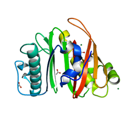
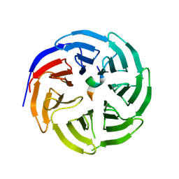
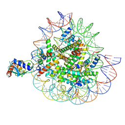
8WQH
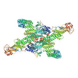 | | cryo-EM structure of neddylated CUL2-RBX1-ELOB-ELOC-FEM1B bound with the C-degron of CCDC89 (conformation 2) | | 分子名称: | Coiled-coil domain-containing protein 89, Cullin-2, E3 ubiquitin-protein ligase RBX1, ... | | 著者 | Chen, X, Zhang, K, Xu, C. | | 登録日 | 2023-10-11 | | 公開日 | 2024-04-03 | | 最終更新日 | 2024-05-08 | | 実験手法 | ELECTRON MICROSCOPY (3.44 Å) | | 主引用文献 | Mechanism of Psi-Pro/C-degron recognition by the CRL2 FEM1B ubiquitin ligase.
Nat Commun, 15, 2024
|
|
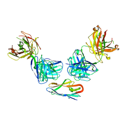
8K6Q
 | | Crystal structure of HOIL-1L LTM domain | | 分子名称: | RanBP-type and C3HC4-type zinc finger-containing protein 1 | | 著者 | Yan, Z, Pan, L.F. | | 登録日 | 2023-07-25 | | 公開日 | 2024-07-03 | | 実験手法 | X-RAY DIFFRACTION (1.59 Å) | | 主引用文献 | Mechanistic insights into the homo-dimerization of HOIL-1L and SHARPIN.
Biochem.Biophys.Res.Commun., 689, 2023
|
|
8R9Y
 | |
8R9W
 | |
6B6U
 | | Pyruvate Kinase M2 mutant - S437Y | | 分子名称: | 1,2-ETHANEDIOL, 2-[3-(2-HYDROXY-1,1-DIHYDROXYMETHYL-ETHYLAMINO)-PROPYLAMINO]-2-HYDROXYMETHYL-PROPANE-1,3-DIOL, CHLORIDE ION, ... | | 著者 | Srivastava, D, Dey, M. | | 登録日 | 2017-10-03 | | 公開日 | 2017-12-20 | | 最終更新日 | 2023-10-04 | | 実験手法 | X-RAY DIFFRACTION (1.35 Å) | | 主引用文献 | Structural Investigation of a Dimeric Variant of Pyruvate Kinase Muscle Isoform 2.
Biochemistry, 56, 2017
|
|
8R9Z
 | |
8R9X
 | |
8QZI
 | | Crystal structure of PptT-ACP from Mycobacterium tuberculosis | | 分子名称: | 4'-phosphopantetheinyl transferase PptT, COENZYME A, ISOPROPYL ALCOHOL, ... | | 著者 | Gavalda, S, Mourey, L, Pedelacq, J.D. | | 登録日 | 2023-10-27 | | 公開日 | 2024-06-19 | | 最終更新日 | 2024-10-23 | | 実験手法 | X-RAY DIFFRACTION (2.5 Å) | | 主引用文献 | Catalytic Cycle of Type II 4'-Phosphopantetheinyl Transferases
Acs Catalysis, 14, 2024
|
|
8QZJ
 | |
8WXT
 | |
8WXQ
 | |
8QZH
 | | Crystal structure of apo-PptT from Mycobacterium tuberculosis | | 分子名称: | 4'-phosphopantetheinyl transferase PptT, ACETATE ION, MAGNESIUM ION | | 著者 | Gavalda, S, Carivenc, C, Mourey, L, Pedelacq, J.D. | | 登録日 | 2023-10-27 | | 公開日 | 2024-06-19 | | 実験手法 | X-RAY DIFFRACTION (1.7 Å) | | 主引用文献 | Catalytic Cycle of Type II 4'-Phosphopantetheinyl Transferases
Acs Catalysis, 14, 2024
|
|
8WXU
 | |
8WXR
 | |
8WXV
 | |
8WXX
 | |
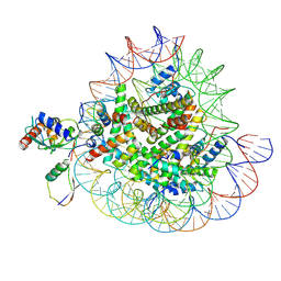
7SUK
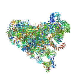 | | Structure of Bfr2-Lcp5 Complex Observed in the Small Subunit Processome Isolated from R2TP-depleted Yeast Cells | | 分子名称: | 18S pre-rRNA, 40S ribosomal protein S11-A, 40S ribosomal protein S13, ... | | 著者 | Rai, J, Zhao, Y, Li, H. | | 登録日 | 2021-11-17 | | 公開日 | 2022-07-06 | | 最終更新日 | 2023-08-16 | | 実験手法 | ELECTRON MICROSCOPY (3.99 Å) | | 主引用文献 | Artificial intelligence-assisted cryoEM structure of Bfr2-Lcp5 complex observed in the yeast small subunit processome.
Commun Biol, 5, 2022
|
|
8QU1
 | | mt-LSU assembly intermediate in GTPBP8 knock-out cells, state 1 | | 分子名称: | 16S ribosomal RNA, 39S ribosomal protein L10, mitochondrial, ... | | 著者 | Valentin Gese, G, Cipullo, M, Rorbach, J, Hallberg, B.M. | | 登録日 | 2023-10-13 | | 公開日 | 2024-06-26 | | 最終更新日 | 2024-07-17 | | 実験手法 | ELECTRON MICROSCOPY (2.74 Å) | | 主引用文献 | GTPBP8 plays a role in mitoribosome formation in human mitochondria.
Nat Commun, 15, 2024
|
|
8X7K
 | | Cryo-EM structures of RNF168/UbcH5c-Ub in complex with H2AK13Ub nucleosomes determined by activity-based chemical trapping strategy (adjacent H2AK13/15 dual-monoubiquitination) | | 分子名称: | DNA (143-MER), E3 ubiquitin-protein ligase RNF168, Histone H2A type 1-B/E, ... | | 著者 | Ai, H.S, Tong, Z.B, Deng, Z.H, Pan, M, Liu, L. | | 登録日 | 2023-11-24 | | 公開日 | 2024-08-07 | | 実験手法 | ELECTRON MICROSCOPY (3.27 Å) | | 主引用文献 | Capturing Snapshots of Nucleosomal H2A K13/K15 Ubiquitination Mediated by the Monomeric E3 Ligase RNF168
Biorxiv, 2024
|
|
8X7J
 | | Cryo-EM structures of RNF168/UbcH5c-Ub/nucleosomes complex determined by activity-based chemical trapping strategy | | 分子名称: | DNA (144-MER), E3 ubiquitin-protein ligase RNF168, Histone H2A type 1-B/E, ... | | 著者 | Ai, H.S, Tong, Z.B, Deng, Z.H, Pan, M, Liu, L. | | 登録日 | 2023-11-24 | | 公開日 | 2024-08-07 | | 実験手法 | ELECTRON MICROSCOPY (3.39 Å) | | 主引用文献 | Capturing Snapshots of Nucleosomal H2A K13/K15 Ubiquitination Mediated by the Monomeric E3 Ligase RNF168
Biorxiv, 2024
|
|
8OO5
 | |
6BWD
 | | 3.7 angstrom cryoEM structure of truncated mouse TRPM7 | | 分子名称: | CHOLESTEROL HEMISUCCINATE, MAGNESIUM ION, Transient receptor potential cation channel subfamily M member 7 | | 著者 | Zhang, J, Li, Z, Duan, J, Li, J, Hulse, R.E, Santa-Cruz, A, Abiria, S.A, Krapivinsky, G, Clapham, D.E. | | 登録日 | 2017-12-14 | | 公開日 | 2018-08-15 | | 最終更新日 | 2024-11-13 | | 実験手法 | ELECTRON MICROSCOPY (3.7 Å) | | 主引用文献 | Structure of the mammalian TRPM7, a magnesium channel required during embryonic development.
Proc. Natl. Acad. Sci. U.S.A., 115, 2018
|
|
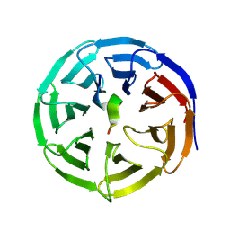
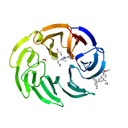
8OG8
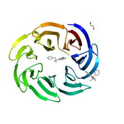 | | Crystal structure of human DCAF1 WD40 repeats (Q1250L) in complex with compound 3 | | 分子名称: | 1,2-ETHANEDIOL, 5-(2-methyl-1-phenyl-propan-2-yl)imidazo[2,1-a]isoquinoline, ACETATE ION, ... | | 著者 | Schroeder, M, Vulpetti, A, Renatus, M. | | 登録日 | 2023-03-19 | | 公開日 | 2023-06-14 | | 最終更新日 | 2024-06-19 | | 実験手法 | X-RAY DIFFRACTION (2.11 Å) | | 主引用文献 | Discovery of New Binders for DCAF1, an Emerging Ligase Target in the Targeted Protein Degradation Field.
Acs Med.Chem.Lett., 14, 2023
|
|
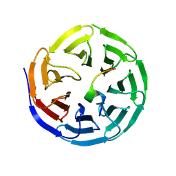
8OG5
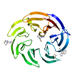 | | Crystal structure of human DCAF1 WD40 repeats (Q1250L) in complex with compound 1 | | 分子名称: | 1,2-ETHANEDIOL, 5-(2-fluorophenyl)-2,3-dihydroimidazo[2,1-a]isoquinoline, ACETATE ION, ... | | 著者 | Schroeder, M, Vulpetti, A, Renatus, M. | | 登録日 | 2023-03-19 | | 公開日 | 2023-06-14 | | 最終更新日 | 2024-06-19 | | 実験手法 | X-RAY DIFFRACTION (2.2 Å) | | 主引用文献 | Discovery of New Binders for DCAF1, an Emerging Ligase Target in the Targeted Protein Degradation Field.
Acs Med.Chem.Lett., 14, 2023
|
|


