Loading
PDBj
メニューPDBj@FacebookPDBj@TwitterPDBj@YouTubewwPDB FoundationwwPDB
RCSB PDBPDBeBMRBAdv. SearchSearch help

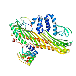
8OMU
DownloadVisualize
BU of 8omu by Molmil
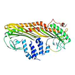
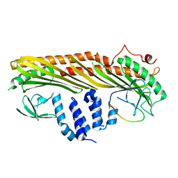
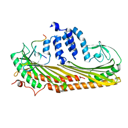
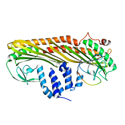
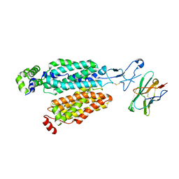
Cryo-EM structure of rat SLC22A6 bound to alpha-ketoglutaric acid in a low occupancy state
分子名称: Solute carrier family 22 member 6, Synthetic nanobody (Sybody)
著者Parker, J.L, Kato, T, Newstead, S.
登録日2023-03-31
公開日2023-07-19
最終更新日2023-11-22
実験手法ELECTRON MICROSCOPY (3.43 Å)
主引用文献Molecular basis for selective uptake and elimination of organic anions in the kidney by OAT1.
Nat.Struct.Mol.Biol., 30, 2023
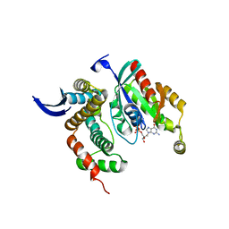
1G4C
DownloadVisualize
BU of 1g4c by Molmil
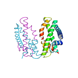
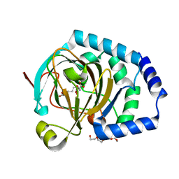
CRYSTAL STRUCTURE OF A COMPLEX OF HPPK(R92A) FROM E.COLI WITH MG2+ AT 1.65 ANGSTROM RESOLUTION
分子名称: 6-HYDROXYMETHYL-7,8-DIHYDROPTERIN PYROPHOSPHOKINASE, CHLORIDE ION, MAGNESIUM ION
著者Blaszczyk, J, Ji, X.
登録日2000-10-26
公開日2003-04-15
最終更新日2023-08-30
実験手法X-RAY DIFFRACTION (1.65 Å)
主引用文献Dynamic Roles of Arginine Residues 82 and 92 of Escherichia coli 6-Hydroxymethyl-7,8-dihydropterin Pyrophosphokinase: Crystallographic Studies
Biochemistry, 42, 2003
6LPG
DownloadVisualize
BU of 6lpg by Molmil
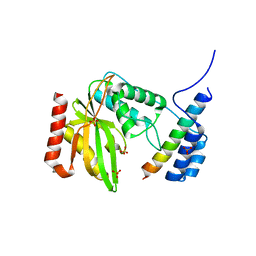
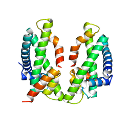
human VASH1-SVBP complex
分子名称: SULFATE ION, Small vasohibin-binding protein, Tubulinyl-Tyr carboxypeptidase 1
著者Ikeda, A, Nishino, T.
登録日2020-01-10
公開日2020-10-21
最終更新日2023-11-29
実験手法X-RAY DIFFRACTION (2.3 Å)
主引用文献The crystal structure of the tetrameric human vasohibin-1-SVBP complex reveals a variable arm region within the structural core.
Acta Crystallogr D Struct Biol, 76, 2020
6NIT
DownloadVisualize
BU of 6nit by Molmil
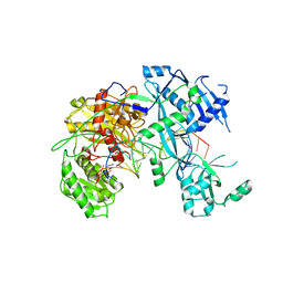
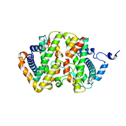
Human Argonaute2-miR-122 bound to a target RNA with four central mismatches (bu4)
分子名称: Protein argonaute-2, RNA (5'-R(P*AP*AP*CP*AP*CP*CP*AP*UP*CP*CP*AP*AP*CP*AP*CP*UP*CP*CP*AP*AP*A)-3'), RNA (5'-R(P*UP*GP*GP*AP*GP*UP*GP*UP*GP*AP*CP*AP*AP*UP*GP*GP*UP*GP*UP*UP*U)-3')
著者Sheu-Gruttadauria, J, MacRae, I.J.
登録日2018-12-31
公開日2019-08-07
最終更新日2024-03-13
実験手法X-RAY DIFFRACTION (3.8 Å)
主引用文献Structural Basis for Target-Directed MicroRNA Degradation.
Mol.Cell, 75, 2019
4TNV
DownloadVisualize
BU of 4tnv by Molmil
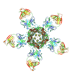
C. elegans glutamate-gated chloride channel (GluCl) in complex with Fab in a non-conducting conformation
分子名称: 2-acetamido-2-deoxy-beta-D-glucopyranose, Avermectin-sensitive glutamate-gated chloride channel GluCl alpha, CHLORIDE ION, ...
著者Althoff, T, Hibbs, R.E, Banerjee, S, Gouaux, E.
登録日2014-06-05
公開日2014-08-13
最終更新日2024-10-16
実験手法X-RAY DIFFRACTION (3.6 Å)
主引用文献X-ray structures of GluCl in apo states reveal a gating mechanism of Cys-loop receptors.
Nature, 512, 2014
7DS8
DownloadVisualize
BU of 7ds8 by Molmil
Crystal structure of actin capping protein in complex with twinflin-1/CD2AP CPI chimera peptide (CDN-TWC)
分子名称: CD2-associated protein,Twinfilin-1, F-actin-capping protein subunit alpha-1, F-actin-capping protein subunit beta isoforms 1
著者Takeda, S.
登録日2020-12-30
公開日2021-03-03
最終更新日2023-11-29
実験手法X-RAY DIFFRACTION (1.95 Å)
主引用文献Structural Insights into the Regulation of Actin Capping Protein by Twinfilin C-terminal Tail.
J.Mol.Biol., 433, 2021
7DS4
DownloadVisualize
BU of 7ds4 by Molmil
Crystal structure of actin capping protein in complex with twinflin-1 C-terminus tail F323K mutant
分子名称: F-actin-capping protein subunit alpha-1, F-actin-capping protein subunit beta isoforms 1, Twinfilin-1
著者Takeda, S.
登録日2020-12-30
公開日2021-03-03
最終更新日2023-11-29
実験手法X-RAY DIFFRACTION (1.85 Å)
主引用文献Structural Insights into the Regulation of Actin Capping Protein by Twinfilin C-terminal Tail.
J.Mol.Biol., 433, 2021
7DS3
DownloadVisualize
BU of 7ds3 by Molmil
Crystal structure of actin capping protein in complex with twinflin-2 C-terminus tail
分子名称: F-actin-capping protein subunit alpha-1, F-actin-capping protein subunit beta isoforms 1, Twinfilin-2
著者Takeda, S.
登録日2020-12-30
公開日2021-03-03
最終更新日2023-11-29
実験手法X-RAY DIFFRACTION (2.09 Å)
主引用文献Structural Insights into the Regulation of Actin Capping Protein by Twinfilin C-terminal Tail.
J.Mol.Biol., 433, 2021
7DS6
DownloadVisualize
BU of 7ds6 by Molmil
Crystal structure of actin capping protein in complex with twinflin-1/CD2AP CPI chimera peptide (TWN-CDC)
分子名称: F-actin-capping protein subunit alpha-1, F-actin-capping protein subunit beta isoforms 1, Twinfilin-1,CD2-associated protein
著者Takeda, S.
登録日2020-12-30
公開日2021-03-03
最終更新日2023-11-29
実験手法X-RAY DIFFRACTION (1.69 Å)
主引用文献Structural Insights into the Regulation of Actin Capping Protein by Twinfilin C-terminal Tail.
J.Mol.Biol., 433, 2021
7DS2
DownloadVisualize
BU of 7ds2 by Molmil
Crystal structure of actin capping protein in complex with twinflin-1 C-terminus tail
分子名称: F-actin-capping protein subunit alpha-1, F-actin-capping protein subunit beta isoforms 1, Twinfilin-1
著者Takeda, S.
登録日2020-12-30
公開日2021-03-03
最終更新日2023-11-29
実験手法X-RAY DIFFRACTION (1.95 Å)
主引用文献Structural Insights into the Regulation of Actin Capping Protein by Twinfilin C-terminal Tail.
J.Mol.Biol., 433, 2021
7DSB
DownloadVisualize
BU of 7dsb by Molmil
Crystal structure of actin capping protein in complex with V-1 and twinfilin C-terminal tail
分子名称: F-actin-capping protein subunit alpha-1, F-actin-capping protein subunit beta isoforms 1, Myotrophin, ...
著者Takeda, S.
登録日2020-12-30
公開日2021-03-03
最終更新日2023-11-29
実験手法X-RAY DIFFRACTION (2.44 Å)
主引用文献Structural Insights into the Regulation of Actin Capping Protein by Twinfilin C-terminal Tail.
J.Mol.Biol., 433, 2021
7DA0
DownloadVisualize
BU of 7da0 by Molmil
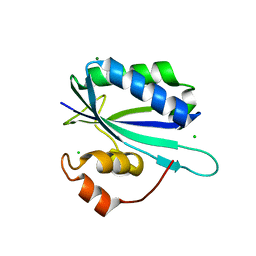
High-resolution crystal structure of the chicken MHF complex
分子名称: Centromere protein S, Centromere protein X
著者Ito, S, Nishino, T.
登録日2020-10-14
公開日2021-03-17
最終更新日2023-11-29
実験手法X-RAY DIFFRACTION (1.25 Å)
主引用文献Structural analysis of the chicken FANCM-MHF complex and its stability.
Acta Crystallogr.,Sect.F, 77, 2021
7DA1
DownloadVisualize
BU of 7da1 by Molmil
Crystal structure of the chicken MHF complex
分子名称: Centromere protein S, Centromere protein X
著者Ito, S, Nishino, T.
登録日2020-10-14
公開日2021-03-17
最終更新日2023-11-29
実験手法X-RAY DIFFRACTION (2.01 Å)
主引用文献Structural analysis of the chicken FANCM-MHF complex and its stability.
Acta Crystallogr.,Sect.F, 77, 2021
7DA2
DownloadVisualize
BU of 7da2 by Molmil
The crystal structure of the chicken FANCM-MHF complex
分子名称: Centromere protein S, Centromere protein X, Fanconi anemia group M protein
著者Nishino, T, Ito, S.
登録日2020-10-14
公開日2021-03-17
最終更新日2023-11-29
実験手法X-RAY DIFFRACTION (2.79 Å)
主引用文献Structural analysis of the chicken FANCM-MHF complex and its stability.
Acta Crystallogr.,Sect.F, 77, 2021
3HW1
DownloadVisualize
BU of 3hw1 by Molmil
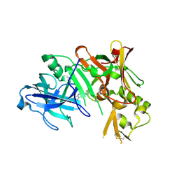
Structure of Bace (beta secretase) in complex with ligand EV2
分子名称: 3-pyrrolidin-1-ylquinoxalin-2-amine, Beta-secretase 1, GLYCEROL
著者Godemann, R, Madden, J, Kramer, J, Smith, M.A, Barker, J, Ebneth, A.
登録日2009-06-17
公開日2009-11-03
最終更新日2023-11-01
実験手法X-RAY DIFFRACTION (2.48 Å)
主引用文献Fragment-Based Discovery of BACE1 Inhibitors Using Functional Assays
Biochemistry, 48, 2009
7BK4
DownloadVisualize
BU of 7bk4 by Molmil
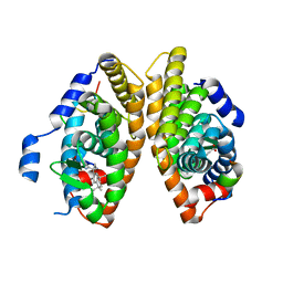
Crystal structure of RXRalpha ligand binding domain in complex with a fragment of the TIF2 coactivator
分子名称: 6-[1-(3,5,5,8,8-PENTAMETHYL-5,6,7,8-TETRAHYDRONAPHTHALEN-2-YL)CYCLOPROPYL]PYRIDINE-3-CARBOXYLIC ACID, Nuclear receptor coactivator 2, Retinoic acid receptor RXR-alpha
著者le Maire, A, Bourguet, W.
登録日2021-01-15
公開日2021-08-04
最終更新日2024-01-31
実験手法X-RAY DIFFRACTION (2.8 Å)
主引用文献Structural Insights into the Interaction of the Intrinsically Disordered Co-activator TIF2 with Retinoic Acid Receptor Heterodimer (RXR/RAR).
J.Mol.Biol., 433, 2021
7K5L
DownloadVisualize
BU of 7k5l by Molmil
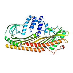
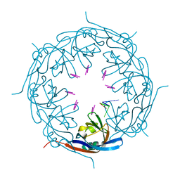
Ebola virus VP40 octameric ring generated by an RNA oligonucleotide
分子名称: HSP RNA oligonucleotide, Matrix protein VP40
著者Landeras-Bueno, S, Wasserman, H, Salie, Z.L, Saphire, E.O.
登録日2020-09-17
公開日2021-04-21
最終更新日2023-10-18
実験手法X-RAY DIFFRACTION (1.38 Å)
主引用文献Cellular mRNA triggers structural transformation of Ebola virus matrix protein VP40 to its essential regulatory form.
Cell Rep, 35, 2021
7K5D
DownloadVisualize
BU of 7k5d by Molmil
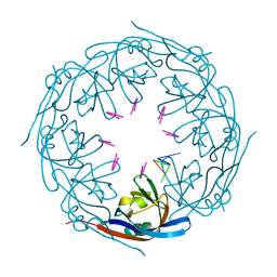
Ebola virus VP40 octameric ring generated by a DNA oligonucleotide
分子名称: HSP DNA oligonucleotide, Matrix protein VP40
著者Landeras-Bueno, S, Wasserman, H, Salie, Z.L, Saphire, E.O.
登録日2020-09-16
公開日2021-04-21
最終更新日2023-10-18
実験手法X-RAY DIFFRACTION (1.78 Å)
主引用文献Cellular mRNA triggers structural transformation of Ebola virus matrix protein VP40 to its essential regulatory form.
Cell Rep, 35, 2021
3LW8
DownloadVisualize
BU of 3lw8 by Molmil
Shigella IpgB2 in complex with human RhoA, GDP and Mg2+ (complex A)
分子名称: GUANOSINE-5'-DIPHOSPHATE, IpgB2, MAGNESIUM ION, ...
著者Klink, B.U, Barden, S, Heidler, T.V, Borchers, C, Ladwein, M, Stradal, T.E.B, Rottner, K, Heinz, D.W.
登録日2010-02-23
公開日2010-03-31
最終更新日2023-11-01
実験手法X-RAY DIFFRACTION (1.85 Å)
主引用文献Structure of Shigella IPGB2 in complex with human RhoA: Implications for the mechanism of bacterial GEF-mimicry
J.Biol.Chem., 285, 2010
7JV3
DownloadVisualize
BU of 7jv3 by Molmil
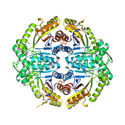
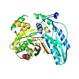
Crystal structure of alkanesulfonate monooxygenase MsuD from Pseudomonas fluorescens
分子名称: Alkanesulfonate monooxygenase
著者Liew, J.J.M, Dowling, D.P.
登録日2020-08-20
公開日2021-05-26
最終更新日2023-10-18
実験手法X-RAY DIFFRACTION (2.8 Å)
主引用文献Structures of the alkanesulfonate monooxygenase MsuD provide insight into C-S bond cleavage, substrate scope, and an unexpected role for the tetramer.
J.Biol.Chem., 297, 2021
7BED
DownloadVisualize
BU of 7bed by Molmil
X-ray structure of WDR5 bound to the WDR5 win motif peptide
分子名称: SULFATE ION, WD repeat-containing protein 5
著者McVey, C.E, Kaye, K.M.
登録日2020-12-23
公開日2021-12-08
最終更新日2024-01-31
実験手法X-RAY DIFFRACTION (1.26 Å)
主引用文献MLL1 is regulated by KSHV LANA and is important for virus latency.
Nucleic Acids Res., 49, 2021
4PEP
DownloadVisualize
BU of 4pep by Molmil
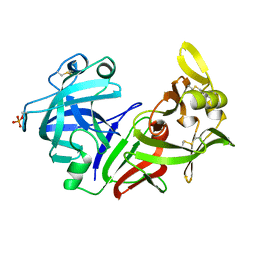
THE MOLECULAR AND CRYSTAL STRUCTURES OF MONOCLINIC PORCINE PEPSIN REFINED AT 1.8 ANGSTROMS RESOLUTION
分子名称: PEPSIN
著者Andreeva, N, Fedorov, A.A, Sielecki, A, James, M.
登録日1989-12-18
公開日1990-04-15
最終更新日2017-11-29
実験手法X-RAY DIFFRACTION (1.8 Å)
主引用文献Molecular and crystal structures of monoclinic porcine pepsin refined at 1.8 A resolution.
J.Mol.Biol., 214, 1990
2CND
DownloadVisualize
BU of 2cnd by Molmil
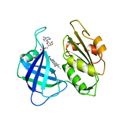
STRUCTURAL STUDIES ON CORN NITRATE REDUCTASE: REFINED STRUCTURE OF THE CYTOCHROME B REDUCTASE FRAGMENT AT 2.5 ANGSTROMS, ITS ADP COMPLEX AND AN ACTIVE SITE MUTANT AND MODELING OF THE CYTOCHROME B DOMAIN
分子名称: FLAVIN-ADENINE DINUCLEOTIDE, NADH-DEPENDENT NITRATE REDUCTASE
著者Lu, G, Lindqvist, Y, Schneider, G.
登録日1995-02-01
公開日1995-04-20
最終更新日2024-02-14
実験手法X-RAY DIFFRACTION (2.5 Å)
主引用文献Structural studies on corn nitrate reductase: refined structure of the cytochrome b reductase fragment at 2.5 A, its ADP complex and an active-site mutant and modeling of the cytochrome b domain.
J.Mol.Biol., 248, 1995
7JSD
DownloadVisualize
BU of 7jsd by Molmil
Hydroxylase homolog of BesD with Fe(II), alpha-ketoglutarate, and lysine
分子名称: 2-OXOGLUTARIC ACID, FE (II) ION, LYSINE, ...
著者Kissman, E.N, Neugebauer, M.E, Chang, M.C.Y.
登録日2020-08-14
公開日2021-08-25
最終更新日2023-10-18
実験手法X-RAY DIFFRACTION (2.5 Å)
主引用文献Reaction pathway engineering converts a radical hydroxylase into a halogenase.
Nat.Chem.Biol., 18, 2022
7JUA
DownloadVisualize
BU of 7jua by Molmil
Crystal structure of the second heterocyclization domain of yersiniabactin synthetase at 2.35 A resolution
分子名称: 2-(2-METHOXYETHOXY)ETHANOL, Irp2 protein, SODIUM ION
著者Xia, Y, Soule, J, Dowling, D.P.
登録日2020-08-19
公開日2021-09-22
最終更新日2023-10-18
実験手法X-RAY DIFFRACTION (2.35 Å)
主引用文献High-resolution structures of a siderophore-producing cyclization domain from Yersinia pestis offer a refined proposal of substrate binding.
J.Biol.Chem., 2022

226262

件を2024-10-16に公開中

PDB statisticsPDBj update infoContact PDBjnumon