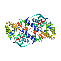
3V1E
 
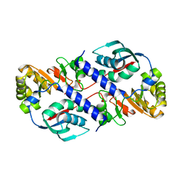
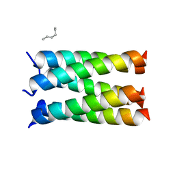
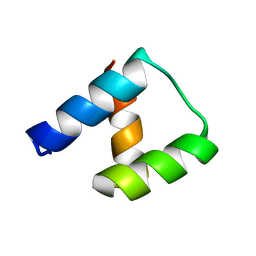
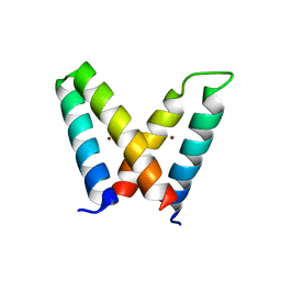 | | Crystal structure of de novo designed MID1-zinc H12E mutant | | 分子名称: | Computational design, MID1-zinc H12E mutant, ZINC ION | | 著者 | Der, B.S, Machius, M, Miley, M.J, Kuhlman, B. | | 登録日 | 2011-12-09 | | 公開日 | 2012-01-11 | | 最終更新日 | 2023-09-13 | | 実験手法 | X-RAY DIFFRACTION (1.073 Å) | | 主引用文献 | Metal-mediated affinity and orientation specificity in a computationally designed protein homodimer.
J.Am.Chem.Soc., 134, 2012
|
|
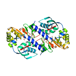
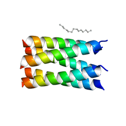
3V1B
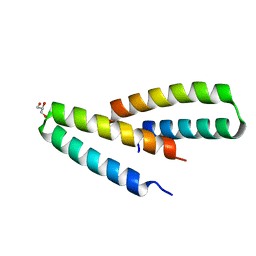 | | Crystal structure of de novo designed MID1-apo2 | | 分子名称: | Computational design, MID1-apo2, GLYCEROL | | 著者 | Der, B.S, Machius, M, Miley, M.J, Kuhlman, B. | | 登録日 | 2011-12-09 | | 公開日 | 2012-01-11 | | 最終更新日 | 2023-09-13 | | 実験手法 | X-RAY DIFFRACTION (1.28 Å) | | 主引用文献 | Metal-mediated affinity and orientation specificity in a computationally designed protein homodimer.
J.Am.Chem.Soc., 134, 2012
|
|
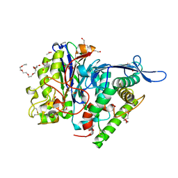
3FGW
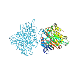 | | One chain form of the 66.3 kDa protein | | 分子名称: | 2-acetamido-2-deoxy-beta-D-glucopyranose, GLYCEROL, IODIDE ION, ... | | 著者 | Lakomek, K, Dickmanns, A, Ficner, R. | | 登録日 | 2008-12-08 | | 公開日 | 2009-09-15 | | 最終更新日 | 2023-11-01 | | 実験手法 | X-RAY DIFFRACTION (2.8 Å) | | 主引用文献 | Initial insight into the function of the lysosomal 66.3 kDa protein from mouse by means of X-ray crystallography
Bmc Struct.Biol., 9, 2009
|
|
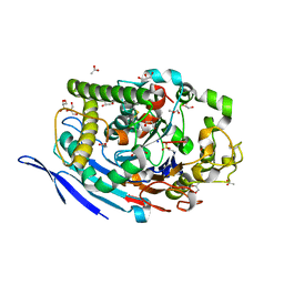
1QPQ
 | | Structure of Quinolinic Acid Phosphoribosyltransferase from Mycobacterium Tuberculosis: A Potential TB Drug Target | | 分子名称: | QUINOLINATE PHOSPHORIBOSYLTRANSFERASE, QUINOLINIC ACID, SULFATE ION | | 著者 | Sharma, V, Grubmeyer, C, Sacchettini, J.C, TB Structural Genomics Consortium (TBSGC) | | 登録日 | 1998-11-20 | | 公開日 | 1998-11-25 | | 最終更新日 | 2023-12-27 | | 実験手法 | X-RAY DIFFRACTION (2.45 Å) | | 主引用文献 | Crystal structure of quinolinic acid phosphoribosyltransferase from Mycobacterium tuberculosis: a potential TB drug target.
Structure, 6, 1998
|
|
1QPR
 | | QUINOLINATE PHOSPHORIBOSYLTRANSFERASE (QAPRTASE) FROM MYCOBACTERIUM TUBERCULOSIS IN COMPLEX WITH PHTHALATE AND PRPCP | | 分子名称: | 1-O-[(R)-hydroxy(phosphonomethyl)phosphoryl]-5-O-phosphono-alpha-D-ribofuranose, MANGANESE (II) ION, PHTHALIC ACID, ... | | 著者 | Sharma, V, Grubmeyer, C, Sacchettini, J.C, TB Structural Genomics Consortium (TBSGC) | | 登録日 | 1998-10-17 | | 公開日 | 1998-10-21 | | 最終更新日 | 2024-02-14 | | 実験手法 | X-RAY DIFFRACTION (2.45 Å) | | 主引用文献 | Crystal structure of quinolinic acid phosphoribosyltransferase from Mycobacterium tuberculosis: a potential TB drug target.
Structure, 6, 1998
|
|
1QPN
 | |
3FGT
 | | Two chain form of the 66.3 kDa protein from mouse lacking the linker peptide | | 分子名称: | 2-(2-(2-(2-(2-(2-ETHOXYETHOXY)ETHOXY)ETHOXY)ETHOXY)ETHOXY)ETHANOL, 2-acetamido-2-deoxy-beta-D-glucopyranose, 2-acetamido-2-deoxy-beta-D-glucopyranose-(1-4)-2-acetamido-2-deoxy-beta-D-glucopyranose, ... | | 著者 | Lakomek, K, Dickmanns, A, Ficner, R. | | 登録日 | 2008-12-08 | | 公開日 | 2009-09-15 | | 最終更新日 | 2023-11-22 | | 実験手法 | X-RAY DIFFRACTION (2.4 Å) | | 主引用文献 | Initial insight into the function of the lysosomal 66.3 kDa protein from mouse by means of X-ray crystallography
Bmc Struct.Biol., 9, 2009
|
|
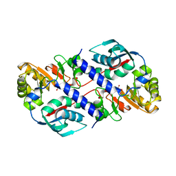
3FGR
 | | Two chain form of the 66.3 kDa protein at 1.8 Angstroem | | 分子名称: | 2-acetamido-2-deoxy-beta-D-glucopyranose, 2-acetamido-2-deoxy-beta-D-glucopyranose-(1-4)-2-acetamido-2-deoxy-beta-D-glucopyranose, ACETATE ION, ... | | 著者 | Lakomek, K, Dickmanns, A, Ficner, R. | | 登録日 | 2008-12-08 | | 公開日 | 2009-09-15 | | 最終更新日 | 2023-11-22 | | 実験手法 | X-RAY DIFFRACTION (1.8 Å) | | 主引用文献 | Initial insight into the function of the lysosomal 66.3 kDa protein from mouse by means of X-ray crystallography
Bmc Struct.Biol., 9, 2009
|
|
1QPO
 | |
7N3T
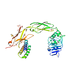 | | TrkA ECD complex with designed miniprotein ligand | | 分子名称: | 1,2-ETHANEDIOL, 2-acetamido-2-deoxy-beta-D-glucopyranose, 2-acetamido-2-deoxy-beta-D-glucopyranose-(1-4)-2-acetamido-2-deoxy-beta-D-glucopyranose, ... | | 著者 | Jude, K.M, Cao, L, Garcia, K.C. | | 登録日 | 2021-06-01 | | 公開日 | 2022-04-20 | | 最終更新日 | 2023-10-18 | | 実験手法 | X-RAY DIFFRACTION (1.84 Å) | | 主引用文献 | Design of protein-binding proteins from the target structure alone.
Nature, 605, 2022
|
|
7N2Y
 | |
7N2Z
 | | Crystal Structure of a de Novo Three-stranded Coiled Coil Peptide Containing Trigonal Pyrmidal Pb(II) complexes in the dual Tris-thiolate Site | | 分子名称: | CHLORIDE ION, LEAD (II) ION, Pb(II)2-(GRAND CoilSerL16CL23C)3, ... | | 著者 | Ruckthong, L, Stuckey, J.A, Pecoraro, V.L. | | 登録日 | 2021-05-30 | | 公開日 | 2022-06-01 | | 最終更新日 | 2023-10-18 | | 実験手法 | X-RAY DIFFRACTION (1.29 Å) | | 主引用文献 | Open Reading Frame 1 Protein of the Human Long Interspersed Nuclear Element 1 Retrotransposon Binds Multiple Equivalents of Lead.
J.Am.Chem.Soc., 143, 2021
|
|
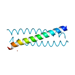
7UDZ
 | | Designed pentameric proton channel LQLL | | 分子名称: | (2R)-2,3-dihydroxypropyl (9Z)-octadec-9-enoate, De novo designed pentameric proton channel LQLL | | 著者 | Kratochvil, H.T, Thomaston, J.L, Mravic, M, Nicoludis, J.M, Liu, L, DeGrado, W.F. | | 登録日 | 2022-03-20 | | 公開日 | 2022-04-06 | | 最終更新日 | 2023-10-25 | | 実験手法 | X-RAY DIFFRACTION (2.48 Å) | | 主引用文献 | Transient water wires mediate selective proton transport in designed channel proteins.
Nat.Chem., 15, 2023
|
|
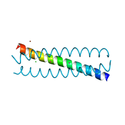
7UDX
 | | Designed pentameric proton channel QLQL | | 分子名称: | (2R)-2,3-dihydroxypropyl (9Z)-octadec-9-enoate, De novo designed pentameric proton channel QLQL | | 著者 | Kratochvil, H.T, Thomaston, J.L, Liu, L, DeGrado, W.F. | | 登録日 | 2022-03-20 | | 公開日 | 2022-04-06 | | 最終更新日 | 2023-10-25 | | 実験手法 | X-RAY DIFFRACTION (2.99 Å) | | 主引用文献 | Transient water wires mediate selective proton transport in designed channel proteins.
Nat.Chem., 15, 2023
|
|
7SMJ
 | |
8FXQ
 | | The Crystal Sturucture of Rhizopuspepsin with a bound modified peptide inhibitor generated by de novo drug design. | | 分子名称: | ALA-CYS-VAL-LYS, CYCLOHEXANE, Rhizopuspepsin, ... | | 著者 | Satyshur, K.A, Rich, D.H, Ripka, A.S. | | 登録日 | 2023-01-25 | | 公開日 | 2023-02-08 | | 実験手法 | X-RAY DIFFRACTION (1.21 Å) | | 主引用文献 | Aspartic protease inhibitors designed from computer-generated templates bind as predicted.
Org Lett, 3, 2001
|
|
2NDL
 | | NMR solution structure of PawS Derived Peptide 22 (PDP-22) | | 分子名称: | PawS derived peptide | | 著者 | Franke, B, Jayasena, A.S, Fisher, M.F, Swedberg, J.E, Taylor, N.L, Mylne, J.S, Rosengren, K. | | 登録日 | 2016-07-17 | | 公開日 | 2016-12-07 | | 最終更新日 | 2023-06-14 | | 実験手法 | SOLUTION NMR | | 主引用文献 | Diverse cyclic seed peptides in the Mexican zinnia (Zinnia haageana).
Biopolymers, 106, 2016
|
|
2N8I
 | | Solution NMR Structure of Designed Protein DA05, Northeast Structural Genomics Consortium (NESG) Target OR626 | | 分子名称: | Designed Protein DA05 | | 著者 | Xu, X, Eletsky, A, Federizon, J.F, Jacobs, T.M, Kuhlman, B, Szyperski, T, Northeast Structural Genomics Consortium (NESG) | | 登録日 | 2015-10-15 | | 公開日 | 2016-01-20 | | 最終更新日 | 2024-05-15 | | 実験手法 | SOLUTION NMR | | 主引用文献 | Design of structurally distinct proteins using strategies inspired by evolution.
Science, 352, 2016
|
|
2N8W
 | | Solution NMR Structure of Designed Protein DA05R1, Northeast Structural Genomics Consortium (NESG) Target OR690 | | 分子名称: | Designed Protein DA05R1 | | 著者 | Eletsky, A, Federizon, J.F, Xu, X, Pulavarti, S, Jacobs, T.M, Kuhlman, B, Szyperski, T, Northeast Structural Genomics Consortium (NESG) | | 登録日 | 2015-10-27 | | 公開日 | 2015-11-25 | | 最終更新日 | 2024-05-15 | | 実験手法 | SOLUTION NMR | | 主引用文献 | Design of structurally distinct proteins using strategies inspired by evolution.
Science, 352, 2016
|
|
2MG4
 | | Computational design and experimental verification of a symmetric protein homodimer | | 分子名称: | Computational designed homodimer | | 著者 | Mou, Y, Huang, P.S, Hsu, F.C, Huang, S.J, Mayo, S.L. | | 登録日 | 2013-10-26 | | 公開日 | 2015-04-08 | | 最終更新日 | 2024-05-15 | | 実験手法 | SOLUTION NMR | | 主引用文献 | Computational design and experimental verification of a symmetric protein homodimer.
Proc.Natl.Acad.Sci.USA, 112, 2015
|
|
4NDL
 | | Computational design and experimental verification of a symmetric homodimer | | 分子名称: | ENH-c2b, computational designed homodimer | | 著者 | Mou, Y, Huang, P.S, Hsu, F.C, Huang, S.J, Mayo, S.L. | | 登録日 | 2013-10-26 | | 公開日 | 2014-11-05 | | 最終更新日 | 2024-02-28 | | 実験手法 | X-RAY DIFFRACTION (3.5 Å) | | 主引用文献 | Computational design and experimental verification of a symmetric protein homodimer.
Proc.Natl.Acad.Sci.USA, 112, 2015
|
|
6Y7N
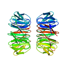 | | The crystal structure of the eight-bladed symmetrical designer protein Tako8 in the presence of tellurotungstic Anderson-Evans (TEW) | | 分子名称: | Tako8 | | 著者 | Vandebroek, L, Noguchi, H, Parac-Vogt, T.N, Van Meervelt, L, Voet, A.R.D. | | 登録日 | 2020-03-02 | | 公開日 | 2020-12-16 | | 最終更新日 | 2024-01-24 | | 実験手法 | X-RAY DIFFRACTION (1.6 Å) | | 主引用文献 | Shape and Size Complementarity-Induced Formation of Supramolecular Protein Assemblies with Metal-Oxo Clusters
Cryst.Growth Des., 2021
|
|
6Y7P
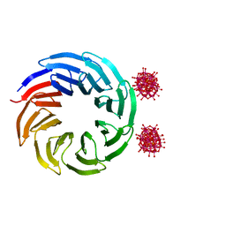 | | The complex between the eight-bladed symmetrical designer protein Tako8 and 1:2 zirconium(IV) Wells-Dawson (ZrWD) | | 分子名称: | Tako8, W-Zr-cluster | | 著者 | Vandebroek, L, Noguchi, H, Parac-Vogt, T.N, Van Meervelt, L, Voet, A.R.D. | | 登録日 | 2020-03-02 | | 公開日 | 2020-12-16 | | 最終更新日 | 2024-01-24 | | 実験手法 | X-RAY DIFFRACTION (1.75 Å) | | 主引用文献 | Shape and Size Complementarity-Induced Formation of Supramolecular Protein Assemblies with Metal-Oxo Clusters
Cryst.Growth Des., 2021
|
|
6Y7O
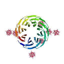 | | The complex between the eight-bladed symmetrical designer protein Tako8 and the silicotungstic acid Keggin (STA) | | 分子名称: | Keggin (STA), Tako8 | | 著者 | Vandebroek, L, Noguchi, H, Parac-Vogt, T.N, Van Meervelt, L, Voet, A.R.D. | | 登録日 | 2020-03-02 | | 公開日 | 2020-12-16 | | 最終更新日 | 2024-01-24 | | 実験手法 | X-RAY DIFFRACTION (2.3 Å) | | 主引用文献 | Shape and Size Complementarity-Induced Formation of Supramolecular Protein Assemblies with Metal-Oxo Clusters
Cryst.Growth Des., 2021
|
|
4OIO
 | | Crystal structure of Thermus thermophilus pre-insertion substrate complex for de novo transcription initiation | | 分子名称: | 5'-D(*CP*CP*TP*GP*CP*AP*TP*CP*CP*GP*TP*GP*AP*GP*TP*CP*GP*AP*GP*GP*G)-3', 5'-D(*TP*AP*TP*AP*AP*TP*GP*GP*GP*AP*GP*CP*TP*GP*TP*CP*AP*CP*GP*GP*AP*TP*GP*CP*AP*GP*G)-3', 5'-O-[(S)-hydroxy{[(S)-hydroxy(phosphonooxy)phosphoryl]methyl}phosphoryl]cytidine, ... | | 著者 | Zhang, Y, Ebright, R.H, Arnold, E. | | 登録日 | 2014-01-20 | | 公開日 | 2014-05-07 | | 最終更新日 | 2023-09-20 | | 実験手法 | X-RAY DIFFRACTION (3.1 Å) | | 主引用文献 | GE23077 binds to the RNA polymerase 'i' and 'i+1' sites and prevents the binding of initiating nucleotides.
Elife, 3, 2014
|
|


