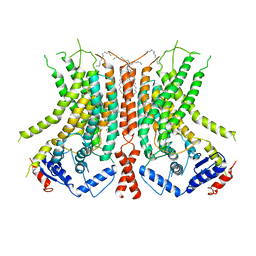
6E1H
 
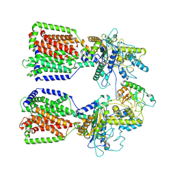 | | Structure of 2:1 human Ptch1-Shh-N complex | | 分子名称: | CALCIUM ION, PALMITIC ACID, Protein patched homolog 1, ... | | 著者 | Qi, X, Li, X. | | 登録日 | 2018-07-09 | | 公開日 | 2018-08-29 | | 最終更新日 | 2024-11-13 | | 実験手法 | ELECTRON MICROSCOPY (3.5 Å) | | 主引用文献 | Two Patched molecules engage distinct sites on Hedgehog yielding a signaling-competent complex.
Science, 362, 2018
|
|
6E1I
 | |
6E1J
 | |
6E1K
 | | Structure of AtTPC1(DDE) reconstituted in saposin A with cat06 Fab | | 分子名称: | CALCIUM ION, PALMITIC ACID, Two pore calcium channel protein 1, ... | | 著者 | Kintzer, A.F, Green, E.M, Cheng, Y, Stroud, R.M. | | 登録日 | 2018-07-10 | | 公開日 | 2018-09-19 | | 最終更新日 | 2024-12-25 | | 実験手法 | ELECTRON MICROSCOPY (3.3 Å) | | 主引用文献 | Structural basis for activation of voltage sensor domains in an ion channel TPC1.
Proc. Natl. Acad. Sci. U.S.A., 115, 2018
|
|
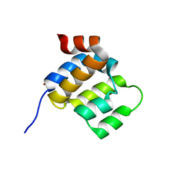
6E1L
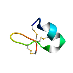 | | GRN3Ala | | 分子名称: | Granulin | | 著者 | Dastpeyman, M, Bansal, P, Wilson, D, Sotillo, J, Brindley, P, Loukas, A, Smout, M, Daly, N. | | 登録日 | 2018-07-10 | | 公開日 | 2019-02-06 | | 最終更新日 | 2024-10-23 | | 実験手法 | SOLUTION NMR | | 主引用文献 | Structural Variants of a Liver Fluke Derived Granulin Peptide Potently Stimulate Wound Healing.
J. Med. Chem., 61, 2018
|
|
6E1M
 | | Structure of AtTPC1(DDE) reconstituted in saposin A | | 分子名称: | 1,2-DIACYL-GLYCEROL-3-SN-PHOSPHATE, CALCIUM ION, PALMITIC ACID, ... | | 著者 | Kintzer, A.F, Green, E.M, Cheng, Y, Stroud, R.M. | | 登録日 | 2018-07-10 | | 公開日 | 2018-09-19 | | 最終更新日 | 2024-12-25 | | 実験手法 | ELECTRON MICROSCOPY (3.3 Å) | | 主引用文献 | Structural basis for activation of voltage sensor domains in an ion channel TPC1.
Proc. Natl. Acad. Sci. U.S.A., 115, 2018
|
|
6E1N
 | | Structure of AtTPC1(DDE) in state 1 | | 分子名称: | 1,2-DIACYL-GLYCEROL-3-SN-PHOSPHATE, CALCIUM ION, PALMITIC ACID, ... | | 著者 | Kintzer, A.F, Green, E.M, Cheng, Y, Stroud, R.M. | | 登録日 | 2018-07-10 | | 公開日 | 2018-09-19 | | 最終更新日 | 2024-12-25 | | 実験手法 | ELECTRON MICROSCOPY (3.7 Å) | | 主引用文献 | Structural basis for activation of voltage sensor domains in an ion channel TPC1.
Proc. Natl. Acad. Sci. U.S.A., 115, 2018
|
|
6E1O
 | |
6E1P
 | | Structure of AtTPC1(DDE) in state 2 | | 分子名称: | CALCIUM ION, PALMITIC ACID, Two pore calcium channel protein 1 | | 著者 | Kintzer, A.F, Green, E.M, Cheng, Y, Stroud, R.M. | | 登録日 | 2018-07-10 | | 公開日 | 2018-09-19 | | 最終更新日 | 2024-12-25 | | 実験手法 | ELECTRON MICROSCOPY (3.7 Å) | | 主引用文献 | Structural basis for activation of voltage sensor domains in an ion channel TPC1.
Proc. Natl. Acad. Sci. U.S.A., 115, 2018
|
|
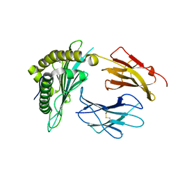
6E1Q
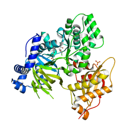 | | AtGH3.15 acyl acid amido synthetase in complex with 2,4-DB | | 分子名称: | (2,4-DICHLOROPHENOXY)ACETIC ACID, AtGH3.15 acyl acid amido synthetase, PHOSPHATE ION | | 著者 | Sharp, A.M, Lee, S.G, Jez, J.M. | | 登録日 | 2018-07-10 | | 公開日 | 2018-10-24 | | 最終更新日 | 2024-11-13 | | 実験手法 | X-RAY DIFFRACTION (2.148 Å) | | 主引用文献 | Modification of auxinic phenoxyalkanoic acid herbicides by the acyl acid amido synthetase GH3.15 from Arabidopsis.
J. Biol. Chem., 293, 2018
|
|
6E1R
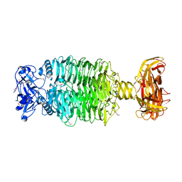 | | Crystal structure of the Acinetobacter phage vB_ApiP_P1 tailspike protein | | 分子名称: | CHLORIDE ION, SODIUM ION, Tailspike protein | | 著者 | Plattner, M, Shneider, M.M, Oliveira, H, Azeredo, J, Leiman, P.G. | | 登録日 | 2018-07-10 | | 公開日 | 2019-07-17 | | 最終更新日 | 2024-10-16 | | 実験手法 | X-RAY DIFFRACTION (2.693 Å) | | 主引用文献 | Crystal structure of the Acinetobacter phage vB_ApiP_P1 tailspike protein
To Be Published
|
|
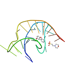
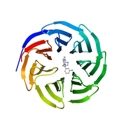
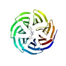
6E1S
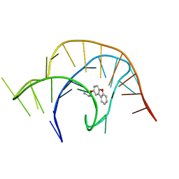 | | Crystal structure of a class I PreQ1 riboswitch complexed with a synthetic compound 1: 2-[(dibenzo[b,d]furan-2-yl)oxy]ethan-1-amine | | 分子名称: | 2-[(dibenzo[b,d]furan-2-yl)oxy]ethan-1-amine, RNA (33-MER) | | 著者 | Numata, T, Connelly, C.M, Schneekloth, J.S, Ferre-D'Amare, A.R. | | 登録日 | 2018-07-10 | | 公開日 | 2019-04-10 | | 最終更新日 | 2023-10-11 | | 実験手法 | X-RAY DIFFRACTION (1.8 Å) | | 主引用文献 | Synthetic ligands for PreQ1riboswitches provide structural and mechanistic insights into targeting RNA tertiary structure.
Nat Commun, 10, 2019
|
|
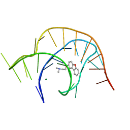
6E1T
 | | Crystal structure of a class I PreQ1 riboswitch complexed with a synthetic compound 1: 2-[(dibenzo[b,d]furan-2-yl)oxy]ethan-1-amine | | 分子名称: | 2-(N-MORPHOLINO)-ETHANESULFONIC ACID, 2-[(dibenzo[b,d]furan-2-yl)oxy]ethan-1-amine, MAGNESIUM ION, ... | | 著者 | Numata, T, Connelly, C.M, Schneekloth, J.S, Ferre-D'Amare, A.R. | | 登録日 | 2018-07-10 | | 公開日 | 2019-04-10 | | 最終更新日 | 2023-10-11 | | 実験手法 | X-RAY DIFFRACTION (1.8 Å) | | 主引用文献 | Synthetic ligands for PreQ1riboswitches provide structural and mechanistic insights into targeting RNA tertiary structure.
Nat Commun, 10, 2019
|
|
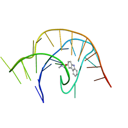
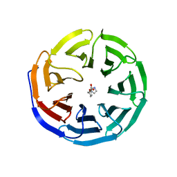
6E1U
 | | Crystal structure of a class I PreQ1 riboswitch complexed with a synthetic compound 2: 2-[(dibenzo[b,d]furan-2-yl)oxy]-N,N-dimethylethan-1-amine | | 分子名称: | 2-[(dibenzo[b,d]furan-2-yl)oxy]-N,N-dimethylethan-1-amine, MAGNESIUM ION, RNA (33-MER) | | 著者 | Numata, T, Connelly, C.M, Schneekloth, J.S, Ferre-D'Amare, A.R. | | 登録日 | 2018-07-10 | | 公開日 | 2019-04-10 | | 最終更新日 | 2023-10-11 | | 実験手法 | X-RAY DIFFRACTION (1.94 Å) | | 主引用文献 | Synthetic ligands for PreQ1riboswitches provide structural and mechanistic insights into targeting RNA tertiary structure.
Nat Commun, 10, 2019
|
|
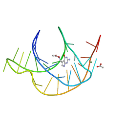
6E1V
 | | Crystal structure of a class I PreQ1 riboswitch complexed with a synthetic compound 3: 2-[(9H-carbazol-3-yl)oxy]-N,N-dimethylethan-1-amine | | 分子名称: | 2-[(9H-carbazol-3-yl)oxy]-N,N-dimethylethan-1-amine, RNA (33-MER) | | 著者 | Numata, T, Connelly, C.M, Schneekloth, J.S, Ferre-D'Amare, A.R. | | 登録日 | 2018-07-10 | | 公開日 | 2019-04-10 | | 最終更新日 | 2023-10-11 | | 実験手法 | X-RAY DIFFRACTION (2.56 Å) | | 主引用文献 | Synthetic ligands for PreQ1riboswitches provide structural and mechanistic insights into targeting RNA tertiary structure.
Nat Commun, 10, 2019
|
|
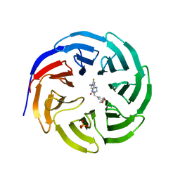
6E1W
 | | Crystal structure of a class I PreQ1 riboswitch complexed with PreQ1 | | 分子名称: | 2-amino-5-(aminomethyl)-1,7-dihydro-4H-pyrrolo[2,3-d]pyrimidin-4-one, ACETATE ION, MAGNESIUM ION, ... | | 著者 | Numata, T, Connelly, C.M, Schneekloth, J.S, Ferre-D'Amare, A.R. | | 登録日 | 2018-07-10 | | 公開日 | 2019-04-10 | | 最終更新日 | 2024-03-13 | | 実験手法 | X-RAY DIFFRACTION (1.69 Å) | | 主引用文献 | Synthetic ligands for PreQ1riboswitches provide structural and mechanistic insights into targeting RNA tertiary structure.
Nat Commun, 10, 2019
|
|
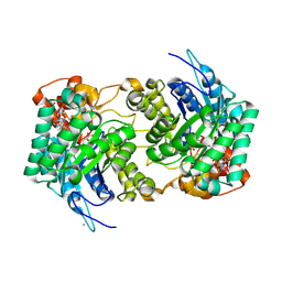
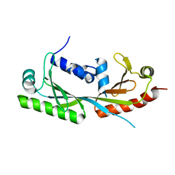
6E1X
 | | Crystal structure of product-bound complex of spermidine/spermine N-acetyltransferase SpeG | | 分子名称: | (4R)-2-METHYLPENTANE-2,4-DIOL, (4S)-2-METHYL-2,4-PENTANEDIOL, 2-AMINO-2-HYDROXYMETHYL-PROPANE-1,3-DIOL, ... | | 著者 | Filippova, E.V, Minasov, G, Kiryukhina, O, Anderson, W.F, Satchell, K.J.F, Joachimiak, A, Center for Structural Genomics of Infectious Diseases (CSGID) | | 登録日 | 2018-07-10 | | 公開日 | 2019-07-10 | | 最終更新日 | 2023-10-11 | | 実験手法 | X-RAY DIFFRACTION (1.35 Å) | | 主引用文献 | Crystal structure of product-bound complex of spermidine/spermine N-acetyltransferase SpeG from Vibrio cholerae.
To Be Published
|
|
6E1Y
 | |
6E1Z
 | |
6E20
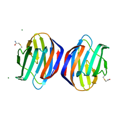 | | Crystal structure of the Dario rerio galectin-1-L2 | | 分子名称: | Galectin, MAGNESIUM ION, beta-D-galactopyranose-(1-4)-2-acetamido-2-deoxy-alpha-D-glucopyranose | | 著者 | Ghosh, A, Bianchet, M.A. | | 登録日 | 2018-07-10 | | 公開日 | 2019-03-20 | | 最終更新日 | 2023-10-11 | | 実験手法 | X-RAY DIFFRACTION (2 Å) | | 主引用文献 | Structure of the zebrafish galectin-1-L2 and model of its interaction with the infectious hematopoietic necrosis virus (IHNV) envelope glycoprotein.
Glycobiology, 29, 2019
|
|
6E21
 | | Joint X-ray/neutron structure of PKAc with products Sr2-ADP and phosphorylated peptide SP20 | | 分子名称: | ADENOSINE-5'-DIPHOSPHATE, STRONTIUM ION, cAMP-dependent protein kinase catalytic subunit alpha, ... | | 著者 | Kovalevsky, A, Gerlits, O.O, Taylor, S. | | 登録日 | 2018-07-10 | | 公開日 | 2019-04-03 | | 最終更新日 | 2024-11-06 | | 実験手法 | NEUTRON DIFFRACTION (2 Å), X-RAY DIFFRACTION | | 主引用文献 | Zooming in on protons: Neutron structure of protein kinase A trapped in a product complex.
Sci Adv, 5, 2019
|
|
6E22
 | |
6E23
 | |
6E24
 | | Ternary structure of c-Myc-TBP-TAF1 | | 分子名称: | Transcription initiation factor TFIID subunit 1,Myc proto-oncogene protein,TATA-box-binding protein | | 著者 | Wei, Y, Dong, A, Sunnerhagen, M, Penn, L, Tong, Y, Edwards, A.M, Arrowsmith, C.H, Structural Genomics Consortium (SGC) | | 登録日 | 2018-07-10 | | 公開日 | 2019-10-02 | | 最終更新日 | 2024-03-13 | | 実験手法 | X-RAY DIFFRACTION (3.001 Å) | | 主引用文献 | Multiple direct interactions of TBP with the MYC oncoprotein.
Nat.Struct.Mol.Biol., 26, 2019
|
|
6E25
 | |


