Loading
PDBj
メニューPDBj@FacebookPDBj@TwitterPDBj@YouTubewwPDB FoundationwwPDB
RCSB PDBPDBeBMRBAdv. SearchSearch help

3NS6
DownloadVisualize
BU of 3ns6 by Molmil
Crystal structure of hte RNA recognition motif of yeast eIF3b residues 76-170
分子名称: Eukaryotic translation initiation factor 3 subunit B, SULFATE ION
著者Khoshnevis, S, Neumann, P, Ficner, R.
登録日2010-07-01
公開日2010-10-06
最終更新日2023-09-06
実験手法X-RAY DIFFRACTION (1.25 Å)
主引用文献Crystal structure of the RNA recognition motif of yeast translation initiation factor eIF3b reveals differences to human eIF3b.
Plos One, 5, 2010
6F8C
DownloadVisualize
BU of 6f8c by Molmil
Crystal structure of cytochrome P450 CYP260A1 (S276I) bound with progesterone
分子名称: Cytochrome P450 CYP260A1, PROGESTERONE, PROTOPORPHYRIN IX CONTAINING FE
著者Jozwik, I.K, Thunnissen, A.M.W.H.
登録日2017-12-12
公開日2018-03-14
最終更新日2024-01-17
実験手法X-RAY DIFFRACTION (1.9 Å)
主引用文献Structure-Based Engineering of Steroidogenic CYP260A1 for Stereo- and Regioselective Hydroxylation of Progesterone.
ACS Chem. Biol., 13, 2018
7E46
DownloadVisualize
BU of 7e46 by Molmil
Structure of the CYP102A1 Haem Domain with N-Carboxybenzyl-L-Prolyl-L-Phenylalanine in complex with (S)-(-)-1-Phenylethylamine
分子名称: (1S)-1-phenylethanamine, (2S)-3-phenyl-2-[[(2S)-1-phenylmethoxycarbonylpyrrolidin-2-yl]carbonylamino]propanoic acid, Bifunctional cytochrome P450/NADPH--P450 reductase, ...
著者Stanfield, J.K, Sugimoto, H, Shoji, O.
登録日2021-02-10
公開日2022-02-16
最終更新日2023-11-29
実験手法X-RAY DIFFRACTION (1.91 Å)
主引用文献Structure of the CYP102A1 Haem Domain with N-Carboxybenzyl-L-Prolyl-L-Phenylalanine in complex with (S)-(-)-1-Phenylethylamine at 1.91 Angstrom Resolution
To Be Published
6EW2
DownloadVisualize
BU of 6ew2 by Molmil
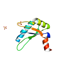
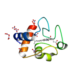
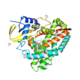
Human myelin protein P2 F57A mutant, tetragonal crystal form
分子名称: CHLORIDE ION, Myelin P2 protein, PALMITIC ACID
著者Laulumaa, S, Lehtimaki, M, Kursula, P.
登録日2017-11-03
公開日2018-07-11
最終更新日2024-01-17
実験手法X-RAY DIFFRACTION (1.588 Å)
主引用文献Structure and dynamics of a human myelin protein P2 portal region mutant indicate opening of the beta barrel in fatty acid binding proteins.
BMC Struct. Biol., 18, 2018
6EW5
DownloadVisualize
BU of 6ew5 by Molmil
Human myelin protein P2 F57A mutant, monoclinic crystal form
分子名称: Myelin P2 protein, PALMITIC ACID
著者Laulumaa, S, Lehtimaki, M, Kursula, P.
登録日2017-11-03
公開日2018-07-11
最終更新日2024-01-17
実験手法X-RAY DIFFRACTION (1.95 Å)
主引用文献Structure and dynamics of a human myelin protein P2 portal region mutant indicate opening of the beta barrel in fatty acid binding proteins.
BMC Struct. Biol., 18, 2018
4ETY
DownloadVisualize
BU of 4ety by Molmil
Crystal structure of a strand-swapped dimer of Mouse Leukocyte-associated immunoglobulin-like receptor 1 (NYSGRC-006047) Extra Cellular Domain
分子名称: 1,2-ETHANEDIOL, Leukocyte-associated immunoglobulin-like receptor 1
著者Sampathkumar, P, Almo, S.C, New York Structural Genomics Research Consortium (NYSGRC), Atoms-to-Animals: The Immune Function Network (IFN)
登録日2012-04-24
公開日2012-06-06
最終更新日2024-04-03
実験手法X-RAY DIFFRACTION (1.9 Å)
主引用文献Crystal structure of a strand-swapped dimer of Mouse Leukocyte-associated immunoglobulin-like receptor 1 Extra Cellular Domain
to be published
3O19
DownloadVisualize
BU of 3o19 by Molmil
Structure-function analysis of human L-Prostaglandin D Synthase bound with fatty acid
分子名称: OLEIC ACID, PALMITIC ACID, Prostaglandin-H2 D-isomerase
著者Zhou, Y, Shaw, N, Li, Y, Zhao, Y, Zhang, R, Liu, Z.-J.
登録日2010-07-21
公開日2010-09-22
最終更新日2023-11-01
実験手法X-RAY DIFFRACTION (1.66 Å)
主引用文献Structure-function analysis of human L-Prostaglandin D Synthase bound with fatty acid
To be Published
4EY1
DownloadVisualize
BU of 4ey1 by Molmil
Human Insulin
分子名称: CHLORIDE ION, Insulin A chain, Insulin B chain, ...
著者Favero-Retto, M.P, Palmieri, L.C, Lima, L.M.T.R.
登録日2012-05-01
公開日2013-05-01
最終更新日2017-11-15
実験手法X-RAY DIFFRACTION (1.471 Å)
主引用文献Structural meta-analysis of regular human insulin in pharmaceutical formulations.
Eur J Pharm Biopharm, 85, 2013
6ETP
DownloadVisualize
BU of 6etp by Molmil
Atomic resolution structure of RNase A (data collection 6)
分子名称: ISOPROPYL ALCOHOL, Ribonuclease pancreatic
著者Caterino, M, Vergara, A, Merlino, A.
登録日2017-10-27
公開日2018-02-21
最終更新日2024-01-17
実験手法X-RAY DIFFRACTION (1.02 Å)
主引用文献Raman-markers of X-ray radiation damage of proteins.
Int. J. Biol. Macromol., 111, 2018
3O1Y
DownloadVisualize
BU of 3o1y by Molmil
Electron transfer complexes: Experimental mapping of the redox-dependent cytochrome c electrostatic surface
分子名称: Cytochrome c, HEME C, NITRATE ION
著者De March, M, De Zorzi, R, Demitri, N, Gabbiani, C, Guerri, A, Casini, A, Messori, L, Geremia, S.
登録日2010-07-22
公開日2012-01-25
最終更新日2023-09-06
実験手法X-RAY DIFFRACTION (1.75 Å)
主引用文献Nitrate as a probe of cytochrome c surface: crystallographic identification of crucial "hot spots" for protein-protein recognition.
J. Inorg. Biochem., 135, 2014
6ETK
DownloadVisualize
BU of 6etk by Molmil
Atomic resolution structure of RNase A (data collection 1)
分子名称: ISOPROPYL ALCOHOL, Ribonuclease pancreatic
著者Caterino, M, Vergara, A, Merlino, A.
登録日2017-10-27
公開日2018-02-21
最終更新日2024-01-17
実験手法X-RAY DIFFRACTION (0.85 Å)
主引用文献Raman-markers of X-ray radiation damage of proteins.
Int. J. Biol. Macromol., 111, 2018
6ETQ
DownloadVisualize
BU of 6etq by Molmil
Atomic resolution structure of RNase A (data collection 7)
分子名称: ISOPROPYL ALCOHOL, Ribonuclease pancreatic
著者Caterino, M, Vergara, A, Merlino, A.
登録日2017-10-27
公開日2018-02-21
最終更新日2024-01-17
実験手法X-RAY DIFFRACTION (1.08 Å)
主引用文献Raman-markers of X-ray radiation damage of proteins.
Int. J. Biol. Macromol., 111, 2018
4EJH
DownloadVisualize
BU of 4ejh by Molmil
Human Cytochrome P450 2A13 in complex with 4-(methylnitrosamino)-1-(3-pyridyl)-1-butanone (NNK)
分子名称: 4-[methyl(nitroso)amino]-1-(pyridin-3-yl)butan-1-one, Cytochrome P450 2A13, GLYCEROL, ...
著者DeVore, N.M, Scott, E.E.
登録日2012-04-06
公開日2012-06-06
最終更新日2023-09-13
実験手法X-RAY DIFFRACTION (2.35 Å)
主引用文献Nicotine and 4-(methylnitrosamino)-1-(3-pyridyl)-1-butanone binding and access channel in human cytochrome P450 2A6 and 2A13 enzymes.
J.Biol.Chem., 287, 2012
7E73
DownloadVisualize
BU of 7e73 by Molmil
Crystal structure of human ERK2 mutant (Y36H)
分子名称: Mitogen-activated protein kinase 1, SULFATE ION
著者Park, Y.S, Kim, M, Ryu, S.E.
登録日2021-02-25
公開日2022-03-02
最終更新日2023-11-29
実験手法X-RAY DIFFRACTION (2.28 Å)
主引用文献Structural mechanism of inhibitor-resistance by ERK2 mutations
Biodesign, 9, 2021
7E75
DownloadVisualize
BU of 7e75 by Molmil
Crystal structure of human ERK2 mutant (G37C)
分子名称: Mitogen-activated protein kinase 1
著者Park, Y.S, Kim, M, Ryu, S.E.
登録日2021-02-25
公開日2022-03-02
最終更新日2023-11-29
実験手法X-RAY DIFFRACTION (2.481 Å)
主引用文献Structural mechanism of inhibitor-resistance by ERK2 mutations
Biodesign, 9, 2021
4EPJ
DownloadVisualize
BU of 4epj by Molmil
Crystal Structure of inactive single chain wild-type HIV-1 Protease in Complex with the substrate p2-NC
分子名称: 1,2-ETHANEDIOL, ACETATE ION, BETA-MERCAPTOETHANOL, ...
著者Schiffer, C.A, Mittal, S.
登録日2012-04-17
公開日2012-06-06
最終更新日2024-02-28
実験手法X-RAY DIFFRACTION (1.69 Å)
主引用文献Structural, kinetic, and thermodynamic studies of specificity designed HIV-1 protease.
Protein Sci., 21, 2012
3O6T
DownloadVisualize
BU of 3o6t by Molmil
Mycobacterium tuberculosis thioredoxin C C40S mutant in Complex with Quinol Inhibitor PMX464
分子名称: 4-(1,3-benzothiazol-2-yl)-4-hydroxycyclohexa-2,5-dien-1-one, TRIETHYLENE GLYCOL, Thioredoxin
著者Hall, G, Emsley, J.
登録日2010-07-29
公開日2010-11-10
最終更新日2023-11-01
実験手法X-RAY DIFFRACTION (2.4 Å)
主引用文献Structure of Mycobacterium tuberculosis thioredoxin in complex with quinol inhibitor PMX464
Protein Sci., 20, 2011
3NQ9
DownloadVisualize
BU of 3nq9 by Molmil
Bovine beta-lactoglobulin complex with caprylic acid
分子名称: Beta-lactoglobulin, CHLORIDE ION, OCTANOIC ACID (CAPRYLIC ACID)
著者Loch, J.I, Lewinski, K.
登録日2010-06-29
公開日2010-07-14
最終更新日2023-11-01
実験手法X-RAY DIFFRACTION (1.9 Å)
主引用文献Two modes of fatty acid binding to bovine beta-lactoglobulin-crystallographic and spectroscopic studies
J.Mol.Recognit., 24, 2011
6F9Z
DownloadVisualize
BU of 6f9z by Molmil
Lysozyme crystallized in presence of 5 mM ammonium sulphate at pH 4.5
分子名称: CHLORIDE ION, Lysozyme C, SULFATE ION
著者Camara-Artigas, A.
登録日2017-12-15
公開日2018-05-09
最終更新日2024-01-17
実験手法X-RAY DIFFRACTION (1.2 Å)
主引用文献Orthorhombic lysozyme crystallization at acidic pH values driven by phosphate binding.
Acta Crystallogr D Struct Biol, 74, 2018
6F2J
DownloadVisualize
BU of 6f2j by Molmil
Crystal structure of Hen Egg-White Lysozyme co-crystallized in presence of 100 mM Tb-Xo4 and 100 mM sodium sulfate
分子名称: CHLORIDE ION, Lysozyme C, SODIUM ION, ...
著者Engilberge, S, Riobe, F, Di Pietro, S, Girard, E, Dumont, E, Maury, O.
登録日2017-11-24
公開日2018-10-03
最終更新日2024-01-17
実験手法X-RAY DIFFRACTION (1.3 Å)
主引用文献Unveiling the Binding Modes of the Crystallophore, a Terbium-based Nucleating and Phasing Molecular Agent for Protein Crystallography.
Chemistry, 24, 2018
6FA4
DownloadVisualize
BU of 6fa4 by Molmil
Antibody derived (Abd-7) small molecule binding to KRAS.
分子名称: 6-(2,3-dihydro-1,4-benzodioxin-5-yl)-~{N}-[4-[(dimethylamino)methyl]phenyl]-2-methoxy-pyridin-3-amine, GTPase KRas, MAGNESIUM ION, ...
著者Quevedo, C.E, Cruz-Migoni, A, Ehebauer, M.T, Carr, S.B, Phillips, S.V.E, Rabbitts, T.H.
登録日2017-12-15
公開日2018-08-22
最終更新日2024-01-17
実験手法X-RAY DIFFRACTION (2.02 Å)
主引用文献Small molecule inhibitors of RAS-effector protein interactions derived using an intracellular antibody fragment.
Nat Commun, 9, 2018
4EFH
DownloadVisualize
BU of 4efh by Molmil
Acanthamoeba Actin complex with Spir domain D
分子名称: ADENOSINE-5'-DIPHOSPHATE, Actin-1, CALCIUM ION, ...
著者Chen, C, Phillips, M, Sawaya, M.R, Quinlan, M.E.
登録日2012-03-29
公開日2012-04-11
最終更新日2023-09-13
実験手法X-RAY DIFFRACTION (2.48 Å)
主引用文献Multiple Forms of Spire-Actin Complexes and their Functional Consequences.
J.Biol.Chem., 287, 2012
3OCP
DownloadVisualize
BU of 3ocp by Molmil
Crystal structure of cAMP bound cGMP-dependent protein kinase(92-227)
分子名称: ADENOSINE-3',5'-CYCLIC-MONOPHOSPHATE, PRKG1 protein
著者Kim, J.J, Huang, G, Kwon, T.K, Zwart, P, Headd, J, Kim, C.
登録日2010-08-10
公開日2011-05-11
最終更新日2023-09-06
実験手法X-RAY DIFFRACTION (2.49 Å)
主引用文献Co-Crystal Structures of PKG Ibeta (92-227) with cGMP and cAMP Reveal the Molecular Details of Cyclic-Nucleotide Binding
Plos One, 6, 2011
4EGO
DownloadVisualize
BU of 4ego by Molmil
The X-ray crystal structure of CYP199A4 in complex with indole-6-carboxylic acid
分子名称: 1H-indole-6-carboxylic acid, CHLORIDE ION, Cytochrome P450, ...
著者Zhou, W, Bell, S.G, Yang, W, Zhou, R.M, Tan, A.B.H, Wong, L.-L.
登録日2012-03-31
公開日2013-02-20
最終更新日2023-11-08
実験手法X-RAY DIFFRACTION (1.76 Å)
主引用文献Investigation of the substrate range of CYP199A4: modification of the partition between hydroxylation and desaturation activities by substrate and protein engineering
Chemistry, 18, 2012
3NWV
DownloadVisualize
BU of 3nwv by Molmil
Human cytochrome c G41S
分子名称: Cytochrome c, HEME C
著者Fagerlund, R.D, Wilbanks, S.M.
登録日2010-07-11
公開日2011-03-09
最終更新日2019-10-02
実験手法X-RAY DIFFRACTION (1.9 Å)
主引用文献The Proapoptotic G41S Mutation to Human Cytochrome c Alters the Heme Electronic Structure and Increases the Electron Self-Exchange Rate.
J.Am.Chem.Soc., 133, 2011

223790

件を2024-08-14に公開中

PDB statisticsPDBj update infoContact PDBjnumon