6VQG
 
 | |
6VQC
 | |
5J9L
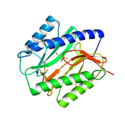
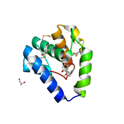
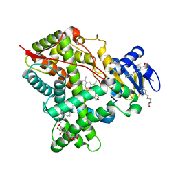
 | | Crystal structure of CPT1691 bound to TAK1-TAB1 | | 分子名称: | Mitogen-activated protein kinase kinase kinase 7,TGF-beta-activated kinase 1 and MAP3K7-binding protein 1, N-(4-((2-((4-(4-methylpiperazin-1-yl)phenyl)amino)-7H-pyrrolo[2,3-d]pyrimidin-4-yl)oxy)phenyl)acrylamide | | 著者 | Gurbani, D, Westover, K.D. | | 登録日 | 2016-04-10 | | 公開日 | 2017-02-15 | | 最終更新日 | 2023-09-27 | | 実験手法 | X-RAY DIFFRACTION (2.7515 Å) | | 主引用文献 | Structure-guided development of covalent TAK1 inhibitors.
Bioorg. Med. Chem., 25, 2017
|
|
1BY1
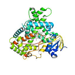
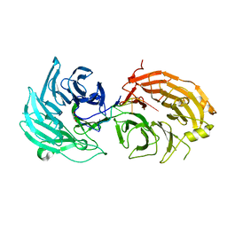
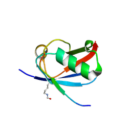
 | | DBL homology domain from beta-PIX | | 分子名称: | PROTEIN (PIX) | | 著者 | Aghazadeh, B, Zhu, K, Kubiseski, T.J, Liu, G.A, Pawson, T, Zheng, Y, Rosen, M.K. | | 登録日 | 1998-10-22 | | 公開日 | 1999-10-24 | | 最終更新日 | 2023-12-27 | | 実験手法 | SOLUTION NMR | | 主引用文献 | Structure and Mutagenesis of the Dbl Homology Domain
Nat.Struct.Biol., 5, 1998
|
|
1C23
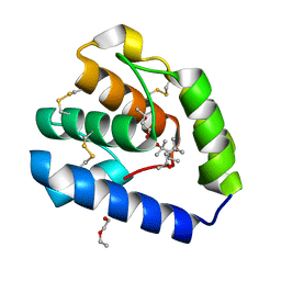
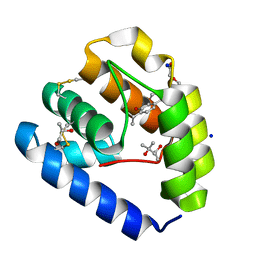
 | | E. COLI METHIONINE AMINOPEPTIDASE: METHIONINE PHOSPHONATE COMPLEX | | 分子名称: | (1-AMINO-3-METHYLSULFANYL-PROPYL)-PHOSPHONIC ACID, COBALT (II) ION, METHIONINE AMINOPEPTIDASE, ... | | 著者 | Lowther, W.T, Zhang, Y, Sampson, P.B, Honek, J.F, Matthews, B.W. | | 登録日 | 1999-07-22 | | 公開日 | 1999-11-17 | | 最終更新日 | 2024-02-07 | | 実験手法 | X-RAY DIFFRACTION (2 Å) | | 主引用文献 | Insights into the mechanism of Escherichia coli methionine aminopeptidase from the structural analysis of reaction products and phosphorus-based transition-state analogues.
Biochemistry, 38, 1999
|
|
5UDA
 | |
8BXU
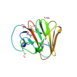
 | | Crystal structure of Odorant Binding Protein 5 from Anopheles gambiae (AgamOBP5) with MPD (2-Methyl-2,4-pentanediol) | | 分子名称: | (4S)-2-METHYL-2,4-PENTANEDIOL, 2-ETHOXYETHANOL, Odorant binding protein, ... | | 著者 | Liggri, P.G.V, Tsitsanou, K.E, Zographos, S.E. | | 登録日 | 2022-12-09 | | 公開日 | 2023-03-22 | | 最終更新日 | 2023-04-12 | | 実験手法 | X-RAY DIFFRACTION (1.35 Å) | | 主引用文献 | The structure of AgamOBP5 in complex with the natural insect repellents Carvacrol and Thymol: Crystallographic, fluorescence and thermodynamic binding studies.
Int.J.Biol.Macromol., 237, 2023
|
|
8BXV
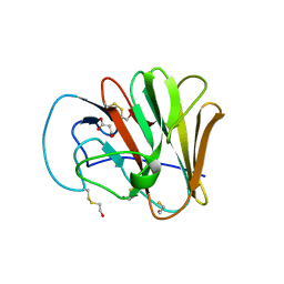
 | | Crystal structure of Odorant Binding Protein 5 from Anopheles gambiae (AgamOBP5) with Thymol | | 分子名称: | (4S)-2-METHYL-2,4-PENTANEDIOL, 5-METHYL-2-(1-METHYLETHYL)PHENOL, DI(HYDROXYETHYL)ETHER, ... | | 著者 | Liggri, P.G.V, Tsitsanou, K.E, Zographos, S.E. | | 登録日 | 2022-12-10 | | 公開日 | 2023-03-22 | | 最終更新日 | 2023-04-12 | | 実験手法 | X-RAY DIFFRACTION (1.3 Å) | | 主引用文献 | The structure of AgamOBP5 in complex with the natural insect repellents Carvacrol and Thymol: Crystallographic, fluorescence and thermodynamic binding studies.
Int.J.Biol.Macromol., 237, 2023
|
|
8BXW
 | | Crystal structure of Odorant Binding Protein 5 from Anopheles gambiae (AgamOBP5) with Carvacrol | | 分子名称: | (4S)-2-METHYL-2,4-PENTANEDIOL, 1-BUTANOL, 2-methyl-5-propan-2-yl-phenol, ... | | 著者 | Liggri, P.G.V, Tsitsanou, K.E, Zographos, S.E. | | 登録日 | 2022-12-10 | | 公開日 | 2023-03-22 | | 最終更新日 | 2023-04-12 | | 実験手法 | X-RAY DIFFRACTION (1.3 Å) | | 主引用文献 | The structure of AgamOBP5 in complex with the natural insect repellents Carvacrol and Thymol: Crystallographic, fluorescence and thermodynamic binding studies.
Int.J.Biol.Macromol., 237, 2023
|
|
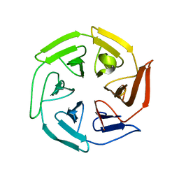
5H1L
 | | Crystal structure of WD40 repeat domains of Gemin5 in complex with 7-nt U4 snRNA fragment | | 分子名称: | GLYCEROL, Gem-associated protein 5, U4 snRNA (5'-R(*AP*UP*UP*UP*UP*UP*G)-3') | | 著者 | Jin, W, Wang, Y, Liu, C.P, Yang, N, Jin, M, Cong, Y, Wang, M, Xu, R.M. | | 登録日 | 2016-10-10 | | 公開日 | 2016-11-23 | | 最終更新日 | 2023-11-08 | | 実験手法 | X-RAY DIFFRACTION (2.1 Å) | | 主引用文献 | Structural basis for snRNA recognition by the double-WD40 repeat domain of Gemin5
Genes Dev., 30, 2016
|
|
6UMA
 | |
6UMB
 | |
5UFG
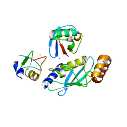
 | | Crystal Structure of CYP2B6 (Y226H/K262R/I114V) in complex with myrtenyl bromide | | 分子名称: | (1S,5R)-2-(bromomethyl)-6,6-dimethylbicyclo[3.1.1]hept-2-ene, 5-CYCLOHEXYL-1-PENTYL-BETA-D-MALTOSIDE, Cytochrome P450 2B6, ... | | 著者 | Shah, M.B, Halpert, J.R. | | 登録日 | 2017-01-04 | | 公開日 | 2017-04-12 | | 最終更新日 | 2023-10-04 | | 実験手法 | X-RAY DIFFRACTION (1.76 Å) | | 主引用文献 | Halogen-pi Interactions in the Cytochrome P450 Active Site: Structural Insights into Human CYP2B6 Substrate Selectivity.
ACS Chem. Biol., 12, 2017
|
|
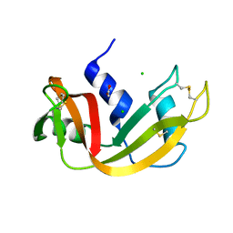
6UYO
 | | Crystal structure of K37-acetylated SUMO1 in complex with PML-SIM | | 分子名称: | Protein PML, Small ubiquitin-related modifier 1 | | 著者 | Wahba, H.M, Gagnon, C, Mascle, X.H, Lussier-Price, M, Sakaguchi, K, Omichinski, J.G. | | 登録日 | 2019-11-14 | | 公開日 | 2019-11-27 | | 最終更新日 | 2023-11-15 | | 実験手法 | X-RAY DIFFRACTION (1.639 Å) | | 主引用文献 | Acetylation of SUMO1 Alters Interactions with the SIMs of PML and Daxx in a Protein-Specific Manner.
Structure, 28, 2020
|
|
6UYX
 | | Crystal structure of K37-acetylated SUMO1 in complex with phosphorylated DAXX | | 分子名称: | Small ubiquitin-related modifier 1, phosphorylated DAXX | | 著者 | Wahba, H.M, Gagnon, C, Mascle, X.H, Lussier-Price, M, Cappadocia, L, Sakaguchi, K, Omichinski, J.G. | | 登録日 | 2019-11-14 | | 公開日 | 2019-11-27 | | 最終更新日 | 2023-11-15 | | 実験手法 | X-RAY DIFFRACTION (1.7 Å) | | 主引用文献 | Acetylation of SUMO1 Alters Interactions with the SIMs of PML and Daxx in a Protein-Specific Manner.
Structure, 28, 2020
|
|
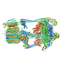
8CEN
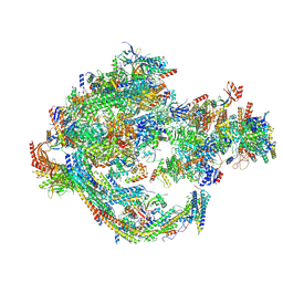 | | Yeast RNA polymerase II transcription pre-initiation complex with core Mediator | | 分子名称: | DNA-directed RNA polymerase II subunit RPB1, DNA-directed RNA polymerase II subunit RPB11, DNA-directed RNA polymerase II subunit RPB2, ... | | 著者 | Wang, H, Schilbach, S, Cramer, P. | | 登録日 | 2023-02-02 | | 公開日 | 2023-03-22 | | 最終更新日 | 2023-04-19 | | 実験手法 | ELECTRON MICROSCOPY (3 Å) | | 主引用文献 | Yeast PIC-Mediator structure with RNA polymerase II C-terminal domain.
Proc.Natl.Acad.Sci.USA, 120, 2023
|
|
8CIA
 | |
8C18
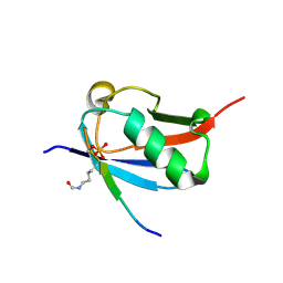
 | | Solution structure of carotenoid-binding protein AstaPo1 in complex with astaxanthin | | 分子名称: | ASTAXANTHIN, Astaxanthin binding fasciclin family protein | | 著者 | Kornilov, F.D, Savitskaya, A.G, Slonimskiy, Y.B, Goncharuk, S.A, Sluchanko, N.N, Mineev, K.S. | | 登録日 | 2022-12-20 | | 公開日 | 2023-04-05 | | 最終更新日 | 2024-06-19 | | 実験手法 | SOLUTION NMR | | 主引用文献 | Structural basis for the ligand promiscuity of the neofunctionalized, carotenoid-binding fasciclin domain protein AstaP.
Commun Biol, 6, 2023
|
|
1C8W
 | | THR45GLY VARIANT OF RIBONUCLEASE A | | 分子名称: | ACETATE ION, CHLORIDE ION, PROTEIN (Ribonuclease A) | | 著者 | Kelemen, B.R, Sweeney, R.T, Schultz, L.W, Raines, R.T. | | 登録日 | 1999-07-30 | | 公開日 | 2002-05-01 | | 最終更新日 | 2018-03-14 | | 実験手法 | X-RAY DIFFRACTION (1.8 Å) | | 主引用文献 | Excavating an active site: the nucleobase specificity of ribonuclease A.
Biochemistry, 39, 2000
|
|
6VQ6
 | |
6W9D
 | |
8C0C
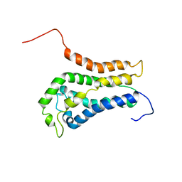
 | | X-ray crystal structure of PPAR gamma ligand binding domain in complex with CZ46 | | 分子名称: | (2~{R})-2-[4-(naphthalen-1-ylmethoxy)phenyl]-4-oxidanyl-3-phenyl-2~{H}-furan-5-one, Peroxisome proliferator-activated receptor gamma | | 著者 | Capelli, D, Montanari, R, Pochetti, G, Villa, S, Meneghetti, F. | | 登録日 | 2022-12-16 | | 公開日 | 2023-04-26 | | 最終更新日 | 2024-06-19 | | 実験手法 | X-RAY DIFFRACTION (2.2 Å) | | 主引用文献 | Biological Screening and Crystallographic Studies of Hydroxy gamma-Lactone Derivatives to Investigate PPAR gamma Phosphorylation Inhibition.
Biomolecules, 13, 2023
|
|
6TTH
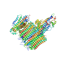
 | | PKM2 in complex with L-threonine | | 分子名称: | 1,6-di-O-phosphono-beta-D-fructofuranose, Pyruvate kinase PKM, THREONINE | | 著者 | Saur, M, Hartshorn, M.J, Dong, J, Reeks, J, Bunkoczi, G, Jhoti, H, Williams, P.A. | | 登録日 | 2019-12-27 | | 公開日 | 2020-01-15 | | 最終更新日 | 2024-05-22 | | 実験手法 | ELECTRON MICROSCOPY (2.6 Å) | | 主引用文献 | Fragment-based drug discovery using cryo-EM.
Drug Discov Today, 25, 2020
|
|
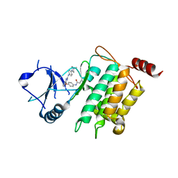
8BLL
 | | Structure of RutB | | 分子名称: | ACETATE ION, Ureidoacrylate amidohydrolase RutB | | 著者 | Rajendran, C. | | 登録日 | 2022-11-09 | | 公開日 | 2023-01-25 | | 最終更新日 | 2023-11-15 | | 実験手法 | X-RAY DIFFRACTION (1.54 Å) | | 主引用文献 | Structural and Functional Characterization of the Ureidoacrylate Amidohydrolase RutB from Escherichia coli .
Biochemistry, 62, 2023
|
|
6TTQ
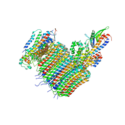
 | | PKM2 in complex with Compound 10 | | 分子名称: | 1,6-di-O-phosphono-beta-D-fructofuranose, 1-propan-2-yl-3-pyridin-4-yl-urea, Pyruvate kinase PKM | | 著者 | Saur, M, Hartshorn, M.J, Dong, J, Reeks, J, Bunkoczi, G, Jhoti, H, Williams, P.A. | | 登録日 | 2019-12-30 | | 公開日 | 2020-01-15 | | 最終更新日 | 2024-05-22 | | 実験手法 | ELECTRON MICROSCOPY (2.7 Å) | | 主引用文献 | Fragment-based drug discovery using cryo-EM.
Drug Discov Today, 25, 2020
|
|


