3VML
 
 | |
2Q59
 | |
3VSQ
 | |
2Q2C
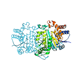
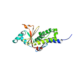
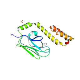
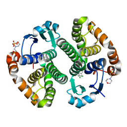
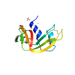
 | | Crystal structures of the arginine-, lysine-, histidine-binding protein ArtJ from the thermophilic bacterium Geobacillus stearothermophilus | | 分子名称: | ArtJ, GLYCEROL, HISTIDINE, ... | | 著者 | Vahedi-Faridi, A, Scheffel, F, Eckey, V, Saenger, W, Schneider, E. | | 登録日 | 2007-05-28 | | 公開日 | 2008-01-15 | | 最終更新日 | 2024-02-21 | | 実験手法 | X-RAY DIFFRACTION (2.35 Å) | | 主引用文献 | Crystal structures and mutational analysis of the arginine-, lysine-, histidine-binding protein ArtJ from Geobacillus stearothermophilus. Implications for interactions of ArtJ with its cognate ATP-binding cassette transporter, Art(MP)2
J.Mol.Biol., 375, 2008
|
|
3VNE
 | |
3DH5
 | | Crystal structure of bovine pancreatic ribonuclease A (wild-type) | | 分子名称: | CHLORIDE ION, Ribonuclease pancreatic, SULFATE ION | | 著者 | Kurpiewska, K, Font, J, Ribo, M, Vilanova, M, Lewinski, K. | | 登録日 | 2008-06-17 | | 公開日 | 2008-07-15 | | 最終更新日 | 2023-11-01 | | 実験手法 | X-RAY DIFFRACTION (1.6 Å) | | 主引用文献 | X-ray crystallographic studies of RNase A variants engineered at the most destabilizing positions of the main hydrophobic core: further insight into protein stability
Proteins, 77, 2009
|
|
3DHD
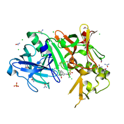
 | | Crystal structure of human NAMPT complexed with nicotinamide mononucleotide and pyrophosphate | | 分子名称: | BETA-NICOTINAMIDE RIBOSE MONOPHOSPHATE, MAGNESIUM ION, Nicotinamide phosphoribosyltransferase, ... | | 著者 | Ho, M, Burgos, E.S, Almo, S.C, Schramm, V.L. | | 登録日 | 2008-06-17 | | 公開日 | 2009-08-18 | | 最終更新日 | 2023-08-30 | | 実験手法 | X-RAY DIFFRACTION (2 Å) | | 主引用文献 | A phosphoenzyme mimic, overlapping catalytic sites and reaction coordinate motion for human NAMPT.
Proc.Natl.Acad.Sci.USA, 106, 2009
|
|
3TPP
 | | Crystal structure of BACE1 complexed with an inhibitor | | 分子名称: | Beta-secretase 1, CHLORIDE ION, N-[(1S,2R)-1-BENZYL-3-(CYCLOPROPYLAMINO)-2-HYDROXYPROPYL]-5-[METHYL(METHYLSULFONYL)AMINO]-N'-[(1R)-1-PHENYLETHYL]ISOPHTHALAMIDE, ... | | 著者 | Xu, Y.C, Li, M.J, Greenblatt, H, Chen, T.T, Silman, I, Sussman, J.L. | | 登録日 | 2011-09-08 | | 公開日 | 2011-11-23 | | 最終更新日 | 2023-11-01 | | 実験手法 | X-RAY DIFFRACTION (1.6 Å) | | 主引用文献 | Flexibility of the flap in the active site of BACE1 as revealed by crystal structures and molecular dynamics simulations
Acta Crystallogr.,Sect.D, 68, 2012
|
|
3DPQ
 | |
2Q62
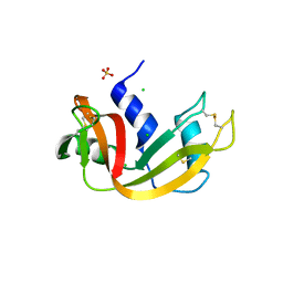
 | | Crystal Structure of ArsH from Sinorhizobium meliloti | | 分子名称: | SULFATE ION, arsH | | 著者 | Ye, J, Yang, H, Bhattacharjee, H, Rosen, B.P. | | 登録日 | 2007-06-04 | | 公開日 | 2007-12-11 | | 最終更新日 | 2024-02-21 | | 実験手法 | X-RAY DIFFRACTION (1.8 Å) | | 主引用文献 | Crystal structure of the flavoprotein ArsH from Sinorhizobium meliloti
FEBS Lett., 581, 2007
|
|
3DIP
 | | Crystal structure of an enolase protein from the environmental genome shotgun sequencing of the Sargasso Sea | | 分子名称: | SULFATE ION, enolase | | 著者 | Bonanno, J.B, Freeman, J, Bain, K.T, Zhang, F, Ozyurt, S, Smith, D, Wasserman, S, Sauder, J.M, Burley, S.K, Almo, S.C, New York SGX Research Center for Structural Genomics (NYSGXRC) | | 登録日 | 2008-06-20 | | 公開日 | 2008-07-29 | | 最終更新日 | 2023-11-15 | | 実験手法 | X-RAY DIFFRACTION (2.5 Å) | | 主引用文献 | Crystal structure of an enolase protein from the environmental genome shotgun sequencing of the Sargasso Sea
To be Published
|
|
3DK6
 | |
2PXT
 | | Variant 15 of Ribonucleoprotein Core of the E. Coli Signal Recognition Particle | | 分子名称: | 4.5 S RNA, COBALT HEXAMMINE(III), Signal recognition particle protein | | 著者 | Keel, A.Y, Rambo, R.P, Batey, R.T, Kieft, J.S. | | 登録日 | 2007-05-14 | | 公開日 | 2007-08-07 | | 最終更新日 | 2021-10-20 | | 実験手法 | X-RAY DIFFRACTION (2.5 Å) | | 主引用文献 | A General Strategy to Solve the Phase Problem in RNA Crystallography.
Structure, 15, 2007
|
|
3VUY
 | | Crystal structure of A20 ZF7 in complex with linear tetraubiquitin | | 分子名称: | POTASSIUM ION, Polyubiquitin-C, Tumor necrosis factor alpha-induced protein 3, ... | | 著者 | Nishimasu, H, Ishitani, R, Nureki, O. | | 登録日 | 2012-07-09 | | 公開日 | 2013-02-13 | | 最終更新日 | 2024-03-20 | | 実験手法 | X-RAY DIFFRACTION (1.981 Å) | | 主引用文献 | Specific recognition of linear polyubiquitin by A20 zinc finger 7 is involved in NF-kappaB regulation
Embo J., 31, 2012
|
|
3DGQ
 | |
3VWL
 | | Crystal structure of 6-aminohexanoate-dimer hydrolase G181D/R187S/H266N/D370Y mutant | | 分子名称: | 2-(N-MORPHOLINO)-ETHANESULFONIC ACID, 6-aminohexanoate-dimer hydrolase, GLYCEROL, ... | | 著者 | Kawashima, Y, Shibata, N, Negoro, S, Higuchi, Y. | | 登録日 | 2012-08-30 | | 公開日 | 2013-10-16 | | 最終更新日 | 2024-03-20 | | 実験手法 | X-RAY DIFFRACTION (1.6 Å) | | 主引用文献 | Structural, kinetic and theoretical analyses of hydrolase mutants altering in the directionality and equilibrium point of reversible amide-synthetic/hydrolytic reaction
To be Published
|
|
2PUK
 | | Crystal structure of the binary complex between ferredoxin: thioredoxin reductase and thioredoxin m | | 分子名称: | Ferredoxin-thioredoxin reductase, catalytic chain, variable chain, ... | | 著者 | Dai, S, Friemann, R, Schurmann, P, Eklund, H. | | 登録日 | 2007-05-09 | | 公開日 | 2007-07-10 | | 最終更新日 | 2021-10-20 | | 実験手法 | X-RAY DIFFRACTION (3 Å) | | 主引用文献 | Structural snapshots along the reaction pathway of ferredoxin-thioredoxin reductase.
Nature, 448, 2007
|
|
3TTW
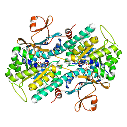
 | | Structure of the F413E variant of E. coli KatE | | 分子名称: | Catalase HPII, PROTOPORPHYRIN IX CONTAINING FE | | 著者 | Loewen, P.C, Jha, V. | | 登録日 | 2011-09-15 | | 公開日 | 2011-10-12 | | 最終更新日 | 2023-12-06 | | 実験手法 | X-RAY DIFFRACTION (1.62 Å) | | 主引用文献 | Mutation of Phe413 to Tyr in catalase KatE from Escherichia coli leads to side chain damage and main chain cleavage.
Arch.Biochem.Biophys., 525, 2012
|
|
3VAE
 | | Crystal Structure of M. tuberculosis LD-transpeptidase type 2 with Modified Catalytic Cysteine (C354) | | 分子名称: | DI(HYDROXYETHYL)ETHER, LD-transpeptidase type 2 | | 著者 | Erdemli, S, Bianchet, M.A, Gupta, R, Lamichhane, G, Amzel, L.M. | | 登録日 | 2011-12-29 | | 公開日 | 2012-12-12 | | 最終更新日 | 2018-04-04 | | 実験手法 | X-RAY DIFFRACTION (2.8 Å) | | 主引用文献 | Targeting the Cell Wall of Mycobacterium tuberculosis: Structure and Mechanism of L,D-Transpeptidase 2.
Structure, 20, 2012
|
|
3DI7
 | | Crystal structure of bovine pancreatic ribonuclease A variant (V54A) | | 分子名称: | CHLORIDE ION, Ribonuclease pancreatic, SULFATE ION | | 著者 | Kurpiewska, K, Font, J, Ribo, M, Vilanova, M, Lewinski, K. | | 登録日 | 2008-06-20 | | 公開日 | 2008-07-15 | | 最終更新日 | 2023-11-01 | | 実験手法 | X-RAY DIFFRACTION (1.6 Å) | | 主引用文献 | X-ray crystallographic studies of RNase A variants engineered at the most destabilizing positions of the main hydrophobic core: further insight into protein stability
Proteins, 77, 2009
|
|
3VAL
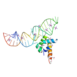
 | | Structure of U2AF65 variant with BrU5C1 DNA | | 分子名称: | 1,4-DIETHYLENE DIOXIDE, DNA (5'-D(*C*UP*UP*UP*(BRU)P*UP*U)-3'), SULFATE ION, ... | | 著者 | Jenkins, J.L, Frato, K.H, Kielkopf, C.L. | | 登録日 | 2011-12-29 | | 公開日 | 2013-02-13 | | 最終更新日 | 2023-09-13 | | 実験手法 | X-RAY DIFFRACTION (2.5 Å) | | 主引用文献 | U2AF65 adapts to diverse pre-mRNA splice sites through conformational selection of specific and promiscuous RNA recognition motifs.
Nucleic Acids Res., 41, 2013
|
|
2Q5E
 | | Crystal structure of human carboxy-terminal domain RNA polymerase II polypeptide A small phosphatase 2 | | 分子名称: | Carboxy-terminal domain RNA polymerase II polypeptide A small phosphatase 2, MAGNESIUM ION | | 著者 | Bonanno, J.B, Dickey, M, Bain, K.T, Lau, C, Romero, R, Smith, D, Wasserman, S, Sauder, J.M, Burley, S.K, Almo, S.C, New York SGX Research Center for Structural Genomics (NYSGXRC) | | 登録日 | 2007-05-31 | | 公開日 | 2007-06-19 | | 最終更新日 | 2024-02-21 | | 実験手法 | X-RAY DIFFRACTION (2.51 Å) | | 主引用文献 | Structural genomics of protein phosphatases.
J.Struct.Funct.Genom., 8, 2007
|
|
3V9I
 | |
2Q5W
 | |
3DKT
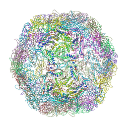 | | Crystal structure of Thermotoga maritima encapsulin | | 分子名称: | Maritimacin, Putative uncharacterized protein | | 著者 | Sutter, M, Boehringer, D, Gutmann, S, Weber-Ban, E, Ban, N. | | 登録日 | 2008-06-26 | | 公開日 | 2008-09-02 | | 最終更新日 | 2024-03-20 | | 実験手法 | X-RAY DIFFRACTION (3.104 Å) | | 主引用文献 | Structural basis of enzyme encapsulation into a bacterial nanocompartment
Nat.Struct.Mol.Biol., 15, 2008
|
|


