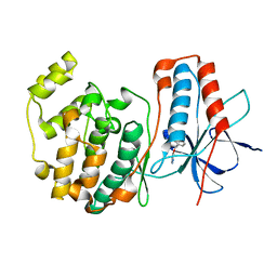
3BSW
 
 | |
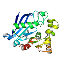
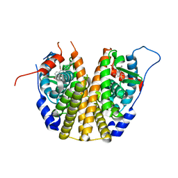
3BSS
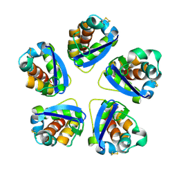
 | | PglD from Campylobacter jejuni, NCTC 11168, with native substrate | | 分子名称: | Acetyltransferase, UDP-2-acetamido-4-amino-2,4,6-trideoxy-alpha-D-glucopyranose | | 著者 | Olivier, N.B, Imperiali, B. | | 登録日 | 2007-12-26 | | 公開日 | 2008-07-22 | | 最終更新日 | 2023-08-30 | | 実験手法 | X-RAY DIFFRACTION (2.3 Å) | | 主引用文献 | Crystal structure and catalytic mechanism of PglD from Campylobacter jejuni.
J.Biol.Chem., 283, 2008
|
|
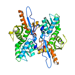
3CDZ
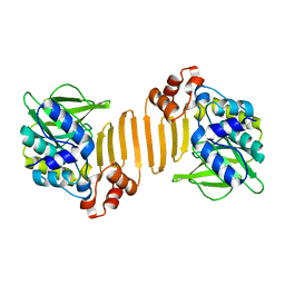
 | | Crystal structure of human factor VIII | | 分子名称: | 2-acetamido-2-deoxy-beta-D-glucopyranose, 2-acetamido-2-deoxy-beta-D-glucopyranose-(1-4)-2-acetamido-2-deoxy-beta-D-glucopyranose, CALCIUM ION, ... | | 著者 | Ngo, J.C, Huang, M, Roth, D.A, Furie, B.C, Furie, B. | | 登録日 | 2008-02-27 | | 公開日 | 2008-04-01 | | 最終更新日 | 2023-08-30 | | 実験手法 | X-RAY DIFFRACTION (3.98 Å) | | 主引用文献 | Crystal structure of human factor VIII: implications for the formation of the factor IXa-factor VIIIa complex.
Structure, 16, 2008
|
|
3SJM
 | |
3TB2
 | | 1-Cys peroxidoxin from Plasmodium Yoelli | | 分子名称: | 1,2-ETHANEDIOL, 1-Cys peroxiredoxin, GLYCEROL, ... | | 著者 | Qiu, W, Artz, J.D, Vedadi, M, Sharma, S, Houston, S, Lew, J, Wasney, G, Amani, M, Xu, X, Bray, J, Sundstrom, M, Arrowsmith, C, Edwards, A, Hui, R, Bochkarev, A, Structural Genomics Consortium (SGC) | | 登録日 | 2011-08-04 | | 公開日 | 2011-10-19 | | 最終更新日 | 2018-01-31 | | 実験手法 | X-RAY DIFFRACTION (2.3 Å) | | 主引用文献 | Genome-scale protein expression and structural biology of Plasmodium falciparum and related Apicomplexan organisms.
Mol.Biochem.Parasitol., 151, 2007
|
|
3S8Y
 | | Bromide soaked structure of an esterase from the oil-degrading bacterium Oleispira antarctica | | 分子名称: | BROMIDE ION, Esterase APC40077 | | 著者 | Petit, P, Dong, A, Kagan, O, Savchenko, A, Yakunin, A.F. | | 登録日 | 2011-05-31 | | 公開日 | 2011-06-15 | | 最終更新日 | 2023-09-13 | | 実験手法 | X-RAY DIFFRACTION (2.1 Å) | | 主引用文献 | Structure and activity of the cold-active and anion-activated carboxyl esterase OLEI01171 from the oil-degrading marine bacterium Oleispira antarctica.
Biochem.J., 445, 2012
|
|
3V62
 | | Structure of the S. cerevisiae Srs2 C-terminal domain in complex with PCNA conjugated to SUMO on lysine 164 | | 分子名称: | ATP-dependent DNA helicase SRS2, N-ETHYLMALEIMIDE, Proliferating cell nuclear antigen, ... | | 著者 | Armstrong, A.A, Mohideen, F, Lima, C.D. | | 登録日 | 2011-12-18 | | 公開日 | 2012-02-29 | | 最終更新日 | 2023-09-13 | | 実験手法 | X-RAY DIFFRACTION (2.9 Å) | | 主引用文献 | Recognition of SUMO-modified PCNA requires tandem receptor motifs in Srs2.
Nature, 483, 2012
|
|
3BSY
 | |
3CLD
 | | Ligand binding domain of the glucocorticoid receptor complexed with fluticazone furoate | | 分子名称: | (6alpha,11alpha,14beta,16alpha,17alpha)-6,9-difluoro-17-{[(fluoromethyl)sulfanyl]carbonyl}-11-hydroxy-16-methyl-3-oxoan drosta-1,4-dien-17-yl furan-2-carboxylate, Glucocorticoid receptor, Tif2 coactivator motif | | 著者 | Shewchuk, L.M, McLay, I, Stewart, E, Biggadike, K.B, Hassell, A.M, Bledsoe, R.K. | | 登録日 | 2008-03-18 | | 公開日 | 2008-06-24 | | 最終更新日 | 2023-08-30 | | 実験手法 | X-RAY DIFFRACTION (2.84 Å) | | 主引用文献 | X-ray crystal structure of the novel enhanced-affinity glucocorticoid agonist fluticasone furoate in the glucocorticoid receptor-ligand binding domain.
J.Med.Chem., 51, 2008
|
|
3DT3
 | |
3DKI
 | |
3DRY
 | |
3DRX
 | |
3DRZ
 | |
1W5R
 | | X-ray crystallographic structure of a C70Q Mycobacterium smegmatis N- arylamine Acetyltransferase | | 分子名称: | ARYLAMINE N-ACETYLTRANSFERASE | | 著者 | Holton, S.J, Sandy, J, Rodrigues-Lima, F, Dupret, J.-M, Bhakta, S, Noble, M.E.M, Sim, E. | | 登録日 | 2004-08-09 | | 公開日 | 2005-05-11 | | 最終更新日 | 2024-05-08 | | 実験手法 | X-RAY DIFFRACTION (1.45 Å) | | 主引用文献 | Investigation of the Catalytic Triad of Arylamine N-Acetyltransferases: Essential Residues Required for Acetyl Transfer to Arylamines.
Biochem.J., 390, 2005
|
|
1WBU
 | | Fragment based lead discovery using crystallography | | 分子名称: | 5-AMINO-1H-PYRIMIDINE-2,4-DIONE, RIBONUCLEASE | | 著者 | Cleasby, A, Hartshorn, M.J, Murray, C.W, Jhoti, H, Tickle, I.J. | | 登録日 | 2004-11-05 | | 公開日 | 2005-01-27 | | 最終更新日 | 2011-07-13 | | 実験手法 | X-RAY DIFFRACTION (1.9 Å) | | 主引用文献 | Fragment-Based Lead Discovery Using X-Ray Crystallography
J.Med.Chem., 48, 2005
|
|
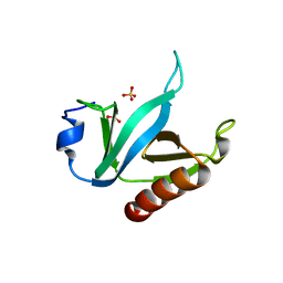
1WAX
 | | Protein tyrosine phosphatase 1B with active site inhibitor | | 分子名称: | MAGNESIUM ION, PROTEIN-TYROSINE PHOSPHATASE, [[4-(AMINOMETHYL)PHENYL]AMINO]OXO-ACETIC ACID, | | 著者 | Hartshorn, M.J, Murray, C.W, Cleasby, A, Frederickson, M, Tickle, I.J, Jhoti, H. | | 登録日 | 2004-10-28 | | 公開日 | 2005-01-27 | | 最終更新日 | 2023-12-13 | | 実験手法 | X-RAY DIFFRACTION (2.2 Å) | | 主引用文献 | Fragment-Based Lead Discovery Using X-Ray Crystallography
J.Med.Chem., 48, 2005
|
|
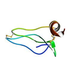
1W7H
 | | p38 Kinase crystal structure in complex with small molecule inhibitor | | 分子名称: | 3-(BENZYLOXY)PYRIDIN-2-AMINE, MITOGEN-ACTIVATED PROTEIN KINASE 14 | | 著者 | Jhoti, H, Gill, A, Cleasby, A, Devine, L. | | 登録日 | 2004-09-02 | | 公開日 | 2005-02-08 | | 最終更新日 | 2023-12-13 | | 実験手法 | X-RAY DIFFRACTION (2.214 Å) | | 主引用文献 | Fragment-Based Lead Discovery Using X-Ray Crystallography
J.Med.Chem., 48, 2005
|
|
1WCC
 | | Screening for fragment binding by X-ray crystallography | | 分子名称: | 2-AMINO-6-CHLOROPYRAZINE, CELL DIVISION PROTEIN KINASE 2 | | 著者 | Cleasby, A, O'Reilly, M, Hartshorn, M.J, Murray, C.W, Tickle, I.J, Jhoti, H, Frederickson, M. | | 登録日 | 2004-11-12 | | 公開日 | 2005-01-27 | | 最終更新日 | 2024-05-08 | | 実験手法 | X-RAY DIFFRACTION (2.2 Å) | | 主引用文献 | Fragment-Based Lead Discovery Using X-Ray Crystallography
J.Med.Chem., 48, 2005
|
|
1WBO
 | | fragment based p38 inhibitors | | 分子名称: | 2-CHLOROPHENOL, MITOGEN-ACTIVATED PROTEIN KINASE 14 | | 著者 | Cleasby, A, Devine, L, Gill, A, Jhoti, H. | | 登録日 | 2004-11-04 | | 公開日 | 2005-01-27 | | 最終更新日 | 2024-05-08 | | 実験手法 | X-RAY DIFFRACTION (2.16 Å) | | 主引用文献 | Fragment-Based Lead Discovery Using X-Ray Crystallography
J.Med.Chem., 48, 2005
|
|
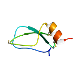
1VHH
 | | A POTENTIAL CATALYTIC SITE WITHIN THE AMINO-TERMINAL SIGNALLING DOMAIN OF SONIC HEDGEHOG | | 分子名称: | SONIC HEDGEHOG, SULFATE ION, ZINC ION | | 著者 | Hall, T.M.T, Porter, J.A, Beachy, P.A, Leahy, D.J. | | 登録日 | 1995-10-03 | | 公開日 | 1996-01-29 | | 最終更新日 | 2024-02-14 | | 実験手法 | X-RAY DIFFRACTION (1.7 Å) | | 主引用文献 | A potential catalytic site revealed by the 1.7-A crystal structure of the amino-terminal signalling domain of Sonic hedgehog.
Nature, 378, 1995
|
|
1TMR
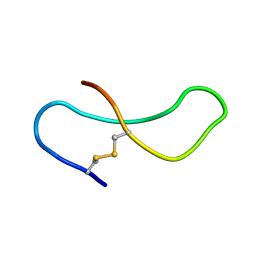 | | THE STRUCTURE OF A 19 RESIDUE FRAGMENT FROM THE C-LOOP OF THE FOURTH EPIDERMAL GROWTH FACTOR-LIKE DOMAIN OF THROMBOMODULIN | | 分子名称: | THROMBOMODULIN PRECURSOR | | 著者 | Adler, M, Seto, M, Nitecki, D, Light, D.R, Morser, J. | | 登録日 | 1994-05-20 | | 公開日 | 1995-06-08 | | 最終更新日 | 2022-03-02 | | 実験手法 | SOLUTION NMR | | 主引用文献 | The structure of a 19-residue fragment from the C-loop of the fourth epidermal growth factor-like domain of thrombomodulin.
J.Biol.Chem., 270, 1995
|
|
1UUA
 | |
1U5F
 | |
1UUB
 | |


