3KYV
 
 | |
8ATG
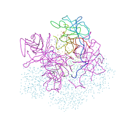
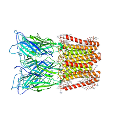 | | Pentameric ligand-gated ion channel GLIC with bound lipids | | 分子名称: | (2S)-3-(hexadecanoyloxy)-2-[(9Z)-octadec-9-enoyloxy]propyl 2-(trimethylammonio)ethyl phosphate, Proton-gated ion channel | | 著者 | Bergh, C, Rovsnik, U, Howard, R.J, Lindahl, E. | | 登録日 | 2022-08-23 | | 公開日 | 2023-09-06 | | 最終更新日 | 2024-03-20 | | 実験手法 | ELECTRON MICROSCOPY (2.9 Å) | | 主引用文献 | Discovery of lipid binding sites in a ligand-gated ion channel by integrating simulations and cryo-EM.
Elife, 12, 2024
|
|
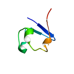
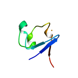
3KQG
 | | Trimeric Structure of Langerin | | 分子名称: | C-type lectin domain family 4 member K, CALCIUM ION | | 著者 | Feinberg, H, Powlesland, A.S, Taylor, M.E, Weis, W.I. | | 登録日 | 2009-11-17 | | 公開日 | 2010-02-23 | | 最終更新日 | 2024-04-03 | | 実験手法 | X-RAY DIFFRACTION (2.3 Å) | | 主引用文献 | Trimeric structure of langerin.
J.Biol.Chem., 285, 2010
|
|
5KGG
 | |
3KYW
 | |
3KYX
 | |
5KGK
 | |
2B83
 | |
3KYY
 | |
2B99
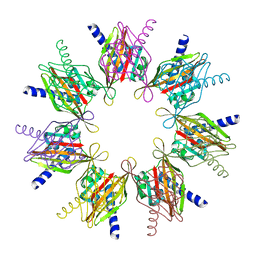
 | | Crystal Structure of an archaeal pentameric riboflavin synthase Complex with a Substrate analog inhibitor | | 分子名称: | 6,7-DIOXO-5H-8-RIBITYLAMINOLUMAZINE, Riboflavin synthase | | 著者 | Ramsperger, A, Augustin, M, Schott, A.K, Gerhardt, S, Krojer, T, Eisenreich, W, Illarionov, B, Cushman, M, Bacher, A, Huber, R, Fischer, M. | | 登録日 | 2005-10-11 | | 公開日 | 2005-11-08 | | 最終更新日 | 2024-02-14 | | 実験手法 | X-RAY DIFFRACTION (2.22 Å) | | 主引用文献 | Crystal Structure of an Archaeal Pentameric Riboflavin Synthase in Complex with a Substrate Analog Inhibitor: stereochemical implications
J.Biol.Chem., 281, 2006
|
|
2BB5
 | | Structure of Human Transcobalamin in complex with Cobalamin | | 分子名称: | COBALAMIN, Transcobalamin II | | 著者 | Wuerges, J, Garau, G, Geremia, S, Fedosov, S.N, Petersen, T.E, Randaccio, L. | | 登録日 | 2005-10-17 | | 公開日 | 2006-04-04 | | 最終更新日 | 2023-08-23 | | 実験手法 | X-RAY DIFFRACTION (3.2 Å) | | 主引用文献 | Structural basis for mammalian vitamin B12 transport by transcobalamin.
Proc.Natl.Acad.Sci.Usa, 103, 2006
|
|
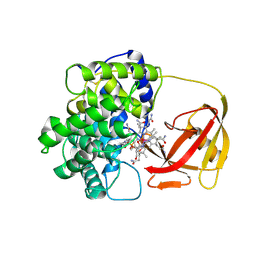
4PUQ
 | | Mus Musculus Tdp2 reaction product complex with 5'-phosphorylated RNA/DNA, glycerol, and Mg2+ | | 分子名称: | ACETATE ION, DNA/RNA hybrid, GLYCEROL, ... | | 著者 | Schellenberg, M.J, Williams, R.S. | | 登録日 | 2014-03-13 | | 公開日 | 2014-05-14 | | 最終更新日 | 2023-09-20 | | 実験手法 | X-RAY DIFFRACTION (1.6 Å) | | 主引用文献 | Proteolytic Degradation of Topoisomerase II (Top2) Enables the Processing of Top2DNA and Top2RNA Covalent Complexes by Tyrosyl-DNA-Phosphodiesterase 2 (TDP2).
J.Biol.Chem., 289, 2014
|
|
2F86
 | |
2XWR
 | |
2F9D
 | | 2.5 angstrom resolution structure of the spliceosomal protein p14 bound to region of SF3b155 | | 分子名称: | Pre-mRNA branch site protein p14, Splicing factor 3B subunit 1 | | 著者 | Schellenberg, M.J, Edwards, R.A, Ritchie, D.B, Glover, J.N.M, Macmillan, A.M. | | 登録日 | 2005-12-05 | | 公開日 | 2006-01-24 | | 最終更新日 | 2017-10-18 | | 実験手法 | X-RAY DIFFRACTION (2.5 Å) | | 主引用文献 | Crystal structure of a core spliceosomal protein interface
Proc.Natl.Acad.Sci.Usa, 103, 2006
|
|
2F4E
 | |
1M6Z
 | | Crystal structure of reduced recombinant cytochrome c4 from Pseudomonas stutzeri | | 分子名称: | 2-AMINO-2-HYDROXYMETHYL-PROPANE-1,3-DIOL, Cytochrome c4, GLYCEROL, ... | | 著者 | Noergaard, A, Harris, P, Larsen, S, Christensen, H.E.M. | | 登録日 | 2002-07-18 | | 公開日 | 2003-09-16 | | 最終更新日 | 2023-10-25 | | 実験手法 | X-RAY DIFFRACTION (1.35 Å) | | 主引用文献 | Structural comparison of recombinant Pseudomonas stutzeri cytochrome c4 in two oxidation states
To be Published
|
|
2F9J
 | |
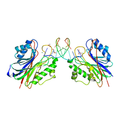
1A54
 | | PHOSPHATE-BINDING PROTEIN MUTANT A197C LABELLED WITH A COUMARIN FLUOROPHORE AND BOUND TO DIHYDROGENPHOSPHATE ION | | 分子名称: | DIHYDROGENPHOSPHATE ION, N-[2-(1-MALEIMIDYL)ETHYL]-7-DIETHYLAMINOCOUMARIN-3-CARBOXAMIDE, Phosphate-binding protein PstS | | 著者 | Hirshberg, M, Henrick, K, Lloyd-Haire, L, Vasisht, N, Brune, M, Corrie, J.E.T, Webb, M.R. | | 登録日 | 1998-02-19 | | 公開日 | 1998-10-14 | | 最終更新日 | 2023-08-02 | | 実験手法 | X-RAY DIFFRACTION (1.6 Å) | | 主引用文献 | Crystal structure of phosphate binding protein labeled with a coumarin fluorophore, a probe for inorganic phosphate.
Biochemistry, 37, 1998
|
|
8OQ3
 | |
1A55
 | | PHOSPHATE-BINDING PROTEIN MUTANT A197C | | 分子名称: | DIHYDROGENPHOSPHATE ION, PHOSPHATE-BINDING PROTEIN | | 著者 | Hirshberg, M, Henrick, K, Lloyd-Haire, L, Vasisht, N, Brune, M, Corrie, J.E.T, Webb, M.R. | | 登録日 | 1998-02-19 | | 公開日 | 1998-10-14 | | 最終更新日 | 2024-05-22 | | 実験手法 | X-RAY DIFFRACTION (2.4 Å) | | 主引用文献 | Crystal structure of phosphate binding protein labeled with a coumarin fluorophore, a probe for inorganic phosphate.
Biochemistry, 37, 1998
|
|
1B4K
 | | High resolution crystal structure of a MG2-dependent 5-aminolevulinic acid dehydratase | | 分子名称: | LAEVULINIC ACID, MAGNESIUM ION, PROTEIN (5-AMINOLEVULINIC ACID DEHYDRATASE), ... | | 著者 | Frankenberg, N, Jahn, D, Heinz, D.W. | | 登録日 | 1998-12-22 | | 公開日 | 1999-07-13 | | 最終更新日 | 2023-08-09 | | 実験手法 | X-RAY DIFFRACTION (1.67 Å) | | 主引用文献 | High resolution crystal structure of a Mg2+-dependent porphobilinogen synthase.
J.Mol.Biol., 289, 1999
|
|
1P6X
 | | Crystal structure of EHV4-TK complexed with Thy and SO4 | | 分子名称: | SULFATE ION, THYMIDINE, Thymidine kinase | | 著者 | Gardberg, A, Shuvalova, L, Monnerjahn, C, Konrad, M, Lavie, A. | | 登録日 | 2003-04-30 | | 公開日 | 2003-11-04 | | 最終更新日 | 2023-08-16 | | 実験手法 | X-RAY DIFFRACTION (2 Å) | | 主引用文献 | Structural basis for the dual thymidine and thymidylate kinase activity of herpes thymidine kinases.
Structure, 11, 2003
|
|
1BIT
 | |
1BGW
 | | TOPOISOMERASE RESIDUES 410-1202, | | 分子名称: | TOPOISOMERASE | | 著者 | Berger, J.M, Gamblin, S.J, Harrison, S.C, Wang, J.C. | | 登録日 | 1996-02-20 | | 公開日 | 1996-07-11 | | 最終更新日 | 2024-02-07 | | 実験手法 | X-RAY DIFFRACTION (2.7 Å) | | 主引用文献 | Structure and mechanism of DNA topoisomerase II.
Nature, 379, 1996
|
|


