4GNT
 
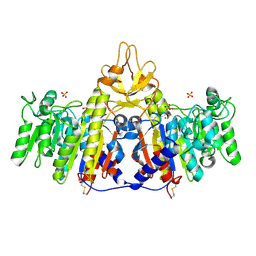
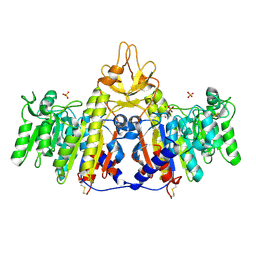
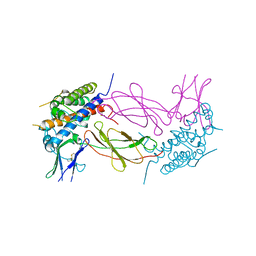
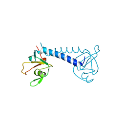
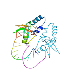
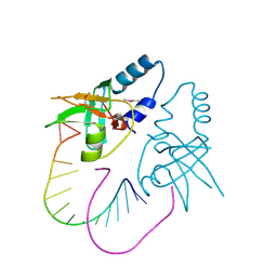
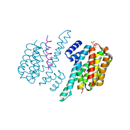 | | Complex of ChREBP and 14-3-3beta | | 分子名称: | 14-3-3 protein beta/alpha, Carbohydrate-responsive element-binding protein, SULFATE ION | | 著者 | Zhang, H, Huang, N. | | 登録日 | 2012-08-17 | | 公開日 | 2012-10-24 | | 最終更新日 | 2024-02-28 | | 実験手法 | X-RAY DIFFRACTION (2.41 Å) | | 主引用文献 | Crystal Structure of Carbohydrate Response Element Binding Protein (ChREBP) in complex with 14-3-3b
To be Published
|
|
5GGE
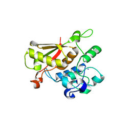
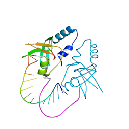
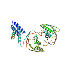
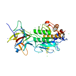
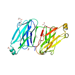
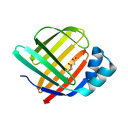 | | Fatty Acid-Binding Protein in Brain Tissue of Drosophila melanogaster | | 分子名称: | CITRIC ACID, Fatty acid bindin protein, isoform B | | 著者 | Cheng, Y.-Y, Huang, Y.-F, Lin, H.-H, Chang, W.W, Lyu, P.-C. | | 登録日 | 2016-06-15 | | 公開日 | 2017-06-21 | | 最終更新日 | 2023-11-08 | | 実験手法 | X-RAY DIFFRACTION (1.861 Å) | | 主引用文献 | The ligand-mediated affinity of brain-type fatty acid-binding protein for membranes determines the directionality of lipophilic cargo transport.
Biochim Biophys Acta Mol Cell Biol Lipids, 1864, 2019
|
|
1HGU
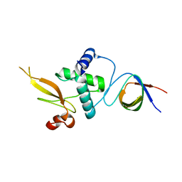
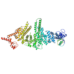
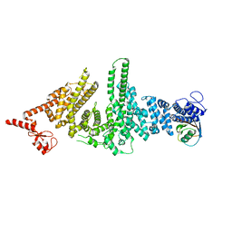
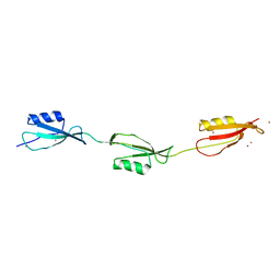
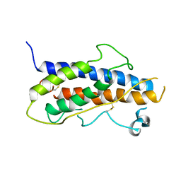 | | HUMAN GROWTH HORMONE | | 分子名称: | HUMAN GROWTH HORMONE | | 著者 | Chantalat, L, Jones, N, Korber, F, Navaza, J, Pavlovsky, A.G. | | 登録日 | 1995-05-11 | | 公開日 | 1995-12-07 | | 最終更新日 | 2019-08-14 | | 実験手法 | X-RAY DIFFRACTION (2.5 Å) | | 主引用文献 | THE CRYSTAL-STRUCTURE OF WILD-TYPE GROWTH-HORMONE AT 2.5 ANGSTROM RESOLUTION.
Protein Pept.Lett., 2, 1995
|
|
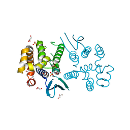
1H1W
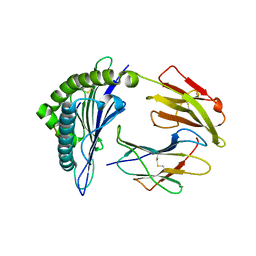
 | | High resolution crystal structure of the human PDK1 catalytic domain | | 分子名称: | 3-PHOSPHOINOSITIDE DEPENDENT PROTEIN KINASE-1, ADENOSINE-5'-TRIPHOSPHATE, GLYCEROL, ... | | 著者 | Biondi, R.M, Komander, D, Thomas, C.C, Lizcano, J.M, Deak, M, Alessi, D.R, Van Aalten, D.M.F. | | 登録日 | 2002-07-23 | | 公開日 | 2003-07-17 | | 最終更新日 | 2023-12-13 | | 実験手法 | X-RAY DIFFRACTION (2 Å) | | 主引用文献 | High Resolution Crystal Structure of the Human Pdk1 Catalytic Domain Defines the Regulatory Phosphopeptide Docking Site
Embo J., 21, 2003
|
|
8BCQ
 | | N-terminal domain of Plasmodium berghei glutamyl-tRNA synthetase (native crystal structure) | | 分子名称: | GLYCEROL, Glutamate--tRNA ligase, SULFATE ION | | 著者 | Benas, P, Jaramillo Ponce, J.R, Frugier, M, Sauter, C. | | 登録日 | 2022-10-17 | | 公開日 | 2023-01-25 | | 最終更新日 | 2024-02-07 | | 実験手法 | X-RAY DIFFRACTION (2.7 Å) | | 主引用文献 | Solution X-ray scattering highlights discrepancies in Plasmodium multi-aminoacyl-tRNA synthetase complexes.
Protein Sci., 32, 2023
|
|
1SHQ
 | | Crystal structure of shrimp alkaline phosphatase with magnesium in M3 | | 分子名称: | 2-acetamido-2-deoxy-beta-D-glucopyranose, MAGNESIUM ION, SULFATE ION, ... | | 著者 | de Backer, M.M.E, McSweeney, S, Lindley, P.F, Hough, E. | | 登録日 | 2004-02-26 | | 公開日 | 2004-08-31 | | 最終更新日 | 2020-07-29 | | 実験手法 | X-RAY DIFFRACTION (2 Å) | | 主引用文献 | Ligand-binding and metal-exchange crystallographic studies on shrimp alkaline phosphatase.
Acta Crystallogr.,Sect.D, 60, 2004
|
|
1SHN
 | | Crystal structure of shrimp alkaline phosphatase with phosphate bound | | 分子名称: | 2-acetamido-2-deoxy-beta-D-glucopyranose, PHOSPHATE ION, SULFATE ION, ... | | 著者 | de Backer, M.M.E, McSweeney, S, Lindley, P.F, Hough, E. | | 登録日 | 2004-02-26 | | 公開日 | 2004-08-31 | | 最終更新日 | 2020-07-29 | | 実験手法 | X-RAY DIFFRACTION (2.15 Å) | | 主引用文献 | Ligand-binding and metal-exchange crystallographic studies on shrimp alkaline phosphatase.
Acta Crystallogr.,Sect.D, 60, 2004
|
|
3TP2
 | |
3REA
 | |
1HWH
 | |
3M63
 | |
3O3I
 | | Crystal Structure of human Hiwi1 PAZ domain (residues 277-399) in complex with 14-mer RNA (12-bp + 2-nt overhang) containing 2'-OH at its 3'-end | | 分子名称: | Piwi-like protein 1, RNA (5'-R(*GP*CP*GP*AP*AP*UP*AP*UP*UP*CP*GP*CP*UP*U)-3') | | 著者 | Tian, Y, Simanshu, D.K, Ma, J.-B, Patel, D.J. | | 登録日 | 2010-07-24 | | 公開日 | 2011-01-12 | | 最終更新日 | 2024-04-03 | | 実験手法 | X-RAY DIFFRACTION (2.801 Å) | | 主引用文献 | Inaugural Article: Structural basis for piRNA 2'-O-methylated 3'-end recognition by Piwi PAZ (Piwi/Argonaute/Zwille) domains.
Proc.Natl.Acad.Sci.USA, 108, 2011
|
|
3M62
 | |
3O7X
 | |
2YMB
 | | Structures of MITD1 | | 分子名称: | CHARGED MULTIVESICULAR BODY PROTEIN 1A, MIT DOMAIN-CONTAINING PROTEIN 1 | | 著者 | Hadders, M.A, Agromayor, M, Obita, T, Perisic, O, Caballe, A, Kloc, M, Lamers, M.H, Williams, R.L, Martin-Serrano, J. | | 登録日 | 2012-10-08 | | 公開日 | 2012-10-24 | | 最終更新日 | 2023-12-20 | | 実験手法 | X-RAY DIFFRACTION (3.404 Å) | | 主引用文献 | Escrt-III Binding Protein Mitd1 is Involved in Cytokinesis and Has an Unanticipated Pld Fold that Binds Membranes.
Proc.Natl.Acad.Sci.USA, 109, 2012
|
|
3M9G
 | | Crystal structure of the three-PASTA-domain of a Ser/Thr kinase from Staphylococcus aureus | | 分子名称: | Protein kinase, ZINC ION | | 著者 | Paracuellos, P, Ballandras, A, Robert, X, Creze, C, Cozzone, A.J, Duclos, B, Gouet, P. | | 登録日 | 2010-03-22 | | 公開日 | 2010-11-03 | | 最終更新日 | 2024-03-20 | | 実験手法 | X-RAY DIFFRACTION (2.9 Å) | | 主引用文献 | The Extended Conformation of the 2.9-A Crystal Structure of the Three-PASTA Domain of a Ser/Thr Kinase from the Human Pathogen Staphylococcus aureus
J.Mol.Biol., 404, 2010
|
|
3NFN
 | | Recognition of peptide-MHC by a V-delta/V-beta TCR | | 分子名称: | 10-mer peptide from Protein Nef, Beta-2-microglobulin, HLA class I histocompatibility antigen, ... | | 著者 | Shi, Y, Qi, J, Gao, F, Gao, G.F. | | 登録日 | 2010-06-10 | | 公開日 | 2011-07-13 | | 最終更新日 | 2013-09-11 | | 実験手法 | X-RAY DIFFRACTION (2.392 Å) | | 主引用文献 | Plasticity of human CD8 alpha alpha binding to peptide-HLA-A*2402
Mol.Immunol., 48, 2011
|
|
3O7V
 | | Crystal Structure of human Hiwi1 (V361M) PAZ domain (residues 277-399) in complex with 14-mer RNA (12-bp + 2-nt overhang) containing 2'-OCH3 at its 3'-end | | 分子名称: | Piwi-like protein 1, RNA (5'-R(*GP*CP*GP*AP*AP*UP*AP*UP*UP*CP*GP*CP*UP*(OMU))-3') | | 著者 | Tian, Y, Simanshu, D.K, Ma, J.-B, Patel, D.J. | | 登録日 | 2010-08-01 | | 公開日 | 2011-01-12 | | 最終更新日 | 2017-11-08 | | 実験手法 | X-RAY DIFFRACTION (2.1 Å) | | 主引用文献 | Inaugural Article: Structural basis for piRNA 2'-O-methylated 3'-end recognition by Piwi PAZ (Piwi/Argonaute/Zwille) domains.
Proc.Natl.Acad.Sci.USA, 108, 2011
|
|
3O6E
 | | Crystal Structure of human Hiwi1 PAZ domain (residues 277-399) in complex with 14-mer RNA (12-bp + 2-nt overhang) containing 2'-OCH3 at its 3'-end | | 分子名称: | Piwi-like protein 1, RNA (5'-R(*GP*CP*GP*AP*AP*UP*AP*UP*UP*CP*GP*CP*UP*(OMU))-3') | | 著者 | Tian, Y, Simanshu, D.K, Ma, J.-B, Patel, D.J. | | 登録日 | 2010-07-28 | | 公開日 | 2011-01-12 | | 最終更新日 | 2023-12-06 | | 実験手法 | X-RAY DIFFRACTION (2.904 Å) | | 主引用文献 | Inaugural Article: Structural basis for piRNA 2'-O-methylated 3'-end recognition by Piwi PAZ (Piwi/Argonaute/Zwille) domains.
Proc.Natl.Acad.Sci.USA, 108, 2011
|
|
3BX1
 | | Complex between the Barley alpha-Amylase/Subtilisin Inhibitor and the subtilisin Savinase | | 分子名称: | Alpha-amylase/subtilisin inhibitor, CALCIUM ION, CHLORIDE ION, ... | | 著者 | Micheelsen, P.O, Vevodova, J, Wilson, K, Skjot, M. | | 登録日 | 2008-01-11 | | 公開日 | 2008-07-08 | | 最終更新日 | 2023-08-30 | | 実験手法 | X-RAY DIFFRACTION (1.85 Å) | | 主引用文献 | Structural and Mutational Analyses of the Interaction between the Barley alpha-Amylase/Subtilisin Inhibitor and the Subtilisin Savinase Reveal a Novel Mode of Inhibition
J.Mol.Biol., 380, 2008
|
|
3L8Q
 | | Structure analysis of the type II cohesin dyad from the adaptor ScaA scaffoldin of Acetivibrio cellulolyticus | | 分子名称: | 1,2-ETHANEDIOL, 1,3-PROPANDIOL, Cellulosomal scaffoldin adaptor protein B, ... | | 著者 | Noach, I, Frolow, F, Bayer, E.A. | | 登録日 | 2010-01-03 | | 公開日 | 2010-05-05 | | 最終更新日 | 2023-11-01 | | 実験手法 | X-RAY DIFFRACTION (1.57 Å) | | 主引用文献 | Modular Arrangement of a Cellulosomal Scaffoldin Subunit Revealed from the Crystal Structure of a Cohesin Dyad
J.Mol.Biol., 399, 2010
|
|
3OS6
 | | Crystal structure of putative 2,3-dihydroxybenzoate-specific isochorismate synthase, DhbC from Bacillus anthracis. | | 分子名称: | GLYCEROL, Isochorismate synthase DhbC, POLYETHYLENE GLYCOL (N=34), ... | | 著者 | Domagalski, M.J, Chruszcz, M, Skarina, T, Onopriyenko, O, Cymborowski, M, Savchenko, A, Edwards, A, Anderson, W, Minor, W, Center for Structural Genomics of Infectious Diseases (CSGID) | | 登録日 | 2010-09-08 | | 公開日 | 2010-10-20 | | 最終更新日 | 2022-04-13 | | 実験手法 | X-RAY DIFFRACTION (2.4 Å) | | 主引用文献 | Structure of isochorismate synthase DhbC from Bacillus anthracis.
Acta Crystallogr.,Sect.F, 69, 2013
|
|
7OKQ
 | | Cryo-EM Structure of the DDB1-DCAF1-CUL4A-RBX1 Complex | | 分子名称: | Cullin-4A, DDB1- and CUL4-associated factor 1, DNA damage-binding protein 1, ... | | 著者 | Mohamed, W.I, Schenk, A.D, Kempf, G, Cavadini, S, Thoma, N.H. | | 登録日 | 2021-05-18 | | 公開日 | 2021-10-13 | | 最終更新日 | 2024-07-10 | | 実験手法 | ELECTRON MICROSCOPY (8.4 Å) | | 主引用文献 | The CRL4 DCAF1 cullin-RING ubiquitin ligase is activated following a switch in oligomerization state.
Embo J., 40, 2021
|
|
7OLJ
 | | Tankyrase 2 in complex with an inhibitor (OUL219) | | 分子名称: | 8-propan-2-yloxy-4~{H}-thieno[2,3-c]isoquinolin-5-one, GLYCEROL, Poly [ADP-ribose] polymerase tankyrase-2, ... | | 著者 | Sowa, S.T, Lehtio, L. | | 登録日 | 2021-05-20 | | 公開日 | 2021-12-08 | | 最終更新日 | 2024-02-07 | | 実験手法 | X-RAY DIFFRACTION (1.8 Å) | | 主引用文献 | Analogs of TIQ-A as inhibitors of human mono-ADP-ribosylating PARPs.
Bioorg.Med.Chem., 52, 2021
|
|
7OMC
 | | Tankyrase 2 in complex with an inhibitor (OUL228) | | 分子名称: | 8-propan-2-yloxy-4~{H}-thieno[2,3-c]isoquinolin-5-one, GLYCEROL, Poly [ADP-ribose] polymerase tankyrase-2, ... | | 著者 | Sowa, S.T, Lehtio, L. | | 登録日 | 2021-05-21 | | 公開日 | 2021-12-08 | | 最終更新日 | 2024-01-31 | | 実験手法 | X-RAY DIFFRACTION (2.1 Å) | | 主引用文献 | Analogs of TIQ-A as inhibitors of human mono-ADP-ribosylating PARPs.
Bioorg.Med.Chem., 52, 2021
|
|


