3DW4
 
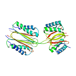
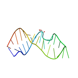 | | Crystal Structure of the Sarcin/Ricin Domain from E. COLI 23 S rRNA, U2650-OCH3 modified | | 分子名称: | GLYCEROL, Sarcin/Ricin Domain from E. Coli 23 S rRNA | | 著者 | Olieric, V, Rieder, U, Lang, K, Serganov, A, Schulze-Briese, C, Micura, R, Dumas, P, Ennifar, E. | | 登録日 | 2008-07-21 | | 公開日 | 2009-03-24 | | 最終更新日 | 2023-08-30 | | 実験手法 | X-RAY DIFFRACTION (0.97 Å) | | 主引用文献 | A fast selenium derivatization strategy for crystallization and phasing of RNA structures.
Rna, 15, 2009
|
|
2Q2H
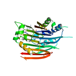
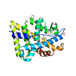
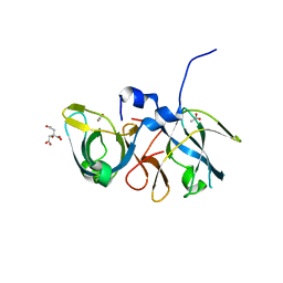 | | Crystal structure of the protein secretion chaperone CsaA from Agrobacterium tumefaciens with a genetically fused phage-display derived peptide substrate at the N-terminus. | | 分子名称: | ACETATE ION, CITRIC ACID, Secretion chaperone, ... | | 著者 | Feldman, A.R, Shapova, Y.A, Paetzel, M. | | 登録日 | 2007-05-28 | | 公開日 | 2008-04-01 | | 最終更新日 | 2023-08-30 | | 実験手法 | X-RAY DIFFRACTION (1.65 Å) | | 主引用文献 | Phage display and crystallographic analysis reveals potential substrate/binding site interactions in the protein secretion chaperone CsaA from Agrobacterium tumefaciens.
J.Mol.Biol., 379, 2008
|
|
3S4P
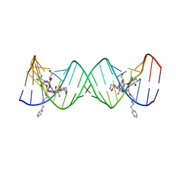 | | Crystal structure of the bacterial ribosomal decoding site complexed with an amphiphilic paromomycin O2''-ether analogue | | 分子名称: | (1R,2R,3S,4R,6S)-4,6-diamino-2-{[3-O-(2,6-diamino-2,6-dideoxy-beta-L-idopyranosyl)-2-O-{2-[(2-phenylethyl)amino]ethyl}-beta-D-ribofuranosyl]oxy}-3-hydroxycyclohexyl 2-amino-2-deoxy-alpha-D-glucopyranoside, RNA (5'-R(P*GP*CP*GP*UP*CP*AP*CP*AP*CP*CP*GP*GP*UP*GP*AP*AP*GP*UP*CP*GP*C)-3') | | 著者 | Szychowski, J, Kondo, J, Zahr, O, Auclair, K, Westhof, E, Hanessian, S, Keillor, J.W. | | 登録日 | 2011-05-20 | | 公開日 | 2011-09-21 | | 最終更新日 | 2023-11-01 | | 実験手法 | X-RAY DIFFRACTION (2.56 Å) | | 主引用文献 | Inhibition of aminoglycoside-deactivating enzymes APH(3')-IIIa and AAC(6')-Ii by amphiphilic paromomycin O2''-ether analogues
Chemmedchem, 6, 2011
|
|
3SDI
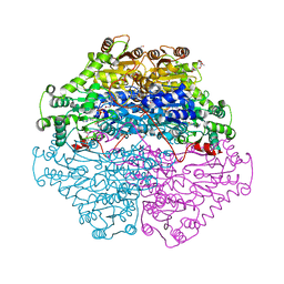
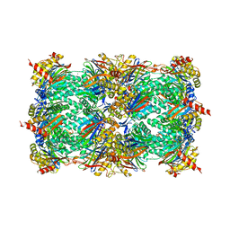 | | Structure of yeast 20S open-gate proteasome with Compound 20 | | 分子名称: | 2-(N-MORPHOLINO)-ETHANESULFONIC ACID, MAGNESIUM ION, N~4~-(2,2-dimethylpropyl)-N~1~-{(2S)-1-[(4-methylbenzyl)amino]-1-oxo-4-phenylbutan-2-yl}-N~2~-[(5-methyl-1,2-oxazol-3-yl)carbonyl]-L-aspartamide, ... | | 著者 | Sintchak, M.D. | | 登録日 | 2011-06-09 | | 公開日 | 2012-06-13 | | 最終更新日 | 2023-09-13 | | 実験手法 | X-RAY DIFFRACTION (2.65 Å) | | 主引用文献 | Optimization of a series of dipeptides with a P3 threonine residue as non-covalent inhibitors of the chymotrypsin-like activity of the human 20S proteasome.
Bioorg.Med.Chem.Lett., 20, 2010
|
|
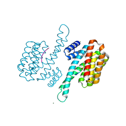
2PG6
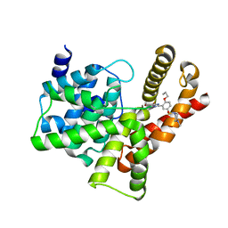
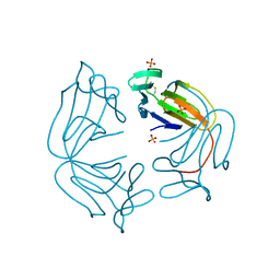
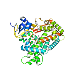 | | Crystal Structure of Human Microsomal P450 2A6 L240C/N297Q | | 分子名称: | Cytochrome P450 2A6, PROTOPORPHYRIN IX CONTAINING FE | | 著者 | Sansen, S, Hsu, M.H, Stout, C.D, Johnson, E.F. | | 登録日 | 2007-04-06 | | 公開日 | 2007-07-24 | | 最終更新日 | 2023-08-30 | | 実験手法 | X-RAY DIFFRACTION (2.53 Å) | | 主引用文献 | Structural insight into the altered substrate specificity of human cytochrome P450 2A6 mutants.
Arch.Biochem.Biophys., 464, 2007
|
|
3DZ4
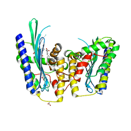
 | | Human AdoMetDC with 5'-[(2-carboxamidoethyl)methylamino]-5'-deoxy-8-methyladenosine | | 分子名称: | 1,4-DIAMINOBUTANE, 3-[{[(2R,3S,4R,5R)-5-(6-amino-8-methyl-9H-purin-9-yl)-3,4-dihydroxytetrahydrofuran-2-yl]methyl}(methyl)amino]propanamid e, S-adenosylmethionine decarboxylase alpha chain, ... | | 著者 | Bale, S, McCloskey, D.E, Pegg, A.E, Secrist III, J.A, Guida, W.C, Ealick, S.E. | | 登録日 | 2008-07-29 | | 公開日 | 2009-03-10 | | 最終更新日 | 2023-11-15 | | 実験手法 | X-RAY DIFFRACTION (1.84 Å) | | 主引用文献 | New Insights into the Design of Inhibitors of Human S-Adenosylmethionine Decarboxylase: Studies of Adenine C8 Substitution in Structural Analogues of S-Adenosylmethionine
J.Med.Chem., 52, 2009
|
|
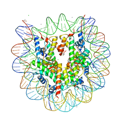
2PH9
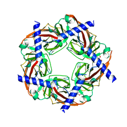 | | Galanthamine bound to an ACh-binding Protein | | 分子名称: | (-)-GALANTHAMINE, Soluble acetylcholine receptor, TETRAETHYLENE GLYCOL | | 著者 | Hansen, S.B, Taylor, P. | | 登録日 | 2007-04-10 | | 公開日 | 2007-07-03 | | 最終更新日 | 2023-08-30 | | 実験手法 | X-RAY DIFFRACTION (2.88 Å) | | 主引用文献 | Galanthamine and non-competitive inhibitor binding to ACh-binding protein: evidence for a binding site on non-alpha-subunit interfaces of heteromeric neuronal nicotinic receptors.
J.Mol.Biol., 369, 2007
|
|
2PL2
 | | Crystal structure of TTC0263: a thermophilic TPR protein in Thermus thermophilus HB27 | | 分子名称: | Hypothetical conserved protein TTC0263 | | 著者 | Lim, H, Kim, K, Han, D, Oh, J. | | 登録日 | 2007-04-18 | | 公開日 | 2008-03-04 | | 最終更新日 | 2024-03-13 | | 実験手法 | X-RAY DIFFRACTION (2.5 Å) | | 主引用文献 | Crystal structure of TTC0263, a thermophilic TPR protein from Thermus thermophilus HB27.
Mol.Cell, 24, 2007
|
|
3SIE
 | |
3DWR
 | |
2Q8H
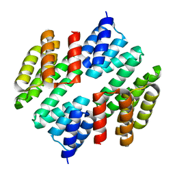
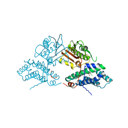 | | Structure of pyruvate dehydrogenase kinase isoform 1 in complex with dichloroacetate (DCA) | | 分子名称: | DICHLORO-ACETIC ACID, POTASSIUM ION, [Pyruvate dehydrogenase [lipoamide]] kinase isozyme 1 | | 著者 | Kato, M, Li, J, Chuang, J.L, Chuang, D.T. | | 登録日 | 2007-06-10 | | 公開日 | 2007-07-24 | | 最終更新日 | 2023-08-30 | | 実験手法 | X-RAY DIFFRACTION (2 Å) | | 主引用文献 | Distinct Structural Mechanisms for Inhibition of Pyruvate Dehydrogenase Kinase Isoforms by AZD7545, Dichloroacetate, and Radicicol.
Structure, 15, 2007
|
|
3DX2
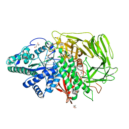 | | Golgi mannosidase II complex with MANNOSTATIN B | | 分子名称: | (1R,2R,3R,4S,5R)-4-amino-5-[(R)-methylsulfinyl]cyclopentane-1,2,3-triol, (4R)-2-METHYLPENTANE-2,4-DIOL, (4S)-2-METHYL-2,4-PENTANEDIOL, ... | | 著者 | Kuntz, D.A, Zhong, W, Guo, J, Rose, D.R, Boons, G.-J. | | 登録日 | 2008-07-23 | | 公開日 | 2009-07-07 | | 最終更新日 | 2023-08-30 | | 実験手法 | X-RAY DIFFRACTION (1.4 Å) | | 主引用文献 | The molecular basis of inhibition of Golgi alpha-mannosidase II by mannostatin A.
Chembiochem, 10, 2009
|
|
2Q8Q
 | |
3SA3
 | | Crystal structure of wild-type HIV-1 protease in complex with AG23 | | 分子名称: | ACETATE ION, N~2~-acetyl-N-[(2S,3R)-4-{(1,3-benzothiazol-6-ylsulfonyl)[(2S)-2-methylbutyl]amino}-3-hydroxy-1-phenylbutan-2-yl]-L-isoleucinamide, PHOSPHATE ION, ... | | 著者 | Schiffer, C.A, Nalam, M.N.L. | | 登録日 | 2011-06-02 | | 公開日 | 2012-06-06 | | 最終更新日 | 2023-09-13 | | 実験手法 | X-RAY DIFFRACTION (1.65 Å) | | 主引用文献 | Protease Inhibitors that protrude out from substrate envelope are more susceptible to developing drug resistance
To be Published
|
|
3DZ7
 | | Human AdoMetDC with 5'-[(carboxamidomethyl)methylamino]-5'-deoxy-8-methyladenosine | | 分子名称: | 1,4-DIAMINOBUTANE, 2-[{[(2R,3S,4R,5R)-5-(6-amino-8-methyl-9H-purin-9-yl)-3,4-dihydroxytetrahydrofuran-2-yl]methyl}(methyl)amino]acetamide, S-adenosylmethionine decarboxylase alpha chain, ... | | 著者 | Bale, S, McCloskey, D.E, Pegg, A.E, Secrist III, J.A, Guida, W.C, Ealick, S.E. | | 登録日 | 2008-07-29 | | 公開日 | 2009-03-10 | | 最終更新日 | 2023-11-15 | | 実験手法 | X-RAY DIFFRACTION (1.91 Å) | | 主引用文献 | New Insights into the Design of Inhibitors of Human S-Adenosylmethionine Decarboxylase: Studies of Adenine C8 Substitution in Structural Analogues of S-Adenosylmethionine
J.Med.Chem., 52, 2009
|
|
3SAB
 | |
2PG1
 | |
2PNU
 | | Crystal structure of human androgen receptor ligand-binding domain in complex with EM-5744 | | 分子名称: | (5S,8R,9S,10S,13R,14S,17S)-13-{2-[(3,5-DIFLUOROBENZYL)OXY]ETHYL}-17-HYDROXY-10-METHYLHEXADECAHYDRO-3H-CYCLOPENTA[A]PHENANTHREN-3-ONE, 2,3-DIHYDROXY-1,4-DITHIOBUTANE, 2-(N-MORPHOLINO)-ETHANESULFONIC ACID, ... | | 著者 | Cantin, L, Faucher, F, Couture, J.F, Pereira de Jesus-Tran, K, Legrand, P, Ciobanu, C.L, Singh, S.M, Labrie, F, Breton, R. | | 登録日 | 2007-04-25 | | 公開日 | 2007-09-11 | | 最終更新日 | 2023-08-30 | | 実験手法 | X-RAY DIFFRACTION (1.65 Å) | | 主引用文献 | Structural Characterization of the Human Androgen Receptor Ligand-binding Domain Complexed with EM5744, a Rationally Designed Steroidal Ligand Bearing a Bulky Chain Directed toward Helix 12.
J.Biol.Chem., 282, 2007
|
|
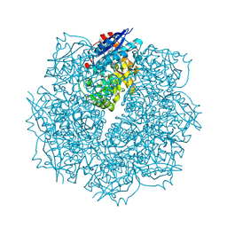
2PGN
 | | The crystal structure of FAD and ThDP-dependent Cyclohexane-1,2-dione Hydrolase in Complex with Cyclohexane-1,2-dione | | 分子名称: | CYCLOHEXANE-1,2-DIONE, Cyclohexane-1,2-dione Hydrolase (Cdh), FLAVIN-ADENINE DINUCLEOTIDE, ... | | 著者 | Fraas, S, Warkentin, E, Ermler, U. | | 登録日 | 2007-04-10 | | 公開日 | 2008-04-22 | | 最終更新日 | 2024-02-21 | | 実験手法 | X-RAY DIFFRACTION (1.2 Å) | | 主引用文献 | The crystal structure of FAD and ThDP-dependent Cyclohexane-1,2-dione Hydrolase in Complex with Cyclohexane-1,2-dione
To be Published
|
|
3DZW
 | |
3SMM
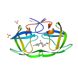
 | | Crystal structure of human 14-3-3 sigma C38N/N166H in complex with task-3 peptide and stabilizer Fusicoccin J aglycone | | 分子名称: | 14-3-3 protein sigma, CHLORIDE ION, Fusicoccin J aglycone, ... | | 著者 | Anders, C, Schumacher, B, Ottmann, C. | | 登録日 | 2011-06-28 | | 公開日 | 2012-07-11 | | 最終更新日 | 2023-09-13 | | 実験手法 | X-RAY DIFFRACTION (2 Å) | | 主引用文献 | A semisynthetic fusicoccane stabilizes a protein-protein interaction and enhances the expression of K+ channels at the cell surface.
Chem. Biol., 20, 2013
|
|
3SN4
 | | Crystal structure of putative L-alanine-DL-glutamate epimerase from Burkholderia xenovorans strain LB400 bound to magnesium and alpha-ketoglutarate | | 分子名称: | 2-OXOGLUTARIC ACID, CHLORIDE ION, MAGNESIUM ION, ... | | 著者 | Bonanno, J.B, Patskovsky, Y, Toro, R, Dickey, M, Bain, K.T, Wu, B, Sauder, J.M, Burley, S.K, Almo, S.C, New York SGX Research Center for Structural Genomics (NYSGXRC) | | 登録日 | 2011-06-28 | | 公開日 | 2011-07-27 | | 最終更新日 | 2023-12-06 | | 実験手法 | X-RAY DIFFRACTION (1.86 Å) | | 主引用文献 | Crystal structure of putative L-alanine-DL-glutamate epimerase from Burkholderia xenovorans strain LB400 bound to magnesium and alpha-ketoglutarate
To be Published
|
|
3DN7
 | | Cyclic nucleotide binding regulatory protein from Cytophaga hutchinsonii. | | 分子名称: | 1,2-ETHANEDIOL, Cyclic nucleotide binding regulatory protein, GLYCEROL | | 著者 | Osipiuk, J, Maltseva, N, Abdullah, J, Joachimiak, A, Midwest Center for Structural Genomics (MCSG) | | 登録日 | 2008-07-01 | | 公開日 | 2008-08-26 | | 最終更新日 | 2017-10-25 | | 実験手法 | X-RAY DIFFRACTION (1.8 Å) | | 主引用文献 | X-ray crystal structure of cyclic nucleotide binding regulatory protein from Cytophaga hutchinsonii.
To be Published
|
|
3SAA
 | |
3X1V
 | |


