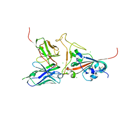
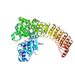
7D8B
 
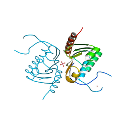
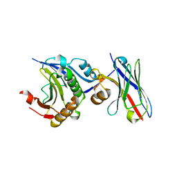 | | Engineering Disulphide-Free Autonomous Antibody VH Domains to modulate intracellular pathways | | 分子名称: | Eukaryotic translation initiation factor 4E, VH-S4 | | 著者 | Frosi, Y, Lin, Y.C, Jiang, S, Brown, C.J. | | 登録日 | 2020-10-07 | | 公開日 | 2021-08-25 | | 最終更新日 | 2023-11-29 | | 実験手法 | X-RAY DIFFRACTION (2.46 Å) | | 主引用文献 | Engineering an autonomous VH domain to modulate intracellular pathways and to interrogate the eIF4F complex.
Nat Commun, 13, 2022
|
|
6LLZ
 | |
2GH2
 | |
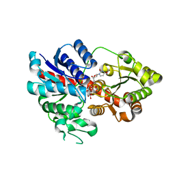
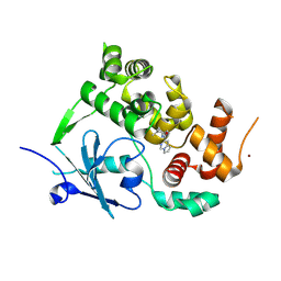
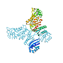
6LMY
 | | Crystal structure of DUSP22 mutant_C88S/S93A | | 分子名称: | Dual specificity protein phosphatase 22, PHOSPHATE ION | | 著者 | Lai, C.H, Lyu, P.C. | | 登録日 | 2019-12-27 | | 公開日 | 2020-10-28 | | 最終更新日 | 2023-11-22 | | 実験手法 | X-RAY DIFFRACTION (1.5 Å) | | 主引用文献 | Structural Insights into the Active Site Formation of DUSP22 in N-loop-containing Protein Tyrosine Phosphatases.
Int J Mol Sci, 21, 2020
|
|
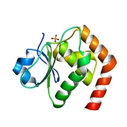
6OQP
 | | U-AITx-Ate1 | | 分子名称: | SER-LYS-TRP-ILE-CYS-ALA-ASN-ARG-SER-VAL-CYS-PRO-ILE | | 著者 | Elnahriry, K.A, Wai, D.C.C, Norton, R.S. | | 登録日 | 2019-04-28 | | 公開日 | 2019-07-31 | | 最終更新日 | 2023-06-14 | | 実験手法 | SOLUTION NMR | | 主引用文献 | Structural and functional characterisation of a novel peptide from the Australian sea anemone Actinia tenebrosa.
Toxicon, 168, 2019
|
|
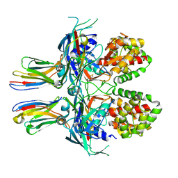
2GHW
 | | Crystal structure of SARS spike protein receptor binding domain in complex with a neutralizing antibody, 80R | | 分子名称: | CHLORIDE ION, Spike glycoprotein, anti-sars scFv antibody, ... | | 著者 | Hwang, W.C, Lin, Y, Santelli, E, Sui, J, Jaroszewski, L, Stec, B, Farzan, M, Marasco, W.A, Liddington, R.C. | | 登録日 | 2006-03-27 | | 公開日 | 2006-09-19 | | 最終更新日 | 2023-08-30 | | 実験手法 | X-RAY DIFFRACTION (2.3 Å) | | 主引用文献 | Structural basis of neutralization by a human anti-severe acute respiratory syndrome spike protein antibody, 80R.
J.Biol.Chem., 281, 2006
|
|
7Z1X
 | |
7YTO
 | |
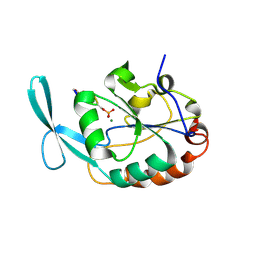
6LOT
 | | Crystal structure of DUSP22 mutant_N128D | | 分子名称: | Dual specificity protein phosphatase 22, SULFATE ION | | 著者 | Lai, C.H, Lyu, P.C. | | 登録日 | 2020-01-07 | | 公開日 | 2020-10-28 | | 最終更新日 | 2023-11-29 | | 実験手法 | X-RAY DIFFRACTION (1.69 Å) | | 主引用文献 | Structural Insights into the Active Site Formation of DUSP22 in N-loop-containing Protein Tyrosine Phosphatases.
Int J Mol Sci, 21, 2020
|
|
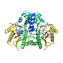
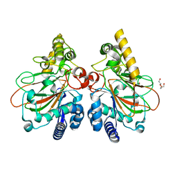
2G8G
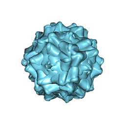 | | Structurally mapping the diverse phenotype of Adeno-Associated Virus serotype 4 | | 分子名称: | 2'-DEOXYADENOSINE-5'-MONOPHOSPHATE, Capsid | | 著者 | Govindasamy, L, Padron, E, McKenna, R, Muzyczka, N, Chiorini, J.A, Agbandje-McKenna, M. | | 登録日 | 2006-03-02 | | 公開日 | 2007-01-02 | | 最終更新日 | 2023-08-30 | | 実験手法 | X-RAY DIFFRACTION (3.2 Å) | | 主引用文献 | Structurally mapping the diverse phenotype of adeno-associated virus serotype 4.
J.Virol., 80, 2006
|
|
6LZG
 | | Structure of novel coronavirus spike receptor-binding domain complexed with its receptor ACE2 | | 分子名称: | 2-acetamido-2-deoxy-beta-D-glucopyranose, Angiotensin-converting enzyme 2, Spike protein S1, ... | | 著者 | Wang, Q.H, Song, H, Qi, J.X. | | 登録日 | 2020-02-19 | | 公開日 | 2020-03-18 | | 最終更新日 | 2023-11-29 | | 実験手法 | X-RAY DIFFRACTION (2.5 Å) | | 主引用文献 | Structural and Functional Basis of SARS-CoV-2 Entry by Using Human ACE2.
Cell, 181, 2020
|
|
2GFP
 | | Structure of the Multidrug Transporter EmrD from Escherichia coli | | 分子名称: | Multidrug resistance protein D | | 著者 | Yin, Y, He, X, Szewczyk, P, Nguyen, T, Chang, G. | | 登録日 | 2006-03-22 | | 公開日 | 2006-05-16 | | 最終更新日 | 2024-02-14 | | 実験手法 | X-RAY DIFFRACTION (3.5 Å) | | 主引用文献 | Structure of the multidrug transporter EmrD from Escherichia coli
Science, 312, 2006
|
|
6LU8
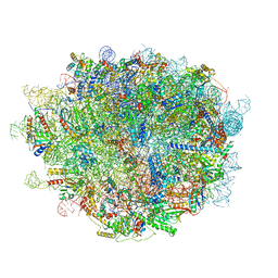 | | Cryo-EM structure of a human pre-60S ribosomal subunit - state A | | 分子名称: | 28S rRNA, 5.8S rRNA, 5S rRNA, ... | | 著者 | Liang, X, Zuo, M, Zhang, Y, Li, N, Ma, C, Dong, M, Gao, N. | | 登録日 | 2020-01-26 | | 公開日 | 2020-08-26 | | 実験手法 | ELECTRON MICROSCOPY (3.13 Å) | | 主引用文献 | Structural snapshots of human pre-60S ribosomal particles before and after nuclear export.
Nat Commun, 11, 2020
|
|
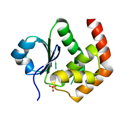
4GGK
 | | Crystal structure of Zucchini from mouse (mZuc / PLD6 / MitoPLD) bound to tungstate | | 分子名称: | Mitochondrial cardiolipin hydrolase, TUNGSTATE(VI)ION, ZINC ION | | 著者 | Ipsaro, J.J, Haase, A.D, Hannon, G.J, Joshua-Tor, L. | | 登録日 | 2012-08-06 | | 公開日 | 2012-10-10 | | 最終更新日 | 2023-09-13 | | 実験手法 | X-RAY DIFFRACTION (2.1 Å) | | 主引用文献 | The structural biochemistry of Zucchini implicates it as a nuclease in piRNA biogenesis.
Nature, 491, 2012
|
|
7ZG3
 | | Structure of the mouse 8-oxoguanine DNA Glycosylase mOGG1 in complex with ligand TH011228 | | 分子名称: | N-glycosylase/DNA lyase, NICKEL (II) ION, ~{N}-[(1~{S})-1,2,2-trimethylcyclopropyl]pyrrolo[1,2-c]pyrimidine-3-carboxamide | | 著者 | Davies, J.R, Scaletti, E, Stenmark, P. | | 登録日 | 2022-04-01 | | 公開日 | 2023-08-16 | | 実験手法 | X-RAY DIFFRACTION (2.3 Å) | | 主引用文献 | Structure of the mouse 8-oxoguanine DNA Glycosylase mOGG1 in complex with ligand TH011228
To Be Published
|
|
7YW9
 | | Crystal structure of the triple mutant CmnC-L136Q,S138G,D249Y in complex with alpha-KG | | 分子名称: | ACETATE ION, CmnC, D-ARGININE, ... | | 著者 | Huang, S.J, Hsiao, Y.H, Lin, E.C, Hsiao, P.Y, Chang, C.Y. | | 登録日 | 2022-08-22 | | 公開日 | 2023-08-30 | | 最終更新日 | 2023-10-04 | | 実験手法 | X-RAY DIFFRACTION (1.76 Å) | | 主引用文献 | Crystal structure of the alpha-ketoglutarate-dependent non-heme iron oxygenase CmnC in capreomycin biosynthesis and its engineering to catalyze hydroxylation of the substrate enantiomer.
Front Chem, 10, 2022
|
|
2GHQ
 | |
5IRN
 | |
4ETU
 | |
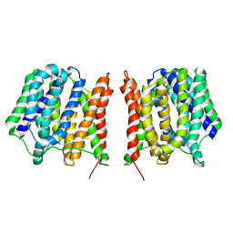
4GJS
 | | Streptavidin-K121H | | 分子名称: | Rhodium, Streptavidin, trichloro{(1,2,3,4,5-eta)-1,2,3,4-tetramethyl-5-[2-({5-[(3aS,4S,6aR)-2-oxohexahydro-1H-thieno[3,4-d]imidazol-4-yl]pentanoyl}amino)ethyl]cyclopentadienyl}rhodium(1+) | | 著者 | Heinisch, T, Schirmer, T. | | 登録日 | 2012-08-10 | | 公開日 | 2013-02-13 | | 最終更新日 | 2024-02-28 | | 実験手法 | X-RAY DIFFRACTION (1.85 Å) | | 主引用文献 | A dual anchoring strategy for the localization and activation of artificial metalloenzymes based on the biotin-streptavidin technology.
J.Am.Chem.Soc., 135, 2013
|
|
5IS2
 | |
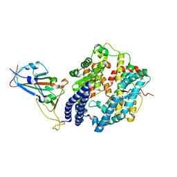
7DDX
 | | Crystal structure of KANK1 S1179F mutant in complex wtih eIF4A1 | | 分子名称: | Eukaryotic initiation factor 4A-I, GLYCEROL, KN motif and ankyrin repeat domains 1, ... | | 著者 | Pan, W, Xu, Y, Wei, Z. | | 登録日 | 2020-10-30 | | 公開日 | 2021-09-08 | | 最終更新日 | 2023-11-29 | | 実験手法 | X-RAY DIFFRACTION (2.5 Å) | | 主引用文献 | Nephrotic-syndrome-associated mutation of KANK2 induces pathologic binding competition with physiological interactor KIF21A.
J.Biol.Chem., 297, 2021
|
|
7Z6O
 | | X-Ray studies of Ku70/80 reveal the binding site for IP6 | | 分子名称: | DNA (5'-D(*GP*TP*TP*TP*TP*TP*AP*GP*TP*TP*TP*AP*T)-3'), DNA (5'-D(P*AP*AP*AP*TP*AP*AP*AP*CP*TP*AP*AP*AP*AP*AP*C)-3'), INOSITOL HEXAKISPHOSPHATE, ... | | 著者 | Varela, P.F, Charbonnier, J.B. | | 登録日 | 2022-03-14 | | 公開日 | 2023-08-30 | | 最終更新日 | 2023-12-06 | | 実験手法 | X-RAY DIFFRACTION (3.7 Å) | | 主引用文献 | Structural and functional basis of inositol hexaphosphate stimulation of NHEJ through stabilization of Ku-XLF interaction.
Nucleic Acids Res., 51, 2023
|
|
2G81
 | | Crystal Structure of the Bowman-Birk Inhibitor from Vigna unguiculata Seeds in Complex with Beta-trypsin at 1.55 Angstrons Resolution | | 分子名称: | 1,2-ETHANEDIOL, ACETIC ACID, Bowman-Birk type seed trypsin and chymotrypsin inhibitor, ... | | 著者 | Freitas, S.M, Barbosa, J.A.R.G, Paulino, L.S, Teles, R.C.L, Esteves, G.F, Ventura, M.M. | | 登録日 | 2006-03-01 | | 公開日 | 2007-01-02 | | 最終更新日 | 2023-10-25 | | 実験手法 | X-RAY DIFFRACTION (1.55 Å) | | 主引用文献 | Crystal Structure of the Bowman-Birk Inhibitor from Vigna unguiculata Seeds in Complex with {beta}-Trypsin at 1.55 A Resolution and Its Structural Properties in Association with Proteinases
Biophys.J., 92, 2007
|
|
5ISK
 | |


