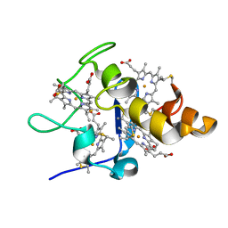
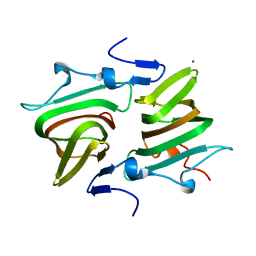
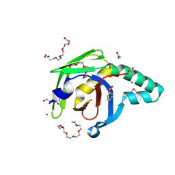
3TRV
 
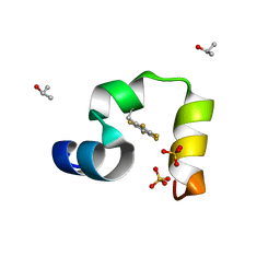 | | Crystal structure of quasiracemic villin headpiece subdomain containing (F5Phe17) substitution | | 分子名称: | D-Villin-1, ISOPROPYL ALCOHOL, L-Villin-1, ... | | 著者 | Mortenson, D.E, Satyshur, K.A, Gellman, S.H, Forest, K.T. | | 登録日 | 2011-09-11 | | 公開日 | 2012-01-25 | | 最終更新日 | 2017-11-08 | | 実験手法 | X-RAY DIFFRACTION (1 Å) | | 主引用文献 | Quasiracemic crystallization as a tool to assess the accommodation of noncanonical residues in nativelike protein conformations.
J.Am.Chem.Soc., 134, 2012
|
|
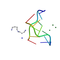
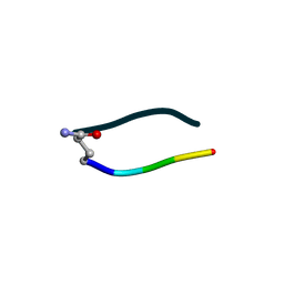
4JRD
 | | Crystal structure of the parallel double-stranded helix of poly(A) RNA | | 分子名称: | AMMONIUM ION, RNA (5'-R(*AP*AP*AP*AP*AP*AP*AP*AP*AP*AP*A)-3') | | 著者 | Safaee, N, Noronha, A.M, Kozlov, G, Rodionov, D, Wilds, C.J, Sheldrick, G.M, Gehring, K. | | 登録日 | 2013-03-21 | | 公開日 | 2013-06-05 | | 最終更新日 | 2024-02-28 | | 実験手法 | X-RAY DIFFRACTION (1 Å) | | 主引用文献 | Structure of the parallel duplex of poly(A) RNA: evaluation of a 50 year-old prediction.
Angew.Chem.Int.Ed.Engl., 52, 2013
|
|
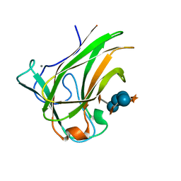
8G22
 | | Crystal Structure of the dTDP-4-dehydrorhamnose Reductase from Streptococcus pneumoniae. | | 分子名称: | dTDP-4-dehydrorhamnose reductase | | 著者 | Minasov, G, Shuvalova, L, Brunzelle, J.S, Kiryukhina, O, Satchell, K.J.F, Center for Structural Biology of Infectious Diseases (CSBID), Center for Structural Genomics of Infectious Diseases (CSGID) | | 登録日 | 2023-02-03 | | 公開日 | 2023-02-22 | | 最終更新日 | 2024-05-22 | | 実験手法 | X-RAY DIFFRACTION (1 Å) | | 主引用文献 | Crystal Structure of the dTDP-4-dehydrorhamnose Reductase from Streptococcus pneumoniae.
To Be Published
|
|
3U7Q
 | |
4UBY
 | |
2EWI
 | |
293D
 | | INTERACTION BETWEEN THE LEFT-HANDED Z-DNA AND POLYAMINE-2: THE CRYSTAL STRUCTURE OF THE D(CG)3 AND SPERMIDINE COMPLEX | | 分子名称: | DNA (5'-D(*CP*GP*CP*GP*CP*G)-3'), MAGNESIUM ION, SODIUM ION, ... | | 著者 | Ohishi, H, Nakanishi, I, Inubushi, K, Van Der Marel, G.A, Van Boom, J.H, Rich, A, Wang, A.H.-J, Hakoshima, T, Tomita, K. | | 登録日 | 1996-10-09 | | 公開日 | 1996-12-02 | | 最終更新日 | 2024-04-03 | | 実験手法 | X-RAY DIFFRACTION (1 Å) | | 主引用文献 | Interaction between the left-handed Z-DNA and polyamine-2. The crystal structure of the d(CG)3 and spermidine complex.
FEBS Lett., 391, 1996
|
|
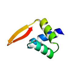
2ERL
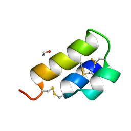 | | PHEROMONE ER-1 FROM | | 分子名称: | ETHANOL, MATING PHEROMONE ER-1 | | 著者 | Anderson, D.H, Weiss, M.S, Eisenberg, D. | | 登録日 | 1995-12-20 | | 公開日 | 1996-07-11 | | 最終更新日 | 2017-11-29 | | 実験手法 | X-RAY DIFFRACTION (1 Å) | | 主引用文献 | A challenging case for protein crystal structure determination: the mating pheromone Er-1 from Euplotes raikovi.
Acta Crystallogr.,Sect.D, 52, 1996
|
|
1OT9
 | | CRYOTRAPPED STATE IN WILD TYPE PHOTOACTIVE YELLOW PROTEIN, INDUCED WITH CONTINUOUS ILLUMINATION AT 110K | | 分子名称: | 4'-HYDROXYCINNAMIC ACID, Photoactive yellow protein | | 著者 | Anderson, S, Crosson, S, Moffat, K. | | 登録日 | 2003-03-21 | | 公開日 | 2004-05-11 | | 最終更新日 | 2019-07-24 | | 実験手法 | X-RAY DIFFRACTION (1 Å) | | 主引用文献 | Short hydrogen bonds in photoactive yellow protein.
Acta Crystallogr.,Sect.D, 60, 2004
|
|
3C1P
 | | Crystal Structure of an alternating D-Alanyl, L-Homoalanyl PNA | | 分子名称: | Peptide Nucleic Acid DLY-HGL-AGD-LHC-AGD-LHC-CUD-LYS | | 著者 | Cuesta-Seijo, J.A, Sheldrick, G.M, Zhang, J, Diederichsen, U. | | 登録日 | 2008-01-23 | | 公開日 | 2009-01-27 | | 最終更新日 | 2023-11-15 | | 実験手法 | X-RAY DIFFRACTION (1 Å) | | 主引用文献 | Continuous beta-turn fold of an alternating alanyl/homoalanyl peptide nucleic acid.
Acta Crystallogr.,Sect.D, 68, 2012
|
|
1KCC
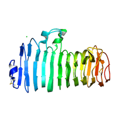 | | Endopolygalacturonase I from Stereum purpureum complexed with a galacturonate at 1.00 A resolution. | | 分子名称: | 2-acetamido-2-deoxy-beta-D-glucopyranose, CHLORIDE ION, ENDOPOLYGALACTURONASE, ... | | 著者 | Shimizu, T, Nakatsu, T, Miyairi, K, Okuno, T, Kato, H. | | 登録日 | 2001-11-08 | | 公開日 | 2002-06-05 | | 最終更新日 | 2023-10-25 | | 実験手法 | X-RAY DIFFRACTION (1 Å) | | 主引用文献 | Active-site architecture of endopolygalacturonase I from Stereum purpureum revealed by crystal structures in native and ligand-bound forms at atomic resolution.
Biochemistry, 41, 2002
|
|
6DIX
 | | NFVFGT segment from Human Immunoglobulin Light-Chain Variable Domain, Residues 98-103, assembled as an amyloid fibril | | 分子名称: | NFVFGT Immunoglobulin Light-Chain Variable Domain | | 著者 | Brumshtein, B, Esswein, S.R, Sawaya, M.R, Eisenberg, D.S. | | 登録日 | 2018-05-23 | | 公開日 | 2018-10-31 | | 最終更新日 | 2024-03-13 | | 実験手法 | X-RAY DIFFRACTION (1 Å) | | 主引用文献 | Identification of two principal amyloid-driving segments in variable domains of Ig light chains in systemic light-chain amyloidosis.
J. Biol. Chem., 293, 2018
|
|
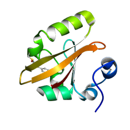
2P5K
 | | Crystal structure of the N-terminal domain of AhrC | | 分子名称: | Arginine repressor | | 著者 | Garnett, J.A, Baumberg, S, Stockley, P.G, Phillips, S.E.V. | | 登録日 | 2007-03-15 | | 公開日 | 2007-10-30 | | 最終更新日 | 2023-08-30 | | 実験手法 | X-RAY DIFFRACTION (1 Å) | | 主引用文献 | A high-resolution structure of the DNA-binding domain of AhrC, the arginine repressor/activator protein from Bacillus subtilis.
Acta Crystallogr.,Sect.F, 63, 2007
|
|
3DW6
 | | Crystal Structure of the Sarcin/Ricin Domain from E. COLI 23 S rRNA, U2650-SECH3 modified | | 分子名称: | GLYCEROL, Sarcin/Ricin Domain from E. Coli 23 S rRNA | | 著者 | Olieric, V, Rieder, U, Lang, K, Serganov, A, Schulze-Briese, C, Micura, R, Dumas, P, Ennifar, E. | | 登録日 | 2008-07-21 | | 公開日 | 2009-03-24 | | 最終更新日 | 2024-02-21 | | 実験手法 | X-RAY DIFFRACTION (1 Å) | | 主引用文献 | A fast selenium derivatization strategy for crystallization and phasing of RNA structures.
Rna, 15, 2009
|
|
4AXO
 | | Structure of the Clostridium difficile EutQ protein | | 分子名称: | ETHANOLAMINE UTILIZATION PROTEIN, MAGNESIUM ION | | 著者 | Pitts, A.C, Tuck, L.R, Faulds-Pain, A, Lewis, R.J, Marles-Wright, J. | | 登録日 | 2012-06-13 | | 公開日 | 2012-06-27 | | 最終更新日 | 2023-12-20 | | 実験手法 | X-RAY DIFFRACTION (1 Å) | | 主引用文献 | Structural Insight Into the Clostridium Difficile Ethanolamine Utilisation Microcompartment.
Plos One, 7, 2012
|
|
6UDR
 | | S2 symmetric peptide design number 3 crystal form 1, Lurch | | 分子名称: | S2-3, Lurch crystal form 1 | | 著者 | Mulligan, V.K, Kang, C.S, Antselovich, I, Sawaya, M.R, Yeates, T.O, Baker, D. | | 登録日 | 2019-09-19 | | 公開日 | 2020-09-23 | | 最終更新日 | 2020-12-02 | | 実験手法 | X-RAY DIFFRACTION (1 Å) | | 主引用文献 | Computational design of mixed chirality peptide macrocycles with internal symmetry.
Protein Sci., 29, 2020
|
|
4BJ0
 | | Xyloglucan binding module (CBM4-2 X2-L110F) in complex with branched xyloses | | 分子名称: | CALCIUM ION, XYLANASE, alpha-D-glucopyranose, ... | | 著者 | Schantz, L, Hakansson, M, Logan, D.T, Nordberg-Karlsson, E, Ohlin, M. | | 登録日 | 2013-04-15 | | 公開日 | 2014-04-23 | | 最終更新日 | 2023-12-20 | | 実験手法 | X-RAY DIFFRACTION (1 Å) | | 主引用文献 | Carbohydrate Binding Module Recognition of Xyloglucan Defined by Polar Contacts with Branching Xyloses and Ch-Pi Interactions.
Proteins, 82, 2014
|
|
4U2W
 | | Atomic resolution crystal structure of HV-BBI protease inhibitor from amphibian skin in complex with bovine trypsin | | 分子名称: | 1,2-ETHANEDIOL, Bowman-Birk trypsin inhibitor, CALCIUM ION, ... | | 著者 | Grudnik, P, Golik, P, Malicki, S, Debowski, D, Legowska, A, Rolka, K, Dubin, G. | | 登録日 | 2014-07-18 | | 公開日 | 2015-01-14 | | 最終更新日 | 2023-12-20 | | 実験手法 | X-RAY DIFFRACTION (1 Å) | | 主引用文献 | Atomic resolution crystal structure of HV-BBI protease inhibitor from amphibian skin in complex with bovine trypsin.
Proteins, 83, 2015
|
|
4ZGF
 | |
2CHH
 | | RALSTONIA SOLANACEARUM HIGH-AFFINITY MANNOSE-BINDING LECTIN | | 分子名称: | CALCIUM ION, PROTEIN RSC3288, UNKNOWN ATOM OR ION, ... | | 著者 | Mitchell, E.P, Wimmerova, M, Imberty, A. | | 登録日 | 2006-03-15 | | 公開日 | 2006-03-16 | | 最終更新日 | 2023-12-13 | | 実験手法 | X-RAY DIFFRACTION (1 Å) | | 主引用文献 | A new Ralstonia solanacearum high-affinity mannose-binding lectin RS-IIL structurally resembling the Pseudomonas aeruginosa fucose-specific lectin PA-IIL.
Mol. Microbiol., 52, 2004
|
|
1HJ8
 | | 1.00 AA Trypsin from Atlantic Salmon | | 分子名称: | BENZAMIDINE, CALCIUM ION, SULFATE ION, ... | | 著者 | Leiros, H.-K.S, Mcsweeney, S.M, Smalas, A.O. | | 登録日 | 2001-01-09 | | 公開日 | 2002-01-04 | | 最終更新日 | 2023-12-13 | | 実験手法 | X-RAY DIFFRACTION (1 Å) | | 主引用文献 | Atomic Resolution Structure of Trypsin Provide Insight Into Structural Radiation Damage
Acta Crystallogr.,Sect.D, 57, 2001
|
|
3BOI
 | | Snow Flea Antifreeze Protein Racemate | | 分子名称: | 6.5 kDa glycine-rich antifreeze protein | | 著者 | Pentelute, B.L, Kent, S.B.H, Gates, Z.P, Tereshko, V, Kossiakoff, A.A, Kurutz, J, Dashnau, J, Vaderkooi, J.M. | | 登録日 | 2007-12-17 | | 公開日 | 2008-09-23 | | 最終更新日 | 2023-08-30 | | 実験手法 | X-RAY DIFFRACTION (1 Å) | | 主引用文献 | X-ray structure of snow flea antifreeze protein determined by racemic crystallization of synthetic protein enantiomers
J.Am.Chem.Soc., 130, 2008
|
|
2CWS
 | | Crystal structure at 1.0 A of alginate lyase A1-II', a member of polysaccharide lyase family-7 | | 分子名称: | GLYCEROL, SULFATE ION, alginate lyase A1-II' | | 著者 | Yamasaki, M, Ogura, K, Hashimoto, W, Mikami, B, Murata, K. | | 登録日 | 2005-06-25 | | 公開日 | 2005-11-15 | | 最終更新日 | 2024-03-13 | | 実験手法 | X-RAY DIFFRACTION (1 Å) | | 主引用文献 | A Structural Basis for Depolymerization of Alginate by Polysaccharide Lyase Family-7
J.Mol.Biol., 352, 2005
|
|
3BWH
 | | Atomic resolution structure of cucurmosin, a novel type 1 RIP from the sarcocarp of Cucurbita moschata | | 分子名称: | 1,2-ETHANEDIOL, PHOSPHATE ION, beta-D-xylopyranose-(1-2)-[alpha-D-mannopyranose-(1-3)][alpha-D-mannopyranose-(1-6)]beta-D-mannopyranose-(1-4)-2-acetamido-2-deoxy-beta-D-glucopyranose-(1-4)-2-acetamido-2-deoxy-beta-D-glucopyranose, ... | | 著者 | Chen, L. | | 登録日 | 2008-01-09 | | 公開日 | 2008-10-07 | | 最終更新日 | 2023-08-30 | | 実験手法 | X-RAY DIFFRACTION (1 Å) | | 主引用文献 | Atomic resolution structure of cucurmosin, a novel type 1 ribosome-inactivating protein from the sarcocarp of Cucurbita moschata.
J.Struct.Biol., 164, 2008
|
|
4EA7
 | | X-ray crystal structure of PerB from Caulobacter crescentus in complex with CoA and GDP-perosamine at 1.0 Angstrom resolution | | 分子名称: | CHLORIDE ION, COENZYME A, GDP-perosamine, ... | | 著者 | Thoden, J.B, Reinhardt, L.A, Cook, P.D, Menden, P, Cleland, W.W, Holden, H.M. | | 登録日 | 2012-03-22 | | 公開日 | 2012-04-04 | | 最終更新日 | 2024-04-03 | | 実験手法 | X-RAY DIFFRACTION (1 Å) | | 主引用文献 | Catalytic Mechanism of Perosamine N-Acetyltransferase Revealed by High-Resolution X-ray Crystallographic Studies and Kinetic Analyses.
Biochemistry, 51, 2012
|
|


