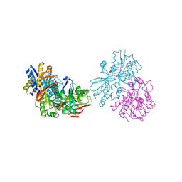
7RK9
 
 | |
6K2Y
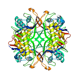
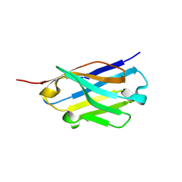
 | | Human Galectin-14 | | 分子名称: | Placental protein 13-like | | 著者 | Su, J. | | 登録日 | 2019-05-15 | | 公開日 | 2020-06-17 | | 最終更新日 | 2021-03-10 | | 実験手法 | X-RAY DIFFRACTION (1.57 Å) | | 主引用文献 | Structure-function studies of galectin-14, an important effector molecule in embryology.
Febs J., 288, 2021
|
|
3QLU
 | |
3QLT
 | |
3OLZ
 | |
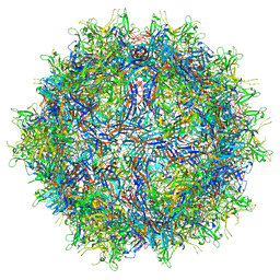
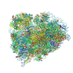
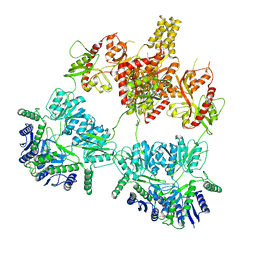
4V8M
 | | High-resolution cryo-electron microscopy structure of the Trypanosoma brucei ribosome | | 分子名称: | 18S RRNA OF THE SMALL RIBOSOMAL SUBUNIT, 40S RIBOSOMAL PROTEIN S10, PUTATIVE, ... | | 著者 | Hashem, Y, des Georges, A, Fu, J, Buss, S.N, Jossinet, F, Jobe, A, Zhang, Q, Liao, H.Y, Grassucci, R.A, Bajaj, C, Westhof, E, Madison-Antenucci, S, Frank, J. | | 登録日 | 2012-12-09 | | 公開日 | 2014-07-09 | | 最終更新日 | 2019-12-11 | | 実験手法 | ELECTRON MICROSCOPY (5.57 Å) | | 主引用文献 | High-Resolution Cryo-Electron Microscopy Structure of the Trypanosoma Brucei Ribosome.
Nature, 494, 2013
|
|
8SCB
 | | Terminating ribosome with SRI-41315 | | 分子名称: | (2S,4aS)-2-cyclobutyl-10-methyl-3-phenyl-2,10-dihydropyrimido[4,5-b]quinoline-4,5(3H,4aH)-dione, 18S_rRNA, 28S_rRNA, ... | | 著者 | Yip, M.C.J, Coelho, J.P.L, Oltion, K, Tauton, J, Shao, S. | | 登録日 | 2023-04-05 | | 公開日 | 2023-12-27 | | 最終更新日 | 2024-07-17 | | 実験手法 | ELECTRON MICROSCOPY (2.5 Å) | | 主引用文献 | The eRF1 degrader SRI-41315 acts as a molecular glue at the ribosomal decoding center.
Nat.Chem.Biol., 20, 2024
|
|
8R6F
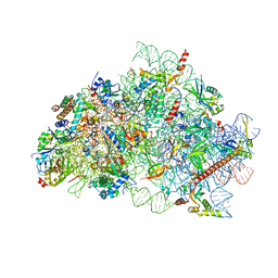 | | CryoEM structure of wheat 40S ribosomal subunit, body domain | | 分子名称: | 30S ribosomal protein S11, chloroplastic, 30S ribosomal protein S4, ... | | 著者 | Kravchenko, O.V, Baymukhametov, T.N, Afonina, Z.A, Vasilenko, K.S. | | 登録日 | 2023-11-22 | | 公開日 | 2023-12-27 | | 最終更新日 | 2024-04-24 | | 実験手法 | ELECTRON MICROSCOPY (2.34 Å) | | 主引用文献 | High-Resolution Structure and Internal Mobility of a Plant 40S Ribosomal Subunit.
Int J Mol Sci, 24, 2023
|
|
3FB4
 | |
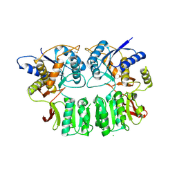
1VAX
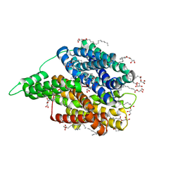
 | | Crystal Structure of Uricase from Arthrobacter globiformis | | 分子名称: | Uric acid oxidase | | 著者 | Hossain, M.T, Suzuki, K, Yamamoto, T, Imamura, S, Sekiguchi, T, Takenaka, A. | | 登録日 | 2004-02-19 | | 公開日 | 2005-06-28 | | 最終更新日 | 2023-10-25 | | 実験手法 | X-RAY DIFFRACTION (1.99 Å) | | 主引用文献 | Crystal Structure of Uricase from Arthrobacter globiformis
To be Published
|
|
7YSJ
 | |
6YV7
 | |
8DMB
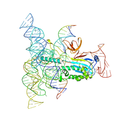 | | Structure of Desulfovirgula thermocuniculi IsrB (DtIsrB) in complex with omega RNA and target DNA | | 分子名称: | MAGNESIUM ION, Ubiquitin-like protein SMT3,IsrB protein,monomeric superfolder Green Fluorescent Protein, non-target DNA, ... | | 著者 | Seiichi, H, Kappel, K, Zhang, F. | | 登録日 | 2022-07-08 | | 公開日 | 2022-10-19 | | 最終更新日 | 2024-06-12 | | 実験手法 | ELECTRON MICROSCOPY (3.1 Å) | | 主引用文献 | Structure of the OMEGA nickase IsrB in complex with omega RNA and target DNA.
Nature, 610, 2022
|
|
6YV9
 | |
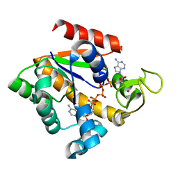
5H7H
 | | Crystal structure of the BCL6 BTB domain in complex with F1324(10-13) | | 分子名称: | 1,2-ETHANEDIOL, B-cell lymphoma 6 protein, F1324 peptide residues 10-13 | | 著者 | Sogabe, S, Ida, K, Lane, W, Snell, G. | | 登録日 | 2016-11-18 | | 公開日 | 2016-12-07 | | 最終更新日 | 2023-11-08 | | 実験手法 | X-RAY DIFFRACTION (1.95 Å) | | 主引用文献 | Discovery of high-affinity BCL6-binding peptide and its structure-activity relationship.
Biochem. Biophys. Res. Commun., 482, 2017
|
|
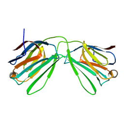
5H8O
 | | Crystal structure of an ASC-binding nanobody in complex with the CARD domain of ASC | | 分子名称: | Apoptosis-associated speck-like protein containing a CARD, VHH nanobody | | 著者 | Lu, A, Schmidt, F.I, Ruan, J, Tang, C, Wu, H, Ploegh, H.L. | | 登録日 | 2015-12-23 | | 公開日 | 2016-04-06 | | 最終更新日 | 2016-05-18 | | 実験手法 | X-RAY DIFFRACTION (4.206 Å) | | 主引用文献 | A single domain antibody fragment that recognizes the adaptor ASC defines the role of ASC domains in inflammasome assembly.
J.Exp.Med., 213, 2016
|
|
5H7G
 | | Crystal structure of the BCL6 BTB domain in complex with F1324 | | 分子名称: | B-cell lymphoma 6 protein, F1324 peptide, SULFATE ION | | 著者 | Sogabe, S, Ida, K, Lane, W, Snell, G. | | 登録日 | 2016-11-18 | | 公開日 | 2016-12-07 | | 最終更新日 | 2023-11-08 | | 実験手法 | X-RAY DIFFRACTION (1.85 Å) | | 主引用文献 | Discovery of high-affinity BCL6-binding peptide and its structure-activity relationship.
Biochem. Biophys. Res. Commun., 482, 2017
|
|
5H8D
 | | Crystal structure of an ASC binding nanobody | | 分子名称: | VHH nanobody | | 著者 | Lu, A, Schmidt, F.I, Ruan, J, Tang, C, Wu, H, Ploegh, H.L. | | 登録日 | 2015-12-23 | | 公開日 | 2016-04-06 | | 最終更新日 | 2016-05-18 | | 実験手法 | X-RAY DIFFRACTION (1.89 Å) | | 主引用文献 | A single domain antibody fragment that recognizes the adaptor ASC defines the role of ASC domains in inflammasome assembly.
J.Exp.Med., 213, 2016
|
|
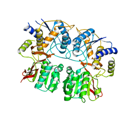
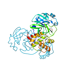
6EI3
 | | Crystal structure of auto inhibited POT family peptide transporter | | 分子名称: | (2S)-2,3-DIHYDROXYPROPYL(7Z)-PENTADEC-7-ENOATE, Proton-dependent oligopeptide transporter family protein | | 著者 | Newstead, S, Brinth, A, Vogeley, L, Caffrey, M. | | 登録日 | 2017-09-17 | | 公開日 | 2017-11-22 | | 最終更新日 | 2024-01-17 | | 実験手法 | X-RAY DIFFRACTION (2.1 Å) | | 主引用文献 | Proton movement and coupling in the POT family of peptide transporters.
Proc. Natl. Acad. Sci. U.S.A., 114, 2017
|
|
6F62
 | |
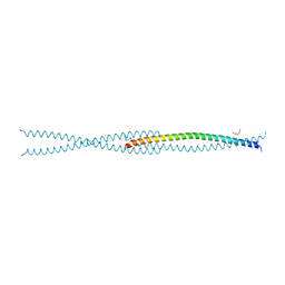
6F64
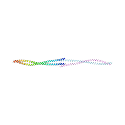
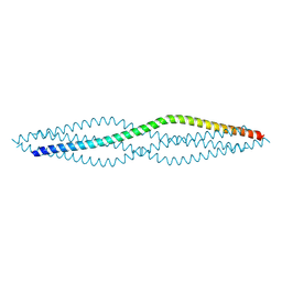 | | Crystal structure of the SYCP1 C-terminal back-to-back assembly | | 分子名称: | ACETATE ION, Synaptonemal complex protein 1 | | 著者 | Dunce, J.M, Millan, C, Uson, I, Davies, O.R. | | 登録日 | 2017-12-04 | | 公開日 | 2018-06-06 | | 最終更新日 | 2020-04-22 | | 実験手法 | X-RAY DIFFRACTION (2.493 Å) | | 主引用文献 | Structural basis of meiotic chromosome synapsis through SYCP1 self-assembly.
Nat. Struct. Mol. Biol., 25, 2018
|
|
6F5X
 | |
7TGR
 | | Structure of SARS-CoV-2 main protease in complex with GC376 | | 分子名称: | (1R,2S)-2-({N-[(benzyloxy)carbonyl]-L-leucyl}amino)-1-hydroxy-3-[(3S)-2-oxopyrrolidin-3-yl]propane-1-sulfonic acid, (1S,2S)-2-({N-[(benzyloxy)carbonyl]-L-leucyl}amino)-1-hydroxy-3-[(3S)-2-oxopyrrolidin-3-yl]propane-1-sulfonic acid, 1,2-ETHANEDIOL, ... | | 著者 | Esler, M.A, Shi, K, Aihara, H, Harris, R.S. | | 登録日 | 2022-01-09 | | 公開日 | 2022-05-04 | | 最終更新日 | 2023-10-18 | | 実験手法 | X-RAY DIFFRACTION (1.68 Å) | | 主引用文献 | Gain-of-Signal Assays for Probing Inhibition of SARS-CoV-2 M pro /3CL pro in Living Cells.
Mbio, 13, 2022
|
|
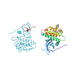
4JQ7
 | | Crystal structure of EGFR kinase domain in complex with compound 2a | | 分子名称: | (2S)-2-[(5,6-diphenylfuro[2,3-d]pyrimidin-4-yl)amino]-2-phenylethanol, Epidermal growth factor receptor | | 著者 | Peng, Y.H, Wu, J.S. | | 登録日 | 2013-03-20 | | 公開日 | 2013-06-19 | | 最終更新日 | 2023-11-08 | | 実験手法 | X-RAY DIFFRACTION (2.73 Å) | | 主引用文献 | Protein Kinase Inhibitor Design by Targeting the Asp-Phe-Gly (DFG) Motif: The Role of the DFG Motif in the Design of Epidermal Growth Factor Receptor Inhibitors
J.Med.Chem., 56, 2013
|
|
4JRV
 | | Crystal structure of EGFR kinase domain in complex with compound 4c | | 分子名称: | 4-(dimethylamino)-N-[3-(4-{[(1S)-2-hydroxy-1-phenylethyl]amino}-6-phenylfuro[2,3-d]pyrimidin-5-yl)phenyl]butanamide, Epidermal growth factor receptor | | 著者 | Peng, Y.H, Wu, J.S. | | 登録日 | 2013-03-22 | | 公開日 | 2013-06-19 | | 最終更新日 | 2023-11-08 | | 実験手法 | X-RAY DIFFRACTION (2.8 Å) | | 主引用文献 | Protein Kinase Inhibitor Design by Targeting the Asp-Phe-Gly (DFG) Motif: The Role of the DFG Motif in the Design of Epidermal Growth Factor Receptor Inhibitors
J.Med.Chem., 56, 2013
|
|


