7SZ2
 
 | |
1XGK
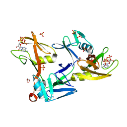
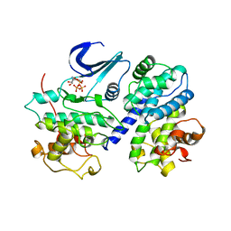
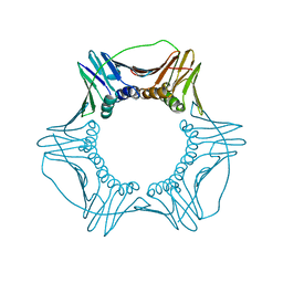
 | | CRYSTAL STRUCTURE OF N12G AND A18G MUTANT NMRA | | 分子名称: | CHLORIDE ION, GLYCEROL, NITROGEN METABOLITE REPRESSION REGULATOR NMRA, ... | | 著者 | Lamb, H.K, Ren, J, Park, A, Johnson, C, Leslie, K, Cocklin, S, Thompson, P, Mee, C, Cooper, A, Stammers, D.K, Hawkins, A.R. | | 登録日 | 2004-09-17 | | 公開日 | 2004-12-14 | | 最終更新日 | 2023-08-23 | | 実験手法 | X-RAY DIFFRACTION (1.4 Å) | | 主引用文献 | Modulation of the ligand binding properties of the transcription repressor NmrA by GATA-containing DNA and site-directed mutagenesis
Protein Sci., 13, 2004
|
|
7SZ3
 | |
1ZME
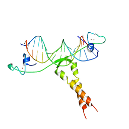 | | CRYSTAL STRUCTURE OF PUT3/DNA COMPLEX | | 分子名称: | DNA (5'-D(*AP*CP*GP*GP*AP*GP*(5IU)P*TP*GP*GP*CP*TP*(5IU)P*CP*CP*CP*G)-3'), DNA (5'-D(*AP*CP*GP*GP*GP*AP*AP*GP*CP*CP*AP*AP*CP*TP*CP*CP*G)-3'), PROLINE UTILIZATION TRANSCRIPTION ACTIVATOR, ... | | 著者 | Swaminathan, K, Marmorstein, R. | | 登録日 | 1997-08-06 | | 公開日 | 1998-09-16 | | 最終更新日 | 2024-02-14 | | 実験手法 | X-RAY DIFFRACTION (2.5 Å) | | 主引用文献 | Crystal structure of a PUT3-DNA complex reveals a novel mechanism for DNA recognition by a protein containing a Zn2Cys6 binuclear cluster.
Nat.Struct.Biol., 4, 1997
|
|
1FIN
 | |
3KAH
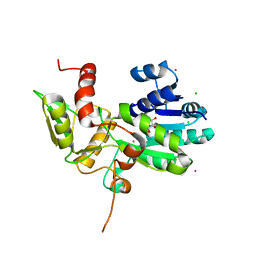
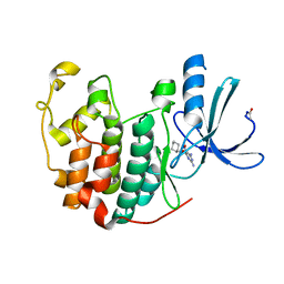
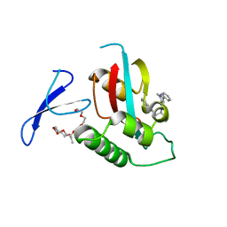 | | Structure-guided design of alpha-amino acid-derived Pin1 inhibitors | | 分子名称: | 3-(1H-benzimidazol-2-yl)-N-[(1-methyl-3-phenyl-1H-pyrazol-5-yl)carbonyl]-D-alanine, DODECAETHYLENE GLYCOL, Peptidyl-prolyl cis-trans isomerase NIMA-interacting 1 | | 著者 | Baker, L.M, Dokurno, P, Robinson, D.A, Surgenor, A.E, Murray, J.B, Potter, A.J, Moore, J.D. | | 登録日 | 2009-10-19 | | 公開日 | 2009-12-22 | | 最終更新日 | 2023-11-01 | | 実験手法 | X-RAY DIFFRACTION (2.3 Å) | | 主引用文献 | Structure-guided design of alpha-amino acid-derived Pin1 inhibitors
Bioorg.Med.Chem.Lett., 20, 2010
|
|
3JVF
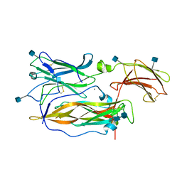 | | Crystal structure of an Interleukin-17 receptor complex | | 分子名称: | 2-acetamido-2-deoxy-beta-D-glucopyranose, 2-acetamido-2-deoxy-beta-D-glucopyranose-(1-4)-2-acetamido-2-deoxy-beta-D-glucopyranose, CALCIUM ION, ... | | 著者 | Ely, L.K, Garcia, K.C. | | 登録日 | 2009-09-16 | | 公開日 | 2009-10-20 | | 最終更新日 | 2020-07-29 | | 実験手法 | X-RAY DIFFRACTION (3.3 Å) | | 主引用文献 | Structural basis of receptor sharing by interleukin 17 cytokines.
Nat.Immunol., 10, 2009
|
|
7EXE
 | |
1VYZ
 | | Structure of CDK2 complexed with PNU-181227 | | 分子名称: | CELL DIVISION PROTEIN KINASE 2, N-(5-CYCLOPROPYL-1H-PYRAZOL-3-YL)BENZAMIDE | | 著者 | Pevarello, P, Brasca, M.G, Amici, R, Orsini, P, Traquandi, G, Corti, L, Piutti, C, Sansonna, P, Villa, M, Pierce, B.S, Pulici, M, Giordano, G, Martina, K, Lfritzen, E, Nugent, R.A, Casale, E, Cameron, A, Ciomei, M, Roletto, F, Isacchi, A, Fogliatto, G, Pesenti, E, Pastori, W, Marsiglio, W, Leach, K.L, Clare, P.M, Fiorentini, F, Varasi, M, Vulpetti, A, Warpehoski, M.A. | | 登録日 | 2004-05-07 | | 公開日 | 2004-06-17 | | 最終更新日 | 2024-05-08 | | 実験手法 | X-RAY DIFFRACTION (2.21 Å) | | 主引用文献 | 3-Aminopyrazole Inhibitors of Cdk2/Cyclin a as Antitumor Agents. 1. Lead Finding
J.Med.Chem., 47, 2004
|
|
3KAI
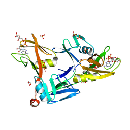
 | | Structure-guided design of alpha-amino acid-derived Pin1 inhibitors | | 分子名称: | (2R)-2-[(2-methyl-5-phenyl-pyrazol-3-yl)carbonylamino]-3-naphthalen-2-yl-propanoic acid, DODECAETHYLENE GLYCOL, Peptidyl-prolyl cis-trans isomerase NIMA-interacting 1 | | 著者 | Baker, L.M, Dokurno, P, Robinson, D.A, Surgenor, A.E, Murray, J.B, Potter, A.J, Moore, J.D. | | 登録日 | 2009-10-19 | | 公開日 | 2009-12-22 | | 最終更新日 | 2023-11-01 | | 実験手法 | X-RAY DIFFRACTION (1.9 Å) | | 主引用文献 | Structure-guided design of alpha-amino acid-derived Pin1 inhibitors
Bioorg.Med.Chem.Lett., 20, 2010
|
|
1W0X
 | |
1FTZ
 | | NUCLEAR MAGNETIC RESONANCE SOLUTION STRUCTURE OF THE FUSHI TARAZU HOMEODOMAIN FROM DROSOPHILA AND COMPARISON WITH THE ANTENNAPEDIA HOMEODOMAIN | | 分子名称: | FUSHI TARAZU PROTEIN | | 著者 | Qian, Y.Q, Furukubo-Tokunaga, K, Resendez-Perez, D, Muller, M, Gehring, W.J, Wuthrich, K. | | 登録日 | 1994-01-07 | | 公開日 | 1994-05-31 | | 最終更新日 | 2024-05-22 | | 実験手法 | SOLUTION NMR | | 主引用文献 | Nuclear magnetic resonance solution structure of the fushi tarazu homeodomain from Drosophila and comparison with the Antennapedia homeodomain.
J.Mol.Biol., 238, 1994
|
|
6CX2
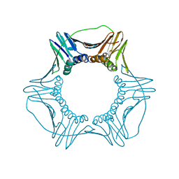 | | S177G Mutant of Yeast PCNA | | 分子名称: | Proliferating cell nuclear antigen | | 著者 | Powers, K.T. | | 登録日 | 2018-04-02 | | 公開日 | 2018-04-11 | | 最終更新日 | 2023-10-04 | | 実験手法 | X-RAY DIFFRACTION (3.101 Å) | | 主引用文献 | Identification of New Mutations at the PCNA Subunit Interface that Block Translesion Synthesis.
PLoS ONE, 11, 2016
|
|
1CLD
 | | DNA-binding protein | | 分子名称: | CADMIUM ION, CD2-LAC9 | | 著者 | Gardner, K.H, Coleman, J.E. | | 登録日 | 1995-06-06 | | 公開日 | 1995-09-15 | | 最終更新日 | 2024-05-22 | | 実験手法 | SOLUTION NMR | | 主引用文献 | Solution structure of the Kluyveromyces lactis LAC9 Cd2 Cys6 DNA-binding domain.
Nat.Struct.Biol., 2, 1995
|
|
1E1X
 | |
1DZ7
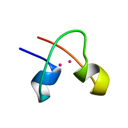
 | | Solution structure of the a-subunit of human chorionic gonadotropin [modeled without carbohydrate residues] | | 分子名称: | CHORIONIC GONADOTROPIN | | 著者 | Erbel, P.J.A, Karimi-Nejad, Y, De Beer, T, Boelens, R, Kamerling, J.P, Vliegenthart, J.F.G. | | 登録日 | 2000-02-18 | | 公開日 | 2000-02-29 | | 最終更新日 | 2019-10-09 | | 実験手法 | SOLUTION NMR | | 主引用文献 | Solution structure of the alpha-subunit of human chorionic gonadotropin.
Eur.J.Biochem., 260, 1999
|
|
5VPB
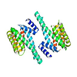
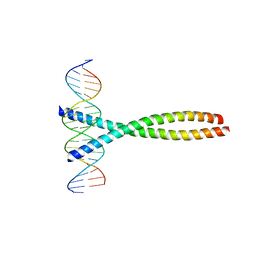
 | | Transcription factor FosB/JunD bZIP domain in its oxidized form, type-I crystal | | 分子名称: | CHLORIDE ION, Protein fosB, Transcription factor jun-D | | 著者 | Yin, Z, Machius, M, Rudenko, G. | | 登録日 | 2017-05-04 | | 公開日 | 2017-09-06 | | 最終更新日 | 2023-10-04 | | 実験手法 | X-RAY DIFFRACTION (2.691 Å) | | 主引用文献 | Activator Protein-1: redox switch controlling structure and DNA-binding.
Nucleic Acids Res., 45, 2017
|
|
1Z4H
 | | The response regulator TorI belongs to a new family of atypical excisionase | | 分子名称: | Tor inhibition protein | | 著者 | Elantak, L, Ansaldi, M, Guerlesquin, F, Mejean, V, Morelli, X. | | 登録日 | 2005-03-16 | | 公開日 | 2005-08-09 | | 最終更新日 | 2024-05-22 | | 実験手法 | SOLUTION NMR | | 主引用文献 | Structural and genetic analyses reveal a key role in prophage excision for the TorI response regulator inhibitor
J.Biol.Chem., 280, 2005
|
|
7CTG
 | | Human Origin Recognition Complex, ORC1-5 State I | | 分子名称: | ADENOSINE-5'-TRIPHOSPHATE, Origin recognition complex subunit 1, Origin recognition complex subunit 2, ... | | 著者 | Cheng, J, Li, N, Wang, X, Hu, J, Zhai, Y, Gao, N. | | 登録日 | 2020-08-18 | | 公開日 | 2021-01-06 | | 最終更新日 | 2024-03-27 | | 実験手法 | ELECTRON MICROSCOPY (5 Å) | | 主引用文献 | Structural insight into the assembly and conformational activation of human origin recognition complex.
Cell Discov, 6, 2020
|
|
5V95
 | |
6CX3
 | | S179T Mutant of Yeast PCNA | | 分子名称: | Proliferating cell nuclear antigen | | 著者 | Powers, K.T. | | 登録日 | 2018-04-02 | | 公開日 | 2018-04-11 | | 最終更新日 | 2023-10-04 | | 実験手法 | X-RAY DIFFRACTION (3.101 Å) | | 主引用文献 | Identification of New Mutations at the PCNA Subunit Interface that Block Translesion Synthesis.
PLoS ONE, 11, 2016
|
|
1F9S
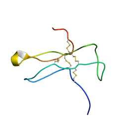
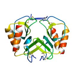 | | CRYSTAL STRUCTURE OF PLATELET FACTOR 4 MUTANT 2 | | 分子名称: | PLATELET FACTOR 4 | | 著者 | Yang, J, Doyle, M, Faulk, T, Visentin, G, Aster, R, Edwards, B. | | 登録日 | 2000-07-11 | | 公開日 | 2003-08-26 | | 最終更新日 | 2024-10-09 | | 実験手法 | X-RAY DIFFRACTION (2.38 Å) | | 主引用文献 | Structure Comparison of Two Platelet Factor 4 Mutants with the Wild-type Reveals the Epitopes for the Heparin-induced Thrombocytopenia Antibodies
To be Published
|
|
1FOS
 | | TWO HUMAN C-FOS:C-JUN:DNA COMPLEXES | | 分子名称: | C-JUN PROTO-ONCOGENE PROTEIN, DNA (5'-D(*AP*AP*TP*GP*GP*AP*TP*GP*AP*GP*TP*CP*AP*TP*AP*GP*GP*AP*GP*A)-3'), DNA (5'-D(*TP*TP*CP*TP*CP*CP*TP*AP*TP*GP*AP*CP*TP*CP*AP*TP*CP*CP*AP*T)-3'), ... | | 著者 | Glover, J.N.M, Harrison, S.C. | | 登録日 | 1995-03-07 | | 公開日 | 1995-07-10 | | 最終更新日 | 2024-02-07 | | 実験手法 | X-RAY DIFFRACTION (3.05 Å) | | 主引用文献 | Crystal structure of the heterodimeric bZIP transcription factor c-Fos-c-Jun bound to DNA.
Nature, 373, 1995
|
|
1YFD
 | | Crystal structure of the Y122H mutant of ribonucleotide reductase R2 protein from E. coli | | 分子名称: | MERCURY (II) ION, MU-OXO-DIIRON, Ribonucleoside-diphosphate reductase 1 beta chain | | 著者 | Kolberg, M, Logan, D.T, Bleifuss, G, Poetsch, S, Sjoeberg, B.M, Graeslund, A, Lubitz, W, Lassmann, G, Lendzian, F. | | 登録日 | 2004-12-31 | | 公開日 | 2005-02-15 | | 最終更新日 | 2023-08-23 | | 実験手法 | X-RAY DIFFRACTION (1.9 Å) | | 主引用文献 | A new tyrosyl radical on Phe208 as ligand to the diiron center in Escherichia coli ribonucleotide reductase, mutant R2-Y122H. Combined x-ray diffraction and EPR/ENDOR studies
J.Biol.Chem., 280, 2005
|
|
1FQ1
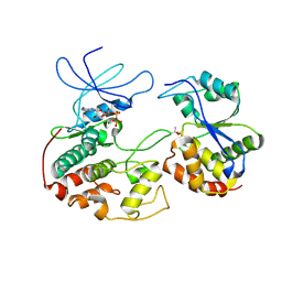 | | CRYSTAL STRUCTURE OF KINASE ASSOCIATED PHOSPHATASE (KAP) IN COMPLEX WITH PHOSPHO-CDK2 | | 分子名称: | ADENOSINE-5'-TRIPHOSPHATE, CELL DIVISION PROTEIN KINASE 2, CYCLIN-DEPENDENT KINASE INHIBITOR 3, ... | | 著者 | Song, H, Hanlon, N, Brown, N.R, Noble, M.E.M, Johnson, L.N, Barford, D. | | 登録日 | 2000-09-01 | | 公開日 | 2001-05-09 | | 最終更新日 | 2018-03-14 | | 実験手法 | X-RAY DIFFRACTION (3 Å) | | 主引用文献 | Phosphoprotein-protein interactions revealed by the crystal structure of kinase-associated phosphatase in complex with phosphoCDK2.
Mol.Cell, 7, 2001
|
|


