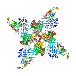
8JU5
 
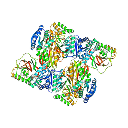
 | | Structure of human TRPV4 with antagonist A1 | | 分子名称: | 4-[(3~{S},4~{S})-4-(aminomethyl)-1-(5-chloranylpyridin-2-yl)sulfonyl-4-oxidanyl-pyrrolidin-3-yl]oxy-2-fluoranyl-benzenecarbonitrile, Transient receptor potential cation channel subfamily V member 4,3C-GFP | | 著者 | Fan, J, Lei, X. | | 登録日 | 2023-06-24 | | 公開日 | 2024-05-08 | | 最終更新日 | 2024-07-24 | | 実験手法 | ELECTRON MICROSCOPY (3.74 Å) | | 主引用文献 | Structural Pharmacology of TRPV4 Antagonists.
Adv Sci, 11, 2024
|
|
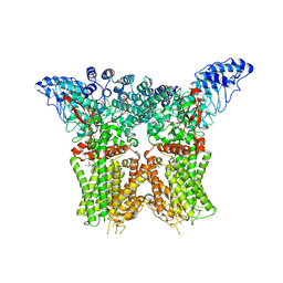
8JVI
 | | Structure of human TRPV4 with antagonist A2 | | 分子名称: | Transient receptor potential cation channel subfamily V member 4,3C-GFP, [6-[[4-(2,4-dimethyl-1,3-thiazol-5-yl)-1,3-thiazol-2-yl]amino]pyridin-3-yl]-[(1~{S},5~{R})-3-[5-(trifluoromethyl)pyrimidin-2-yl]-3,8-diazabicyclo[3.2.1]octan-8-yl]methanone | | 著者 | Fan, J, Lei, X. | | 登録日 | 2023-06-28 | | 公開日 | 2024-05-08 | | 最終更新日 | 2024-07-24 | | 実験手法 | ELECTRON MICROSCOPY (3.21 Å) | | 主引用文献 | Structural Pharmacology of TRPV4 Antagonists.
Adv Sci, 11, 2024
|
|
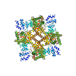
8JVJ
 | | Structure of human TRPV4 with antagonist A2 and RhoA | | 分子名称: | Transforming protein RhoA, Transient receptor potential cation channel subfamily V member 4,3C-GFP, [6-[[4-(2,4-dimethyl-1,3-thiazol-5-yl)-1,3-thiazol-2-yl]amino]pyridin-3-yl]-[(1~{S},5~{R})-3-[5-(trifluoromethyl)pyrimidin-2-yl]-3,8-diazabicyclo[3.2.1]octan-8-yl]methanone | | 著者 | Fan, J, Lei, X. | | 登録日 | 2023-06-28 | | 公開日 | 2024-05-08 | | 最終更新日 | 2024-07-24 | | 実験手法 | ELECTRON MICROSCOPY (3.44 Å) | | 主引用文献 | Structural Pharmacology of TRPV4 Antagonists.
Adv Sci, 11, 2024
|
|
8JU6
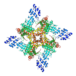 | | Structure of human TRPV4 with antagonist GSK279 | | 分子名称: | 1-({(5S,7S)-3-[5-(2-hydroxypropan-2-yl)pyrazin-2-yl]-7-methyl-2-oxo-1-oxa-3-azaspiro[4.5]decan-7-yl}methyl)-1H-benzimidazole-6-carbonitrile, Transient receptor potential cation channel subfamily V member 4,3C-GFP | | 著者 | Fan, J, Lei, X. | | 登録日 | 2023-06-24 | | 公開日 | 2024-05-08 | | 最終更新日 | 2024-07-24 | | 実験手法 | ELECTRON MICROSCOPY (3.45 Å) | | 主引用文献 | Structural Pharmacology of TRPV4 Antagonists.
Adv Sci, 11, 2024
|
|
3VZD
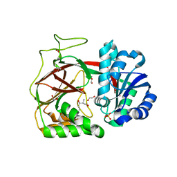
 | | Crystal structure of Sphingosine Kinase 1 with inhibitor and ADP | | 分子名称: | 4-{[4-(4-chlorophenyl)-1,3-thiazol-2-yl]amino}phenol, ADENOSINE-5'-DIPHOSPHATE, CHLORIDE ION, ... | | 著者 | Min, X, Walker, N.P, Wang, Z. | | 登録日 | 2012-10-11 | | 公開日 | 2013-05-01 | | 最終更新日 | 2024-03-20 | | 実験手法 | X-RAY DIFFRACTION (2.3 Å) | | 主引用文献 | Molecular basis of sphingosine kinase 1 substrate recognition and catalysis.
Structure, 21, 2013
|
|
8OTZ
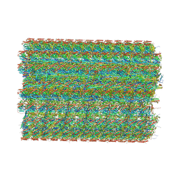 | | 48-nm repeat of the native axonemal doublet microtubule from bovine sperm | | 分子名称: | ATP6V1F neighbor, CFAP97 domain containing 1, Chromosome 13 C20orf85 homolog, ... | | 著者 | Leung, M.R, Zeng, J, Zhang, R, Zeev-Ben-Mordehai, T. | | 登録日 | 2023-04-21 | | 公開日 | 2023-11-22 | | 実験手法 | ELECTRON MICROSCOPY (3.6 Å) | | 主引用文献 | Structural specializations of the sperm tail.
Cell, 186, 2023
|
|
3VZB
 | | Crystal structure of Sphingosine Kinase 1 | | 分子名称: | (2S,3R,4E)-2-aminooctadec-4-ene-1,3-diol, 1,2-ETHANEDIOL, SULFATE ION, ... | | 著者 | Min, X, Walker, N.P, Wang, Z. | | 登録日 | 2012-10-10 | | 公開日 | 2013-05-08 | | 最終更新日 | 2024-05-29 | | 実験手法 | X-RAY DIFFRACTION (2 Å) | | 主引用文献 | Molecular basis of sphingosine kinase 1 substrate recognition and catalysis.
Structure, 21, 2013
|
|
8IYJ
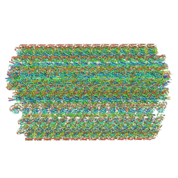 | | Cryo-EM structure of the 48-nm repeat doublet microtubule from mouse sperm | | 分子名称: | Cilia and flagella-associated protein 77, Cilia- and flagella-associated protein 107, Cilia- and flagella-associated protein 141, ... | | 著者 | Zhou, L.N, Gui, M, Wu, J.P. | | 登録日 | 2023-04-05 | | 公開日 | 2023-07-05 | | 最終更新日 | 2024-07-03 | | 実験手法 | ELECTRON MICROSCOPY (3.5 Å) | | 主引用文献 | Structures of sperm flagellar doublet microtubules expand the genetic spectrum of male infertility.
Cell, 186, 2023
|
|
3VZC
 | | Crystal structure of Sphingosine Kinase 1 with inhibitor | | 分子名称: | 1,2-ETHANEDIOL, 4-{[4-(4-chlorophenyl)-1,3-thiazol-2-yl]amino}phenol, Sphingosine kinase 1 | | 著者 | Min, X, Walker, N.P, Wang, Z. | | 登録日 | 2012-10-11 | | 公開日 | 2013-05-08 | | 最終更新日 | 2024-03-20 | | 実験手法 | X-RAY DIFFRACTION (2.3 Å) | | 主引用文献 | Molecular basis of sphingosine kinase 1 substrate recognition and catalysis.
Structure, 21, 2013
|
|
1ALW
 | | INHIBITOR AND CALCIUM BOUND DOMAIN VI OF PORCINE CALPAIN | | 分子名称: | 3-(4-IODO-PHENYL)-2-MERCAPTO-PROPIONIC ACID, CALCIUM ION, CALPAIN | | 著者 | Narayana, S.V.L, Lin, G. | | 登録日 | 1997-06-04 | | 公開日 | 1998-06-10 | | 最終更新日 | 2024-04-03 | | 実験手法 | X-RAY DIFFRACTION (2.03 Å) | | 主引用文献 | Crystal structure of calcium bound domain VI of calpain at 1.9 A resolution and its role in enzyme assembly, regulation, and inhibitor binding.
Nat.Struct.Biol., 4, 1997
|
|
1ALV
 | | CALCIUM BOUND DOMAIN VI OF PORCINE CALPAIN | | 分子名称: | CALCIUM ION, CALPAIN | | 著者 | Narayana, S.V.L, Lin, G, Chattopadhyay, D, Maki, M. | | 登録日 | 1997-06-03 | | 公開日 | 1998-06-03 | | 最終更新日 | 2024-02-07 | | 実験手法 | X-RAY DIFFRACTION (1.9 Å) | | 主引用文献 | Crystal structure of calcium bound domain VI of calpain at 1.9 A resolution and its role in enzyme assembly, regulation, and inhibitor binding.
Nat.Struct.Biol., 4, 1997
|
|
1A2X
 | | COMPLEX OF TROPONIN C WITH A 47 RESIDUE (1-47) FRAGMENT OF TROPONIN I | | 分子名称: | CALCIUM ION, TROPONIN C, TROPONIN I | | 著者 | Vassylyev, D.G, Takeda, S, Wakatsuki, S, Maeda, K, Maeda, Y. | | 登録日 | 1998-01-13 | | 公開日 | 1998-07-15 | | 最終更新日 | 2024-02-07 | | 実験手法 | X-RAY DIFFRACTION (2.3 Å) | | 主引用文献 | Crystal structure of troponin C in complex with troponin I fragment at 2.3-A resolution.
Proc.Natl.Acad.Sci.USA, 95, 1998
|
|
1B9A
 | | PARVALBUMIN (MUTATION;D51A, F102W) | | 分子名称: | CALCIUM ION, PROTEIN (PARVALBUMIN) | | 著者 | Cates, M.S, Berry, M.B, Ho, E.L, Li, Q, Potter, J.D, Phillips Jr, G.N. | | 登録日 | 1999-02-10 | | 公開日 | 1999-02-15 | | 最終更新日 | 2023-08-09 | | 実験手法 | X-RAY DIFFRACTION (2 Å) | | 主引用文献 | Metal-ion affinity and specificity in EF-hand proteins: coordination geometry and domain plasticity in parvalbumin.
Structure Fold.Des., 7, 1999
|
|
1B8L
 | | Calcium-bound D51A/E101D/F102W Triple Mutant of Beta Carp Parvalbumin | | 分子名称: | CALCIUM ION, CARBONATE ION, PROTEIN (PARVALBUMIN) | | 著者 | Cates, M.S, Berry, M.B, Ho, E, Li, Q, Potter, J.D, Phillips Jr, G.N. | | 登録日 | 1999-02-01 | | 公開日 | 1999-10-05 | | 最終更新日 | 2023-08-09 | | 実験手法 | X-RAY DIFFRACTION (1.7 Å) | | 主引用文献 | Metal-ion affinity and specificity in EF-hand proteins: coordination geometry and domain plasticity in parvalbumin.
Structure Fold.Des., 7, 1999
|
|
1B8R
 | | PARVALBUMIN | | 分子名称: | CALCIUM ION, PROTEIN (PARVALBUMIN) | | 著者 | Cates, M.S, Berry, M.B, Ho, E.L, Li, Q, Potter, J.D, Phillips Jr, G.N. | | 登録日 | 1999-02-02 | | 公開日 | 1999-02-09 | | 最終更新日 | 2023-08-09 | | 実験手法 | X-RAY DIFFRACTION (1.9 Å) | | 主引用文献 | Metal-ion affinity and specificity in EF-hand proteins: coordination geometry and domain plasticity in parvalbumin.
Structure Fold.Des., 7, 1999
|
|
2B1O
 | | Solution Structure of Ca2+-bound DdCAD-1 | | 分子名称: | CALCIUM ION, Calcium-dependent cell adhesion molecule-1 | | 著者 | Lin, Z, Sriskanthadevan, S, Huang, H.B, Siu, C.H, Yang, D.W. | | 登録日 | 2005-09-16 | | 公開日 | 2006-09-26 | | 最終更新日 | 2024-05-29 | | 実験手法 | SOLUTION NMR | | 主引用文献 | Solution structures of the adhesion molecule DdCAD-1 reveal new insights into Ca(2+)-dependent cell-cell adhesion
Nat.Struct.Mol.Biol., 13, 2006
|
|
2AQX
 | | Crystal Structure of the Catalytic and CaM-Binding domains of Inositol 1,4,5-Trisphosphate 3-Kinase B | | 分子名称: | ADENOSINE-5'-TRIPHOSPHATE, MAGNESIUM ION, PREDICTED: inositol 1,4,5-trisphosphate 3-kinase B | | 著者 | Chamberlain, P.P, Sandberg, M.L, Sauer, K, Cooke, M.P, Lesley, S.A, Spraggon, G. | | 登録日 | 2005-08-18 | | 公開日 | 2005-12-06 | | 最終更新日 | 2024-03-13 | | 実験手法 | X-RAY DIFFRACTION (2.5 Å) | | 主引用文献 | Structural insights into enzyme regulation for inositol 1,4,5-trisphosphate 3-kinase B
Biochemistry, 44, 2005
|
|
1DTL
 | | CRYSTAL STRUCTURE OF CALCIUM-SATURATED (3CA2+) CARDIAC TROPONIN C COMPLEXED WITH THE CALCIUM SENSITIZER BEPRIDIL AT 2.15 A RESOLUTION | | 分子名称: | 1-ISOBUTOXY-2-PYRROLIDINO-3[N-BENZYLANILINO] PROPANE, CALCIUM ION, CARDIAC TROPONIN C | | 著者 | Li, Y, Love, M.L, Putkey, J.A, Cohen, C. | | 登録日 | 2000-01-12 | | 公開日 | 2000-05-12 | | 最終更新日 | 2023-08-09 | | 実験手法 | X-RAY DIFFRACTION (2.15 Å) | | 主引用文献 | Bepridil opens the regulatory N-terminal lobe of cardiac troponin C.
Proc.Natl.Acad.Sci.USA, 97, 2000
|
|
2C32
 | |
2BL0
 | | Physarum polycephalum myosin II regulatory domain | | 分子名称: | CALCIUM ION, MAJOR PLASMODIAL MYOSIN HEAVY CHAIN, MYOSIN REGULATORY LIGHT CHAIN | | 著者 | Debreczeni, J.E, Farkas, L, Harmat, V, Nyitray, L. | | 登録日 | 2005-02-23 | | 公開日 | 2005-10-13 | | 最終更新日 | 2024-05-08 | | 実験手法 | X-RAY DIFFRACTION (1.75 Å) | | 主引用文献 | Structural Evidence for Non-Canonical Binding of Ca2+ to a Canonical EF-Hand of a Conventional Myosin.
J.Biol.Chem., 280, 2005
|
|
2B6P
 | | X-ray structure of lens Aquaporin-0 (AQP0) (lens MIP) in an open pore state | | 分子名称: | Lens fiber major intrinsic protein | | 著者 | Gonen, T, Cheng, Y, Sliz, P, Hiroaki, Y, Fujiyoshi, Y, Harrison, S.C, Walz, T. | | 登録日 | 2005-10-03 | | 公開日 | 2005-12-06 | | 最終更新日 | 2024-02-14 | | 実験手法 | X-RAY DIFFRACTION (2.4 Å) | | 主引用文献 | Lipid-protein interactions in double-layered two-dimensional AQP0 crystals.
Nature, 438, 2005
|
|
1BK2
 | |
1E6H
 | |
1E6G
 | | A-SPECTRIN SH3 DOMAIN A11V, V23L, M25I, V53I, V58L MUTANT | | 分子名称: | SPECTRIN ALPHA CHAIN, SULFATE ION | | 著者 | Vega, M.C, Serrano, L. | | 登録日 | 2000-08-15 | | 公開日 | 2002-05-23 | | 最終更新日 | 2023-12-13 | | 実験手法 | X-RAY DIFFRACTION (2.3 Å) | | 主引用文献 | Conformational Strain in the Hydrophobic Core and its Implications for Protein Folding and Design
Nat.Struct.Biol., 9, 2002
|
|
2B6O
 | | Electron crystallographic structure of lens Aquaporin-0 (AQP0) (lens MIP) at 1.9A resolution, in a closed pore state | | 分子名称: | 1,2-DIMYRISTOYL-RAC-GLYCERO-3-PHOSPHOCHOLINE, Lens fiber major intrinsic protein | | 著者 | Gonen, T, Cheng, Y, Sliz, P, Hiroaki, Y, Fujiyoshi, Y, Harrison, S.C, Walz, T. | | 登録日 | 2005-10-03 | | 公開日 | 2005-12-06 | | 最終更新日 | 2023-08-23 | | 実験手法 | ELECTRON CRYSTALLOGRAPHY (1.9 Å) | | 主引用文献 | Lipid-protein interactions in double-layered two-dimensional AQP0 crystals.
Nature, 438, 2005
|
|


