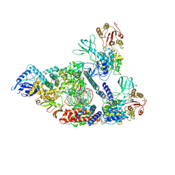
7BWD
 
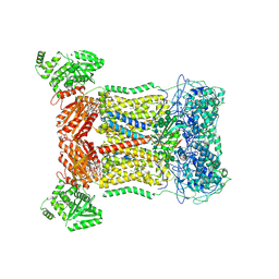
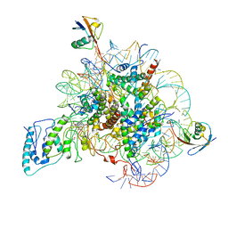 | | Structure of Dot1L-H2BK34ub Nucleosome Complex | | 分子名称: | DNA (146-MER), Histone H2A type 1-B/E, Histone H2B type 1-K, ... | | 著者 | Lou, Z.Y, Liu, L, Cao, L, Ai, H.S, Sun, Z.X. | | 登録日 | 2020-04-14 | | 公開日 | 2021-04-14 | | 実験手法 | ELECTRON MICROSCOPY (4.32 Å) | | 主引用文献 | Structure of the Dot1L-H2BK34ub Nucleosome Complex Reveals an Unprecedented Crosstalk Activation Mechanism through Nucleosome Shape Distortion
To Be Published
|
|
6TRL
 | |
6TLE
 | |
6CBI
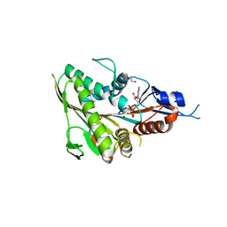
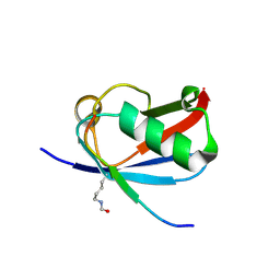
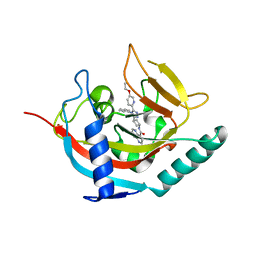
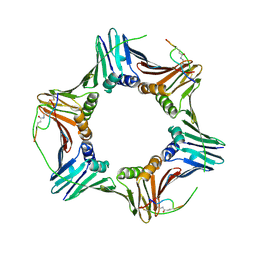 | | PCNA in complex with inhibitor | | 分子名称: | GLY-ARG-LYS-ARG-ARG-GLN-DAB-SER-MET-THR-GLU-PHE-TYR-HIS, Proliferating cell nuclear antigen, SULFATE ION | | 著者 | Bruning, J.B, Wegener, K.L. | | 登録日 | 2018-02-03 | | 公開日 | 2018-07-04 | | 最終更新日 | 2020-01-29 | | 実験手法 | X-RAY DIFFRACTION (2.75 Å) | | 主引用文献 | Rational Design of a 310-Helical PIP-Box Mimetic Targeting PCNA, the Human Sliding Clamp.
Chemistry, 24, 2018
|
|
6CCD
 | |
8A8N
 | |
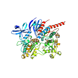
6CF6
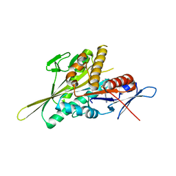
 | | RNF146 TBM-Tankyrase ARC2-3 complex | | 分子名称: | RNF146, Tankyrase-1 | | 著者 | Da Rosa, P.A, Xu, W. | | 登録日 | 2018-02-13 | | 公開日 | 2018-04-18 | | 最終更新日 | 2023-10-04 | | 実験手法 | X-RAY DIFFRACTION (1.93 Å) | | 主引用文献 | Structural basis for tankyrase-RNF146 interaction reveals noncanonical tankyrase-binding motifs.
Protein Sci., 27, 2018
|
|
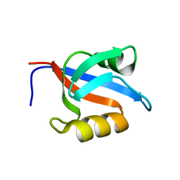
6CPI
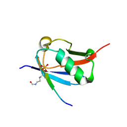
 | | Solution structure of SH3 domain from Shank1 | | 分子名称: | SH3 and multiple ankyrin repeat domains protein 1 | | 著者 | Ishida, H, Vogel, H.J. | | 登録日 | 2018-03-13 | | 公開日 | 2018-08-15 | | 最終更新日 | 2024-05-01 | | 実験手法 | SOLUTION NMR | | 主引用文献 | Solution structures of the SH3 domains from Shank scaffold proteins and their interactions with Cav1.3 calcium channels.
FEBS Lett., 592, 2018
|
|
6UMA
 | |
6UMB
 | |
8BXU
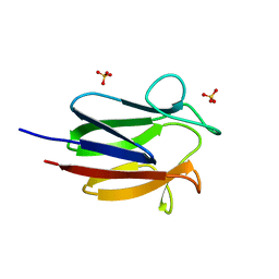
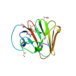
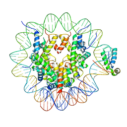
 | | Crystal structure of Odorant Binding Protein 5 from Anopheles gambiae (AgamOBP5) with MPD (2-Methyl-2,4-pentanediol) | | 分子名称: | (4S)-2-METHYL-2,4-PENTANEDIOL, 2-ETHOXYETHANOL, Odorant binding protein, ... | | 著者 | Liggri, P.G.V, Tsitsanou, K.E, Zographos, S.E. | | 登録日 | 2022-12-09 | | 公開日 | 2023-03-22 | | 最終更新日 | 2023-04-12 | | 実験手法 | X-RAY DIFFRACTION (1.35 Å) | | 主引用文献 | The structure of AgamOBP5 in complex with the natural insect repellents Carvacrol and Thymol: Crystallographic, fluorescence and thermodynamic binding studies.
Int.J.Biol.Macromol., 237, 2023
|
|
8BXV
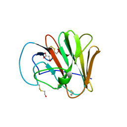
 | | Crystal structure of Odorant Binding Protein 5 from Anopheles gambiae (AgamOBP5) with Thymol | | 分子名称: | (4S)-2-METHYL-2,4-PENTANEDIOL, 5-METHYL-2-(1-METHYLETHYL)PHENOL, DI(HYDROXYETHYL)ETHER, ... | | 著者 | Liggri, P.G.V, Tsitsanou, K.E, Zographos, S.E. | | 登録日 | 2022-12-10 | | 公開日 | 2023-03-22 | | 最終更新日 | 2023-04-12 | | 実験手法 | X-RAY DIFFRACTION (1.3 Å) | | 主引用文献 | The structure of AgamOBP5 in complex with the natural insect repellents Carvacrol and Thymol: Crystallographic, fluorescence and thermodynamic binding studies.
Int.J.Biol.Macromol., 237, 2023
|
|
7D3E
 | |
6UYO
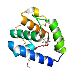
 | | Crystal structure of K37-acetylated SUMO1 in complex with PML-SIM | | 分子名称: | Protein PML, Small ubiquitin-related modifier 1 | | 著者 | Wahba, H.M, Gagnon, C, Mascle, X.H, Lussier-Price, M, Sakaguchi, K, Omichinski, J.G. | | 登録日 | 2019-11-14 | | 公開日 | 2019-11-27 | | 最終更新日 | 2023-11-15 | | 実験手法 | X-RAY DIFFRACTION (1.639 Å) | | 主引用文献 | Acetylation of SUMO1 Alters Interactions with the SIMs of PML and Daxx in a Protein-Specific Manner.
Structure, 28, 2020
|
|
6UYX
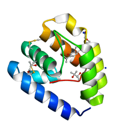
 | | Crystal structure of K37-acetylated SUMO1 in complex with phosphorylated DAXX | | 分子名称: | Small ubiquitin-related modifier 1, phosphorylated DAXX | | 著者 | Wahba, H.M, Gagnon, C, Mascle, X.H, Lussier-Price, M, Cappadocia, L, Sakaguchi, K, Omichinski, J.G. | | 登録日 | 2019-11-14 | | 公開日 | 2019-11-27 | | 最終更新日 | 2023-11-15 | | 実験手法 | X-RAY DIFFRACTION (1.7 Å) | | 主引用文献 | Acetylation of SUMO1 Alters Interactions with the SIMs of PML and Daxx in a Protein-Specific Manner.
Structure, 28, 2020
|
|
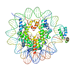
6T7A
 | | Structure of human Sox11 transcription factor in complex with a nucleosome | | 分子名称: | DNA (147-MER), Histone H2A type 1-B/E, Histone H2B type 1-K, ... | | 著者 | Dodonova, S.O, Zhu, F, Dienemann, C, Taipale, J, Cramer, P. | | 登録日 | 2019-10-21 | | 公開日 | 2020-04-29 | | 最終更新日 | 2024-05-22 | | 実験手法 | ELECTRON MICROSCOPY (3.7 Å) | | 主引用文献 | Nucleosome-bound SOX2 and SOX11 structures elucidate pioneer factor function.
Nature, 580, 2020
|
|
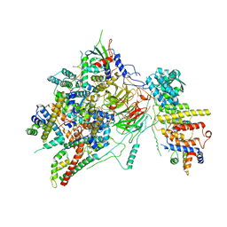
7CXN
 | | Architecture of a SARS-CoV-2 mini replication and transcription complex | | 分子名称: | Helicase, Non-structural protein 7, Non-structural protein 8, ... | | 著者 | Yan, L, Zhang, Y, Ge, J, Zheng, L, Gao, Y, Wang, T, Jia, Z, Wang, H, Huang, Y, Li, M, Wang, Q, Rao, Z, Lou, Z. | | 登録日 | 2020-09-02 | | 公開日 | 2020-11-04 | | 最終更新日 | 2024-03-27 | | 実験手法 | ELECTRON MICROSCOPY (3.84 Å) | | 主引用文献 | Architecture of a SARS-CoV-2 mini replication and transcription complex.
Nat Commun, 11, 2020
|
|
6T7B
 | | Structure of human Sox2 transcription factor in complex with a nucleosome | | 分子名称: | DNA (147-MER), Histone H2A type 1-B/E, Histone H2B type 1-K, ... | | 著者 | Dodonova, S.O, Zhu, F, Dienemann, C, Taipale, J, Cramer, P. | | 登録日 | 2019-10-21 | | 公開日 | 2020-04-29 | | 最終更新日 | 2024-05-22 | | 実験手法 | ELECTRON MICROSCOPY (5.1 Å) | | 主引用文献 | Nucleosome-bound SOX2 and SOX11 structures elucidate pioneer factor function.
Nature, 580, 2020
|
|
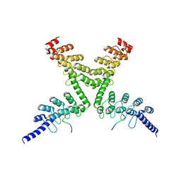
6T9K
 | | SAGA Core module | | 分子名称: | Protein SPT3, SAGA-associated factor 73, Transcription factor SPT20, ... | | 著者 | Wang, H, Cheung, A, Cramer, P. | | 登録日 | 2019-10-28 | | 公開日 | 2020-01-29 | | 最終更新日 | 2024-05-22 | | 実験手法 | ELECTRON MICROSCOPY (3.3 Å) | | 主引用文献 | Structure of the transcription coactivator SAGA.
Nature, 577, 2020
|
|
6TKQ
 | | Tankyrase 2 in complex with an inhibitor (OM-2700) | | 分子名称: | Tankyrase-2, ZINC ION, ~{N}-[3-[5-(5-ethoxypyridin-2-yl)-4-phenyl-1,2,4-triazol-3-yl]cyclobutyl]-1,5-naphthyridine-4-carboxamide | | 著者 | Sowa, S.T, Lehtio, L. | | 登録日 | 2019-11-28 | | 公開日 | 2020-07-08 | | 最終更新日 | 2024-01-24 | | 実験手法 | X-RAY DIFFRACTION (2.5 Å) | | 主引用文献 | Preclinical Lead Optimization of a 1,2,4-Triazole Based Tankyrase Inhibitor.
J.Med.Chem., 63, 2020
|
|
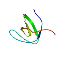
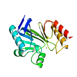
1Y7N
 | | Solution structure of the second PDZ domain of the human neuronal adaptor X11alpha | | 分子名称: | Amyloid beta A4 precursor protein-binding family A member 1 | | 著者 | Duquesne, A.E, de Ruijter, M, Brouwer, J, Drijfhout, J.W, Nabuurs, S.B, Spronk, C.A.E.M, Vuister, G.W, Ubbink, M, Canters, G.W. | | 登録日 | 2004-12-09 | | 公開日 | 2005-11-22 | | 最終更新日 | 2024-05-29 | | 実験手法 | SOLUTION NMR | | 主引用文献 | Solution structure of the second PDZ domain of the neuronal adaptor X11alpha and its interaction with the C-terminal peptide of the human copper chaperone for superoxide dismutase
J.Biomol.Nmr, 32, 2005
|
|
6TCA
 | | Phosphorylated p38 and MAPKAPK2 complex with inhibitor | | 分子名称: | MAP kinase-activated protein kinase 2, Mitogen-activated protein kinase 14, N-[5-(dimethylsulfamoyl)-2-methylphenyl]-1-phenyl-5-propyl-1H-pyrazole-4-carboxamide | | 著者 | Sok, P, Remenyi, A. | | 登録日 | 2019-11-05 | | 公開日 | 2020-07-22 | | 最終更新日 | 2024-01-24 | | 実験手法 | X-RAY DIFFRACTION (3.7 Å) | | 主引用文献 | MAP Kinase-Mediated Activation of RSK1 and MK2 Substrate Kinases.
Structure, 28, 2020
|
|
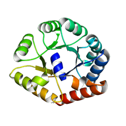
1BMC
 | | STRUCTURE OF A ZINC METALLO-BETA-LACTAMASE FROM BACILLUS CEREUS | | 分子名称: | METALLO-BETA-LACTAMASE, ZINC ION | | 著者 | Carfi, A, Pares, S, Duee, E, Dideberg, O. | | 登録日 | 1995-06-16 | | 公開日 | 1996-08-28 | | 最終更新日 | 2024-02-07 | | 実験手法 | X-RAY DIFFRACTION (2.5 Å) | | 主引用文献 | The 3-D structure of a zinc metallo-beta-lactamase from Bacillus cereus reveals a new type of protein fold.
EMBO J., 14, 1995
|
|
7YFV
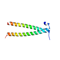 | | Structure of Rpgrip1l CC1 | | 分子名称: | Protein fantom | | 著者 | He, R, Chen, G, Li, Z, Li, J. | | 登録日 | 2022-07-09 | | 公開日 | 2023-05-17 | | 最終更新日 | 2024-05-29 | | 実験手法 | X-RAY DIFFRACTION (2.2 Å) | | 主引用文献 | Structure of the N-terminal coiled-coil domains of the ciliary protein Rpgrip1l.
Iscience, 26, 2023
|
|
7YFU
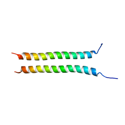 | | Structure of Rpgrip1l CC2 | | 分子名称: | Protein fantom | | 著者 | He, R, Chen, G, Li, Z, Li, J. | | 登録日 | 2022-07-09 | | 公開日 | 2023-05-17 | | 最終更新日 | 2024-05-29 | | 実験手法 | X-RAY DIFFRACTION (1.5 Å) | | 主引用文献 | Structure of the N-terminal coiled-coil domains of the ciliary protein Rpgrip1l.
Iscience, 26, 2023
|
|


