6GSN
 
 | |
6OFM
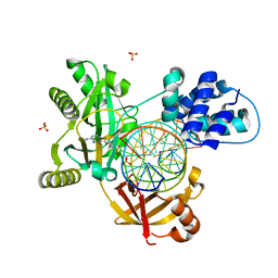
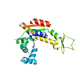
 | | Crystal structure of green fluorescent protein (GFP); S65T, Y66(3-CH3Y); ih circular permutant (50-51) | | 分子名称: | Green fluorescent protein (GFP); S65T, Y66(3-CH3Y); ih circular permutant (50-51) | | 著者 | Lin, C.-Y, Romei, M.G, Mathews, I.I, Boxer, S.G. | | 登録日 | 2019-03-31 | | 公開日 | 2019-07-10 | | 最終更新日 | 2023-11-15 | | 実験手法 | X-RAY DIFFRACTION (1.48 Å) | | 主引用文献 | Unified Model for Photophysical and Electro-Optical Properties of Green Fluorescent Proteins.
J.Am.Chem.Soc., 141, 2019
|
|
6OFK
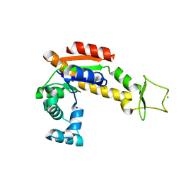
 | | Crystal structure of green fluorescent protein (GFP); S65T; ih circular permutant (50-51) | | 分子名称: | ACETATE ION, Green Fluorescent Protein (GFP); S65T; ih circular permutant (50-51) | | 著者 | Lin, C.-Y, Romei, M.G, Mathews, I.I, Boxer, S.G. | | 登録日 | 2019-03-30 | | 公開日 | 2019-07-10 | | 最終更新日 | 2023-11-15 | | 実験手法 | X-RAY DIFFRACTION (1.15 Å) | | 主引用文献 | Unified Model for Photophysical and Electro-Optical Properties of Green Fluorescent Proteins.
J.Am.Chem.Soc., 141, 2019
|
|
6OFN
 | | Crystal structure of green fluorescent protein (GFP); S65T, T203(3-OMeY); ih circular permutant (50-51) | | 分子名称: | Green fluorescent protein (GFP); S65T, T203(3-OMeY); ih circular permutant (50-51) | | 著者 | Lin, C.-Y, Romei, M.G, Mathews, I.I, Boxer, S.G. | | 登録日 | 2019-03-31 | | 公開日 | 2019-07-10 | | 最終更新日 | 2023-11-15 | | 実験手法 | X-RAY DIFFRACTION (1.649 Å) | | 主引用文献 | Unified Model for Photophysical and Electro-Optical Properties of Green Fluorescent Proteins.
J.Am.Chem.Soc., 141, 2019
|
|
6OFL
 | | Crystal structure of green fluorescent protein (GFP); S65T, Y66(3-ClY); ih circular permutant (50-51) | | 分子名称: | Green fluorescent protein (GFP); S65T, Y66(3-ClY); ih circular permutant (50-51) | | 著者 | Lin, C.-Y, Romei, M.G, Mathews, I.I, Boxer, S.G. | | 登録日 | 2019-03-31 | | 公開日 | 2019-07-10 | | 最終更新日 | 2023-11-15 | | 実験手法 | X-RAY DIFFRACTION (1.25 Å) | | 主引用文献 | Unified Model for Photophysical and Electro-Optical Properties of Green Fluorescent Proteins.
J.Am.Chem.Soc., 141, 2019
|
|
6OFO
 | | Crystal structure of split green fluorescent protein (GFP); s10 circular permutant (194-195) | | 分子名称: | Green fluorescent protein (GFP); s10 circular permutant (194-195) | | 著者 | Lin, C.-Y, Romei, M.G, Deller, M.C, Doukov, T.I, Boxer, S.G. | | 登録日 | 2019-03-31 | | 公開日 | 2019-07-10 | | 最終更新日 | 2023-11-15 | | 実験手法 | X-RAY DIFFRACTION (2.603 Å) | | 主引用文献 | Unified Model for Photophysical and Electro-Optical Properties of Green Fluorescent Proteins.
J.Am.Chem.Soc., 141, 2019
|
|
6RAS
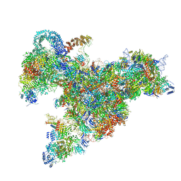
 | | Pmar-Lig_Pre. | | 分子名称: | ADENOSINE MONOPHOSPHATE, ATP-dependent DNA ligase, DNA, ... | | 著者 | Leiros, H.K.S, Williamson, A. | | 登録日 | 2019-04-07 | | 公開日 | 2019-07-10 | | 最終更新日 | 2024-05-01 | | 実験手法 | X-RAY DIFFRACTION (2.75 Å) | | 主引用文献 | Structural intermediates of a DNA-ligase complex illuminate the role of the catalytic metal ion and mechanism of phosphodiester bond formation.
Nucleic Acids Res., 47, 2019
|
|
6R6P
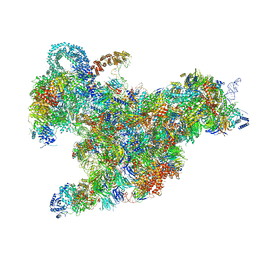
 | | Structure of XBP1u-paused ribosome nascent chain complex (rotated state) | | 分子名称: | 18S rRNA, 28S ribosomal RNA, 40S ribosomal protein S12, ... | | 著者 | Shanmuganathan, V, Cheng, J, Berninghausen, O, Beckmann, R. | | 登録日 | 2019-03-27 | | 公開日 | 2019-07-10 | | 最終更新日 | 2024-05-22 | | 実験手法 | ELECTRON MICROSCOPY (3.1 Å) | | 主引用文献 | Structural and mutational analysis of the ribosome-arresting human XBP1u.
Elife, 8, 2019
|
|
6R6G
 | | Structure of XBP1u-paused ribosome nascent chain complex with SRP. | | 分子名称: | 18S ribosomal RNA, 28S ribosomal RNA, 40S ribosomal protein S12, ... | | 著者 | Shanmuganathan, V, Cheng, J, Braunger, K, Berninghausen, O, Beatrix, B, Beckmann, R. | | 登録日 | 2019-03-27 | | 公開日 | 2019-07-10 | | 最終更新日 | 2019-10-30 | | 実験手法 | ELECTRON MICROSCOPY (3.7 Å) | | 主引用文献 | Structural and mutational analysis of the ribosome-arresting human XBP1u.
Elife, 8, 2019
|
|
6RAU
 | | PostS3_Pmar_lig4_WT | | 分子名称: | ADENOSINE MONOPHOSPHATE, ATP-dependent DNA ligase, DNA (5'-D(*AP*TP*TP*GP*CP*GP*AP*CP*CP*CP*CP*AP*CP*TP*AP*TP*CP*GP*GP*AP*A)-3'), ... | | 著者 | Leiros, H.K.S, Williamson, A. | | 登録日 | 2019-04-08 | | 公開日 | 2019-07-10 | | 最終更新日 | 2024-05-01 | | 実験手法 | X-RAY DIFFRACTION (1.99 Å) | | 主引用文献 | Structural intermediates of a DNA-ligase complex illuminate the role of the catalytic metal ion and mechanism of phosphodiester bond formation.
Nucleic Acids Res., 47, 2019
|
|
6R7Q
 | | Structure of XBP1u-paused ribosome nascent chain complex with Sec61. | | 分子名称: | 18S ribosomal RNA, 28S ribosomal RNA, 40S ribosomal protein S12, ... | | 著者 | Shanmuganathan, V, Cheng, J, Braunger, K, Berninghausen, O, Beatrix, B, Beckmann, R. | | 登録日 | 2019-03-29 | | 公開日 | 2019-07-10 | | 最終更新日 | 2024-05-22 | | 実験手法 | ELECTRON MICROSCOPY (3.9 Å) | | 主引用文献 | Structural and mutational analysis of the ribosome-arresting human XBP1u.
Elife, 8, 2019
|
|
6R5Q
 | | Structure of XBP1u-paused ribosome nascent chain complex (post-state) | | 分子名称: | 18S ribosomal RNA, 28S ribosomal RNA, 40S ribosomal protein S12, ... | | 著者 | Shanmuganathan, V, Cheng, J, Berninghausen, O, Beckmann, R. | | 登録日 | 2019-03-25 | | 公開日 | 2019-07-10 | | 最終更新日 | 2019-10-30 | | 実験手法 | ELECTRON MICROSCOPY (3 Å) | | 主引用文献 | Structural and mutational analysis of the ribosome-arresting human XBP1u.
Elife, 8, 2019
|
|
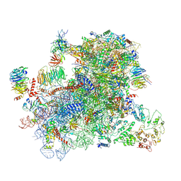
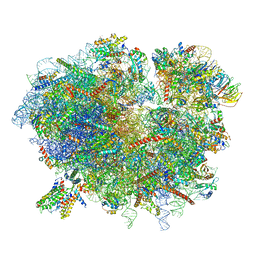
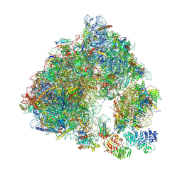
6RM3
 | | Evolutionary compaction and adaptation visualized by the structure of the dormant microsporidian ribosome | | 分子名称: | 16S rRNA, 23S rRNA, 5S rRNA, ... | | 著者 | Barandun, J, Hunziker, M, Vossbrinck, C.R, Klinge, S. | | 登録日 | 2019-05-05 | | 公開日 | 2019-07-10 | | 最終更新日 | 2024-07-10 | | 実験手法 | ELECTRON MICROSCOPY (3.4 Å) | | 主引用文献 | Evolutionary compaction and adaptation visualized by the structure of the dormant microsporidian ribosome.
Nat Microbiol, 4, 2019
|
|
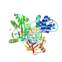
6OHC
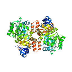 | | E. coli Guanine Deaminase | | 分子名称: | GLYCEROL, Guanine deaminase, ZINC ION | | 著者 | Shek, R.S, French, J.B. | | 登録日 | 2019-04-05 | | 公開日 | 2019-07-24 | | 最終更新日 | 2023-10-11 | | 実験手法 | X-RAY DIFFRACTION (2.3 Å) | | 主引用文献 | Structural Determinants for Substrate Selectivity in Guanine Deaminase Enzymes of the Amidohydrolase Superfamily.
Biochemistry, 58, 2019
|
|
6OHB
 | | E. coli Guanine Deaminase | | 分子名称: | Guanine deaminase, ZINC ION | | 著者 | Shek, R.S, French, J.B. | | 登録日 | 2019-04-05 | | 公開日 | 2019-07-24 | | 最終更新日 | 2023-10-11 | | 実験手法 | X-RAY DIFFRACTION (2.3 Å) | | 主引用文献 | Structural Determinants for Substrate Selectivity in Guanine Deaminase Enzymes of the Amidohydrolase Superfamily.
Biochemistry, 58, 2019
|
|
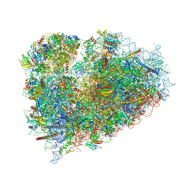
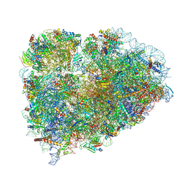
6S47
 | | Saccharomyces cerevisiae 80S ribosome bound with ABCF protein New1 | | 分子名称: | 18S rRNA (1707-MER), 28S ribosomal RNA, 40S ribosomal protein S0-A, ... | | 著者 | Kasari, V, Pochopien, A.A, Margus, T, Murina, V, Turnbull, K, Zhou, Y, Nissan, T, Graf, M, Novacek, J, Atkinson, G.C, Johansson, M.J.O, Wilson, D.N, Hauryliuk, V. | | 登録日 | 2019-06-26 | | 公開日 | 2019-07-24 | | 最終更新日 | 2019-10-23 | | 実験手法 | ELECTRON MICROSCOPY (3.28 Å) | | 主引用文献 | A role for the Saccharomyces cerevisiae ABCF protein New1 in translation termination/recycling.
Nucleic Acids Res., 47, 2019
|
|
6GSM
 | | Structure of a partial yeast 48S preinitiation complex in open conformation. | | 分子名称: | 18S ribosomal RNA, 40S ribosomal protein S0, 40S ribosomal protein S1, ... | | 著者 | Llacer, J.L, Hussain, T, Gordiyenko, Y, Ramakrishnan, V. | | 登録日 | 2018-06-14 | | 公開日 | 2019-07-31 | | 最終更新日 | 2024-07-10 | | 実験手法 | ELECTRON MICROSCOPY (5.15 Å) | | 主引用文献 | Large-scale movement of eIF3 domains during translation initiation modulate start codon selection.
Nucleic Acids Res., 2021
|
|
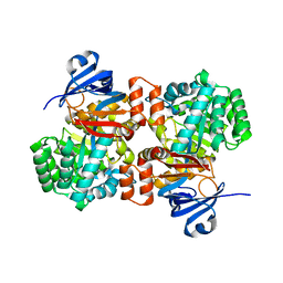
6S36
 | | Crystal structure of E. coli Adenylate kinase R119K mutant | | 分子名称: | Adenylate kinase, CHLORIDE ION, MAGNESIUM ION, ... | | 著者 | Grundstrom, C, Rogne, P, Wolf-Watz, M, Sauer-Eriksson, A.E. | | 登録日 | 2019-06-24 | | 公開日 | 2019-08-07 | | 最終更新日 | 2024-01-24 | | 実験手法 | X-RAY DIFFRACTION (1.6 Å) | | 主引用文献 | Nucleation of an Activating Conformational Change by a Cation-pi Interaction.
Biochemistry, 58, 2019
|
|
6PFU
 | |
6PFR
 | |
6PFS
 | |
6PFT
 | |
6RZE
 | | Crystal structure of E. coli Adenylate kinase R119A mutant | | 分子名称: | Adenylate kinase, CHLORIDE ION, SODIUM ION | | 著者 | Grundstrom, C, Rogne, P, Wolf-Watz, M, Sauer-Eriksson, A.E. | | 登録日 | 2019-06-13 | | 公開日 | 2019-08-07 | | 最終更新日 | 2024-01-24 | | 実験手法 | X-RAY DIFFRACTION (1.69 Å) | | 主引用文献 | Nucleation of an Activating Conformational Change by a Cation-pi Interaction.
Biochemistry, 58, 2019
|
|
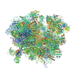
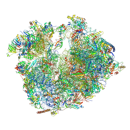
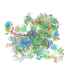
6RXZ
 | | Cryo-EM structure of the 90S pre-ribosome (Kre33-Noc4) from Chaetomium thermophilum, state b | | 分子名称: | 35S ribosomal RNA, 40S ribosomal protein S11-like protein, 40S ribosomal protein S13-like protein, ... | | 著者 | Cheng, J, Kellner, N, Griesel, S, Berninghausen, O, Beckmann, R, Hurt, E. | | 登録日 | 2019-06-10 | | 公開日 | 2019-08-14 | | 最終更新日 | 2024-05-22 | | 実験手法 | ELECTRON MICROSCOPY (4.4 Å) | | 主引用文献 | Thermophile 90S Pre-ribosome Structures Reveal the Reverse Order of Co-transcriptional 18S rRNA Subdomain Integration.
Mol.Cell, 75, 2019
|
|
6RXX
 | | Cryo-EM structure of the 90S pre-ribosome (Kre33-Noc4) from Chaetomium thermophilum, state C, Poly-Ala | | 分子名称: | 35S ribosomal RNA, 40S ribosomal protein S1, 40S ribosomal protein S11-like protein, ... | | 著者 | Cheng, J, Kellner, N, Griesel, S, Berninghausen, O, Beckmann, R, Hurt, E. | | 登録日 | 2019-06-10 | | 公開日 | 2019-08-14 | | 最終更新日 | 2024-05-22 | | 実験手法 | ELECTRON MICROSCOPY (7.1 Å) | | 主引用文献 | Thermophile 90S Pre-ribosome Structures Reveal the Reverse Order of Co-transcriptional 18S rRNA Subdomain Integration.
Mol.Cell, 75, 2019
|
|


