3AJW
 
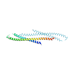 | | Structure of FliJ, a soluble component of flagellar type III export apparatus | | 分子名称: | Flagellar fliJ protein, MERCURY (II) ION | | 著者 | Imada, K, Ibuki, T, Minamino, T, Namba, K. | | 登録日 | 2010-06-23 | | 公開日 | 2011-02-02 | | 最終更新日 | 2024-03-13 | | 実験手法 | X-RAY DIFFRACTION (2.1 Å) | | 主引用文献 | Common architecture of the flagellar type III protein export apparatus and F- and V-type ATPases
Nat.Struct.Mol.Biol., 18, 2011
|
|
3AJ1
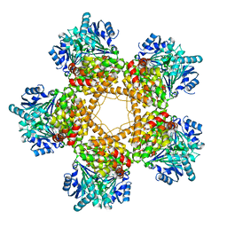
 | | The structure of AxCeSD octamer (N-terminal HIS-tag) from Acetobacter xylinum | | 分子名称: | Cellulose synthase operon protein D | | 著者 | Hu, S.Q, Tajima, K, Zhou, Y, Tanaka, I, Yao, M. | | 登録日 | 2010-05-20 | | 公開日 | 2010-10-06 | | 最終更新日 | 2011-07-13 | | 実験手法 | X-RAY DIFFRACTION (2.5 Å) | | 主引用文献 | Structure of bacterial cellulose synthase subunit D octamer with four inner passageways
Proc.Natl.Acad.Sci.USA, 107, 2010
|
|
3AKC
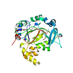
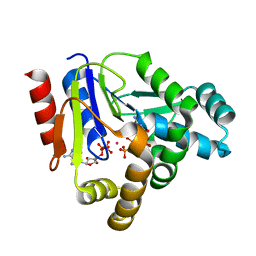 | | Crystal structure of CMP kinase in complex with CDP and ADP from Thermus thermophilus HB8 | | 分子名称: | ADENOSINE-5'-DIPHOSPHATE, CYTIDINE-5'-DIPHOSPHATE, Cytidylate kinase, ... | | 著者 | Mega, R, Nakagawa, N, Kuramitsu, S, Masui, R. | | 登録日 | 2010-07-12 | | 公開日 | 2011-07-06 | | 最終更新日 | 2023-11-01 | | 実験手法 | X-RAY DIFFRACTION (1.65 Å) | | 主引用文献 | The crystal structure of the tertiary complex of CMP kinase with a phosphoryl group acceptor and a donor from Thermus thermophilus HB8
To be Published
|
|
3ARW
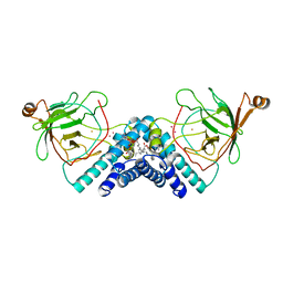
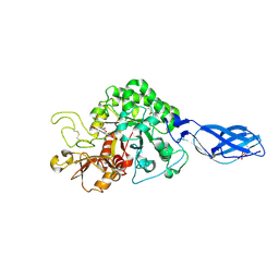 | | Crystal Structure Analysis of Chitinase A from Vibrio harveyi with novel inhibitors - complex structure with chelerythrine | | 分子名称: | 1,2-dimethoxy-12-methyl[1,3]benzodioxolo[5,6-c]phenanthridin-12-ium, Chitinase A, GLYCEROL | | 著者 | Pantoom, S, Vetter, I.R, Prinz, H, Suginta, W. | | 登録日 | 2010-12-09 | | 公開日 | 2011-04-20 | | 最終更新日 | 2023-11-01 | | 実験手法 | X-RAY DIFFRACTION (1.5 Å) | | 主引用文献 | Potent family-18 chitinase inhibitors: x-ray structures, affinities, and binding mechanisms
J.Biol.Chem., 286, 2011
|
|
5RAD
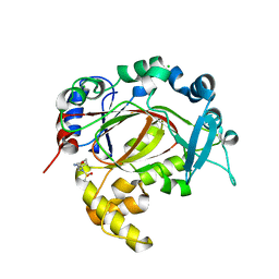 | | PanDDA analysis group deposition -- Crystal Structure of JMJD1B in complex with FM001568a | | 分子名称: | 2-[cyclohexyl(methylsulfonyl)amino]ethanamide, CHLORIDE ION, Lysine-specific demethylase 3B, ... | | 著者 | Snee, M, Nowak, R, Johansson, C, Burgess-Brown, N.A, Arrowsmith, C.H, Bountra, C, Edwards, A.M, Oppermann, U. | | 登録日 | 2020-03-16 | | 公開日 | 2020-04-22 | | 最終更新日 | 2024-03-06 | | 実験手法 | X-RAY DIFFRACTION (1.9 Å) | | 主引用文献 | PanDDA analysis group deposition of Human JMJD1B screened against the DSPL Fragment Library
To Be Published
|
|
5RAY
 | | PanDDA analysis group deposition -- Crystal Structure of JMJD1B in complex with FM001469a | | 分子名称: | 2,4-difluoro-6-[(3S)-pyrazolidin-3-yl]phenol, CHLORIDE ION, Lysine-specific demethylase 3B, ... | | 著者 | Snee, M, Nowak, R, Johansson, C, Burgess-Brown, N.A, Arrowsmith, C.H, Bountra, C, Edwards, A.M, Oppermann, U. | | 登録日 | 2020-03-16 | | 公開日 | 2020-04-22 | | 最終更新日 | 2024-03-06 | | 実験手法 | X-RAY DIFFRACTION (1.63 Å) | | 主引用文献 | PanDDA analysis group deposition of Human JMJD1B screened against the DSPL Fragment Library
To Be Published
|
|
5RAB
 | | PanDDA analysis group deposition -- Crystal Structure of JMJD1B in complex with FM001726a | | 分子名称: | (2R,5S)-5-(4-chlorophenyl)oxolane-2-carbohydrazide, CHLORIDE ION, Lysine-specific demethylase 3B, ... | | 著者 | Snee, M, Nowak, R, Johansson, C, Burgess-Brown, N.A, Arrowsmith, C.H, Bountra, C, Edwards, A.M, Oppermann, U. | | 登録日 | 2020-03-16 | | 公開日 | 2020-04-22 | | 最終更新日 | 2024-03-06 | | 実験手法 | X-RAY DIFFRACTION (1.52 Å) | | 主引用文献 | PanDDA analysis group deposition of Human JMJD1B screened against the DSPL Fragment Library
To Be Published
|
|
5RAS
 | | PanDDA analysis group deposition -- Crystal Structure of JMJD1B in complex with XS036302b | | 分子名称: | 2-(4-phenylpiperidin-1-yl)ethanoic acid, CHLORIDE ION, Lysine-specific demethylase 3B, ... | | 著者 | Snee, M, Nowak, R, Johansson, C, Burgess-Brown, N.A, Arrowsmith, C.H, Bountra, C, Edwards, A.M, Oppermann, U. | | 登録日 | 2020-03-16 | | 公開日 | 2020-04-22 | | 最終更新日 | 2024-03-06 | | 実験手法 | X-RAY DIFFRACTION (1.58 Å) | | 主引用文献 | PanDDA analysis group deposition of Human JMJD1B screened against the DSPL Fragment Library
To Be Published
|
|
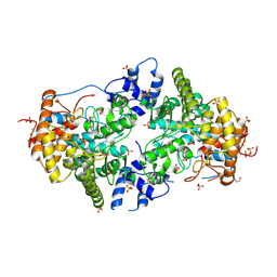
1DLQ
 | | STRUCTURE OF CATECHOL 1,2-DIOXYGENASE FROM ACINETOBACTER SP. ADP1 INHIBITED BY BOUND MERCURY | | 分子名称: | CATECHOL 1,2-DIOXYGENASE, FE (III) ION, MERCURY (II) ION, ... | | 著者 | Vetting, M.W, Ohlendorf, D.H. | | 登録日 | 1999-12-11 | | 公開日 | 2000-05-23 | | 最終更新日 | 2024-02-07 | | 実験手法 | X-RAY DIFFRACTION (2.3 Å) | | 主引用文献 | The 1.8 A crystal structure of catechol 1,2-dioxygenase reveals a novel hydrophobic helical zipper as a subunit linker.
Structure Fold.Des., 8, 2000
|
|
3A8E
 | | The structure of AxCesD octamer complexed with cellopentaose | | 分子名称: | Cellulose synthase operon protein D, beta-D-glucopyranose-(1-4)-beta-D-glucopyranose-(1-4)-beta-D-glucopyranose-(1-4)-beta-D-glucopyranose-(1-4)-beta-D-glucopyranose | | 著者 | Hu, S.Q, Tajima, K, Zhou, Y, Yao, M, Tanaka, I. | | 登録日 | 2009-10-05 | | 公開日 | 2010-09-22 | | 最終更新日 | 2023-11-01 | | 実験手法 | X-RAY DIFFRACTION (3 Å) | | 主引用文献 | Structure of bacterial cellulose synthase subunit D octamer with four inner passageways
Proc.Natl.Acad.Sci.USA, 107, 2010
|
|
2FLQ
 | |
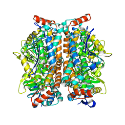
3BFJ
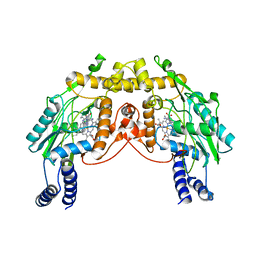
 | | Crystal structure analysis of 1,3-propanediol oxidoreductase | | 分子名称: | 1,3-propanediol oxidoreductase, FE (II) ION | | 著者 | Marcal, D, Enguita, F.J, Carrondo, M.A, Structural Proteomics in Europe (SPINE) | | 登録日 | 2007-11-21 | | 公開日 | 2008-11-25 | | 最終更新日 | 2024-02-21 | | 実験手法 | X-RAY DIFFRACTION (2.7 Å) | | 主引用文献 | 1,3-propanediol dehydrogenase from Klebsiella pneumoniae: decameric quaternary structure and possible subunit cooperativity
J.Bacteriol., 191, 2009
|
|
2JJB
 | | Family 37 trehalase from Escherichia coli in complex with casuarine-6- O-alpha-glucopyranose | | 分子名称: | 1,2-ETHANEDIOL, CASUARINE, PERIPLASMIC TREHALASE, ... | | 著者 | Gloster, T.M, Roberts, S, Davies, G.J, Cardona, F, Parmeggiani, C, Bonaccini, C, Gratteri, P, Sim, L, Rose, D.R, Goti, A. | | 登録日 | 2008-03-28 | | 公開日 | 2009-01-13 | | 最終更新日 | 2023-12-13 | | 実験手法 | X-RAY DIFFRACTION (1.9 Å) | | 主引用文献 | Total Syntheses of Casuarine and its 6-O-Alpha-Glucoside: Complementary Inhibition Towards Glycoside Hydrolases of the Gh31 and Gh37 Families.
Chemistry, 15, 2009
|
|
2JGN
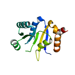 | | DDX3 helicase domain | | 分子名称: | ATP-DEPENDENT RNA HELICASE DDX3X | | 著者 | Rodamilans, B, Montoya, G. | | 登録日 | 2007-02-13 | | 公開日 | 2008-05-13 | | 最終更新日 | 2023-12-13 | | 実験手法 | X-RAY DIFFRACTION (1.91 Å) | | 主引用文献 | Expression, Purification, Crystallization and Preliminary X-Ray Diffraction Analysis of the Ddx3 RNA Helicase Domain.
Acta Crystallogr.,Sect.F, 63, 2007
|
|
2JTT
 | |
2FF0
 | | Solution Structure of Steroidogenic Factor 1 DNA Binding Domain Bound to its Target Sequence in the Inhibin alpha-subunit Promoter | | 分子名称: | CTGTGGCCCTGAGCC, GGCTCAGGGCCACAG, Steroidogenic factor 1, ... | | 著者 | Little, T.H, Zhang, Y, Matulis, C.K, Weck, J, Zhang, Z, Ramachandran, A, Mayo, K.E, Radhakrishnan, I. | | 登録日 | 2005-12-17 | | 公開日 | 2006-04-11 | | 最終更新日 | 2024-05-29 | | 実験手法 | SOLUTION NMR | | 主引用文献 | Sequence-specific deoxyribonucleic Acid (DNA) recognition by steroidogenic factor 1: a helix at the carboxy terminus of the DNA binding domain is necessary for complex stability.
Mol.Endocrinol., 20, 2006
|
|
2ZQR
 | | Crystal structure of AUH without RNA | | 分子名称: | Methylglutaconyl-CoA hydratase | | 著者 | Kurimoto, K, Kuwasako, K, Muto, Y, Nureki, O, Yokoyama, S, RIKEN Structural Genomics/Proteomics Initiative (RSGI) | | 登録日 | 2008-08-16 | | 公開日 | 2009-06-30 | | 最終更新日 | 2023-11-01 | | 実験手法 | X-RAY DIFFRACTION (2.5 Å) | | 主引用文献 | AU-rich RNA-binding induces changes in the quaternary structure of AUH
Proteins, 75, 2009
|
|
2GET
 | | Pantothenate kinase from Mycobacterium tuberculosis (MtPanK) in complex with a coenzyme A derivative, Form-I (LT) | | 分子名称: | GLYCEROL, Pantothenate kinase, [(2R,3S,4R,5R)-5-(6-AMINO-9H-PURIN-9-YL)-4-HYDROXY-3-(PHOSPHONOOXY)TETRAHYDROFURAN-2-YL]METHYL (3R)-3-HYDROXY-4-{[3-({2-[(2-HYDROXYETHYL)DITHIO]ETHYL}AMINO)-3-OXOPROPYL]AMINO}-2,2-DIMETHYL-4-OXOBUTYL DIHYDROGEN DIPHOSPHATE | | 著者 | Das, S, Kumar, P, Bhor, V, Surolia, A, Vijayan, M. | | 登録日 | 2006-03-20 | | 公開日 | 2006-06-06 | | 最終更新日 | 2023-10-25 | | 実験手法 | X-RAY DIFFRACTION (2.35 Å) | | 主引用文献 | Invariance and variability in bacterial PanK: a study based on the crystal structure of Mycobacterium tuberculosis PanK.
Acta Crystallogr.,Sect.D, 62, 2006
|
|
2GEU
 | | Pantothenate kinase from Mycobacterium tuberculosis (MtPanK) in complex with a coenzyme A derivative, Form-II (RT) | | 分子名称: | Pantothenate kinase, [(2R,3S,4R,5R)-5-(6-AMINO-9H-PURIN-9-YL)-4-HYDROXY-3-(PHOSPHONOOXY)TETRAHYDROFURAN-2-YL]METHYL (3R)-3-HYDROXY-4-{[3-({2-[(2-HYDROXYETHYL)DITHIO]ETHYL}AMINO)-3-OXOPROPYL]AMINO}-2,2-DIMETHYL-4-OXOBUTYL DIHYDROGEN DIPHOSPHATE | | 著者 | Das, S, Kumar, P, Bhor, V, Surolia, A, Vijayan, M. | | 登録日 | 2006-03-20 | | 公開日 | 2006-06-06 | | 最終更新日 | 2023-10-25 | | 実験手法 | X-RAY DIFFRACTION (2.9 Å) | | 主引用文献 | Invariance and variability in bacterial PanK: a study based on the crystal structure of Mycobacterium tuberculosis PanK.
Acta Crystallogr.,Sect.D, 62, 2006
|
|
2GEV
 | | Pantothenate kinase from Mycobacterium tuberculosis (MtPanK) in complex with a coenzyme A derivative, Form-II (LT) | | 分子名称: | 2-AMINO-2-HYDROXYMETHYL-PROPANE-1,3-DIOL, GLYCEROL, Pantothenate kinase, ... | | 著者 | Das, S, Kumar, P, Bhor, V, Surolia, A, Vijayan, M. | | 登録日 | 2006-03-20 | | 公開日 | 2006-06-06 | | 最終更新日 | 2023-10-25 | | 実験手法 | X-RAY DIFFRACTION (2.35 Å) | | 主引用文献 | Invariance and variability in bacterial PanK: a study based on the crystal structure of Mycobacterium tuberculosis PanK.
Acta Crystallogr.,Sect.D, 62, 2006
|
|
2GLZ
 | |
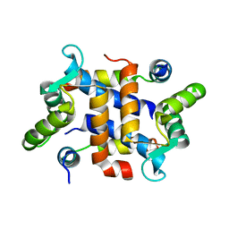
2FGT
 | | Crystal Structure of YycH from Bacillus subtilis | | 分子名称: | Two-component system yycF/yycG regulatory protein yycH | | 著者 | Szurmant, H, Zhao, H, Mohan, M.A, James, H.A, Varughese, K.I. | | 登録日 | 2005-12-22 | | 公開日 | 2006-04-25 | | 最終更新日 | 2024-02-14 | | 実験手法 | X-RAY DIFFRACTION (2.3 Å) | | 主引用文献 | The crystal structure of YycH involved in the regulation of the essential YycFG two-component system in Bacillus subtilis reveals a novel tertiary structure.
Protein Sci., 15, 2006
|
|
1G64
 | | THE THREE-DIMENSIONAL STRUCTURE OF ATP:CORRINOID ADENOSYLTRANSFERASE FROM SALMONELLA TYPHIMURIUM. COBALAMIN/ATP TERNARY COMPLEX | | 分子名称: | ADENOSINE-5'-TRIPHOSPHATE, COB(I)ALAMIN ADENOSYLTRANSFERASE, COBALAMIN, ... | | 著者 | Rayment, I, Escalante-Semerena, J.C, Bauer, C.B. | | 登録日 | 2000-11-03 | | 公開日 | 2000-12-20 | | 最終更新日 | 2024-02-07 | | 実験手法 | X-RAY DIFFRACTION (2.1 Å) | | 主引用文献 | Three-dimensional structure of ATP:corrinoid adenosyltransferase from Salmonella typhimurium in its free state, complexed with MgATP, or complexed with hydroxycobalamin and MgATP.
Biochemistry, 40, 2001
|
|
2JA1
 | | Thymidine kinase from B. cereus with TTP bound as phosphate donor. | | 分子名称: | (4S)-2-METHYL-2,4-PENTANEDIOL, THYMIDINE KINASE, THYMIDINE-5'-TRIPHOSPHATE, ... | | 著者 | Kosinska, U, Carnrot, C, Sandrini, M.P.B, Clausen, A.R, Wang, L, Piskur, J, Eriksson, S, Eklund, H. | | 登録日 | 2006-11-17 | | 公開日 | 2007-01-23 | | 最終更新日 | 2023-12-13 | | 実験手法 | X-RAY DIFFRACTION (2.8 Å) | | 主引用文献 | Structural Studies of Thymidine Kinases from Bacillus Anthracis and Bacillus Cereus Provide Insights Into Quaternary Structure and Conformational Changes Upon Substrate Binding
FEBS J., 274, 2007
|
|
2J9R
 | | Thymidine kinase from B. anthracis in complex with dT. | | 分子名称: | PHOSPHATE ION, THYMIDINE, THYMIDINE KINASE, ... | | 著者 | Kosinska, U, Carnrot, C, Sandrini, M.P.B, Clausen, A.R, Wang, L, Piskur, J, Eriksson, S, Eklund, H. | | 登録日 | 2006-11-15 | | 公開日 | 2007-01-23 | | 最終更新日 | 2023-12-13 | | 実験手法 | X-RAY DIFFRACTION (2.7 Å) | | 主引用文献 | Structural Studies of Thymidine Kinases from Bacillus Anthracis and Bacillus Cereus Provide Insights Into Quaternary Structure and Conformational Changes Upon Substrate Binding
FEBS J., 274, 2007
|
|


