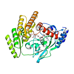
8OEK
 
 | |
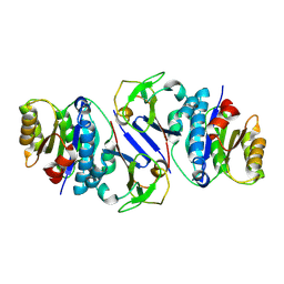
8OEP
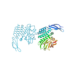
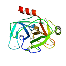
 | | Crystal structure of the PTPN3 PDZ domain bound to the HPV18 E6 oncoprotein C-terminal peptide | | 分子名称: | Protein E6, SODIUM ION, Tyrosine-protein phosphatase non-receptor type 3 | | 著者 | Genera, M, Colcombet-Cazenave, B, Croitoru, A, Raynal, B, Mechaly, A, Caillet, J, Haouz, A, Wolff, N, Caillet-Saguy, C. | | 登録日 | 2023-03-11 | | 公開日 | 2023-05-10 | | 最終更新日 | 2024-06-19 | | 実験手法 | X-RAY DIFFRACTION (1.87 Å) | | 主引用文献 | Interactions of the protein tyrosine phosphatase PTPN3 with viral and cellular partners through its PDZ domain: insights into structural determinants and phosphatase activity.
Front Mol Biosci, 10, 2023
|
|
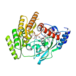
8ODR
 | | Mimetic of UBC9-SUMO1 | | 分子名称: | SULFATE ION, SUMO-conjugating enzyme UBC9, Small ubiquitin-related modifier 1 | | 著者 | Coste, F, Goffinont, S, Suskiewicz, M.J. | | 登録日 | 2023-03-09 | | 公開日 | 2023-06-07 | | 最終更新日 | 2024-06-19 | | 実験手法 | X-RAY DIFFRACTION (2.85 Å) | | 主引用文献 | Structural insights into the regulation of the human E2∼SUMO conjugate through analysis of its stable mimetic.
J.Biol.Chem., 299, 2023
|
|
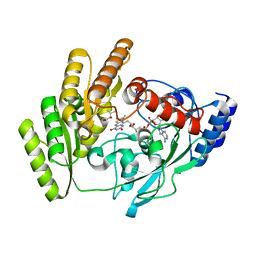
5JXE
 | |
8OFD
 | |
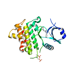
5JYI
 | | Trypsin bound with succinic acid at 1.9A | | 分子名称: | CALCIUM ION, Cationic trypsin, SODIUM ION, ... | | 著者 | Manohar, R, Kutumbarao, N.H.V, KarthiK, L, Malathy, P, Velmurugan, D, Gunasekaran, K. | | 登録日 | 2016-05-14 | | 公開日 | 2016-07-06 | | 最終更新日 | 2023-11-08 | | 実験手法 | X-RAY DIFFRACTION (1.914 Å) | | 主引用文献 | Trypsin bound with succinic acid at 1.9A
To Be Published
|
|
5JQR
 | | The Structure of Ascorbate Peroxidase Compound II formed by reaction with m-CPBA | | 分子名称: | Ascorbate peroxidase, POTASSIUM ION, PROTOPORPHYRIN IX CONTAINING FE, ... | | 著者 | Kwon, H, Raven, E.L, Moody, P.C.E. | | 登録日 | 2016-05-05 | | 公開日 | 2016-12-21 | | 最終更新日 | 2024-01-10 | | 実験手法 | X-RAY DIFFRACTION (1.81 Å) | | 主引用文献 | Direct visualization of a Fe(IV)-OH intermediate in a heme enzyme.
Nat Commun, 7, 2016
|
|
5JRM
 | | Crystal Structure of a Xylanase at 1.56 Angstroem resolution | | 分子名称: | Endo-1,4-beta-xylanase, GLYCEROL, SULFATE ION | | 著者 | Gomez, S, Payne, A.M, Savko, M, Fox, G.C, Shepard, W.E, Fernandez, F.J, Vega, M.C. | | 登録日 | 2016-05-06 | | 公開日 | 2017-05-24 | | 最終更新日 | 2024-01-10 | | 実験手法 | X-RAY DIFFRACTION (1.56 Å) | | 主引用文献 | Crystal Structure of a Xylanase at 1.56 Angstroem resolution
To Be Published
|
|
5JZB
 | | Crystal structure of HsaD bound to 3,5-dichlorobenzene sulphonamide | | 分子名称: | 3,5-dichlorobenzene-1-sulfonamide, 4,5:9,10-diseco-3-hydroxy-5,9,17-trioxoandrosta-1(10),2-diene-4-oate hydrolase, PHOSPHATE ION | | 著者 | Ryan, A, Polycarpou, E, Lack, N.A, Evangelopoulos, D, Sieg, C, Halman, A, Bhakta, S, Sinclair, A, Eleftheriadou, O, McHugh, T.D, Keany, S, Lowe, E, Ballet, R, Abihammad, A, Ciulli, A, Sim, E. | | 登録日 | 2016-05-16 | | 公開日 | 2017-04-05 | | 最終更新日 | 2024-05-08 | | 実験手法 | X-RAY DIFFRACTION (2.102 Å) | | 主引用文献 | Investigation of the mycobacterial enzyme HsaD as a potential novel target for anti-tubercular agents using a fragment-based drug design approach.
Br. J. Pharmacol., 174, 2017
|
|
5JZN
 | | Crystal structure of DCLK1-KD in complex with NVP-TAE684 | | 分子名称: | 5-CHLORO-N-[2-METHOXY-4-[4-(4-METHYLPIPERAZIN-1-YL)PIPERIDIN-1-YL]PHENYL]-N'-(2-PROPAN-2-YLSULFONYLPHENYL)PYRIMIDINE-2,4-DIAMINE, SULFATE ION, Serine/threonine-protein kinase DCLK1 | | 著者 | Patel, O, Lucet, I. | | 登録日 | 2016-05-17 | | 公開日 | 2016-08-24 | | 最終更新日 | 2023-09-27 | | 実験手法 | X-RAY DIFFRACTION (2.85 Å) | | 主引用文献 | Biochemical and Structural Insights into Doublecortin-like Kinase Domain 1.
Structure, 24, 2016
|
|
5JS5
 | | Nitric oxide complex of the L16F mutant of cytochrome c prime from Alcaligenes xylosoxidans | | 分子名称: | ASCORBIC ACID, Cytochrome c', HEME C, ... | | 著者 | Kekilli, D, Strange, R.W, Hough, M.A. | | 登録日 | 2016-05-07 | | 公開日 | 2017-03-08 | | 最終更新日 | 2024-01-10 | | 実験手法 | X-RAY DIFFRACTION (1.7 Å) | | 主引用文献 | Engineering proximal vs. distal heme-NO coordination via dinitrosyl dynamics: implications for NO sensor design.
Chem Sci, 8, 2017
|
|
8JTL
 | | Structure of OY phytoplasma SAP05 binding with AtRpn10 | | 分子名称: | 26S proteasome non-ATPase regulatory subunit 4 homolog, Sequence-variable mosaic (SVM) signal sequence domain-containing protein | | 著者 | Du, Y.X, Zhang, L.Y, Zheng, Q.Y. | | 登録日 | 2023-06-22 | | 公開日 | 2023-07-12 | | 最終更新日 | 2024-07-17 | | 実験手法 | X-RAY DIFFRACTION (1.78 Å) | | 主引用文献 | Structure basis for recognition of plant Rpn10 by phytoplasma SAP05 in ubiquitin-independent protein degradation.
Iscience, 27, 2024
|
|
8JUK
 | |
5K1L
 | |
8JU8
 | | de novo designed protein | | 分子名称: | de novo designed protein | | 著者 | Zhang, L. | | 登録日 | 2023-06-25 | | 公開日 | 2023-07-26 | | 最終更新日 | 2024-05-29 | | 実験手法 | X-RAY DIFFRACTION (1.85 Å) | | 主引用文献 | de novo designed Rossmann fold protein
To Be Published
|
|
8JTK
 | | Structure of AYWB phytoplasma SAP05 recognizing AtRpn10 | | 分子名称: | 26S proteasome non-ATPase regulatory subunit 4 homolog, Sequence-variable mosaic (SVM) signal sequence domain-containing protein | | 著者 | Du, Y.X, Zhang, L.Y, Zheng, Q.Y. | | 登録日 | 2023-06-22 | | 公開日 | 2023-07-19 | | 最終更新日 | 2024-07-17 | | 実験手法 | X-RAY DIFFRACTION (1.57 Å) | | 主引用文献 | Structure basis for recognition of plant Rpn10 by phytoplasma SAP05 in ubiquitin-independent protein degradation.
Iscience, 27, 2024
|
|
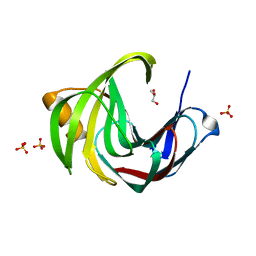
8OI7
 | | Trichomonas vaginalis riboside hydrolase | | 分子名称: | CALCIUM ION, DI(HYDROXYETHYL)ETHER, Inosine-uridine preferring nucleoside hydrolase family protein, ... | | 著者 | Patrone, M, Stockman, B.J, Degano, M. | | 登録日 | 2023-03-22 | | 公開日 | 2023-08-02 | | 最終更新日 | 2023-09-06 | | 実験手法 | X-RAY DIFFRACTION (1.85 Å) | | 主引用文献 | A riboside hydrolase that salvages both nucleobases and nicotinamide in the auxotrophic parasite Trichomonas vaginalis.
J.Biol.Chem., 299, 2023
|
|
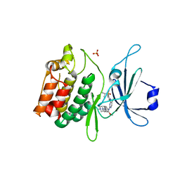
8OIZ
 | | Crystal structure of human CRBN-DDB1 in complex with Pomalidomide | | 分子名称: | 1,2-ETHANEDIOL, DNA damage-binding protein 1, Protein cereblon, ... | | 著者 | Le Bihan, Y.-V, Cabry, M.P, van Montfort, R.L.M. | | 登録日 | 2023-03-23 | | 公開日 | 2023-07-19 | | 最終更新日 | 2023-12-13 | | 実験手法 | X-RAY DIFFRACTION (2.5 Å) | | 主引用文献 | A Degron Blocking Strategy Towards Improved CRL4 CRBN Recruiting PROTAC Selectivity.
Chembiochem, 24, 2023
|
|
8K4Y
 | | Structure of a triple-helix region of human ReCol 3 from Trautec | | 分子名称: | SULFATE ION, triple-helix region of human ReCol 3 | | 著者 | Chu, Y, Zhai, Y, Fan, X, Li, J, Wang, L, Fu, S, Feng, P, Qian, S. | | 登録日 | 2023-07-20 | | 公開日 | 2023-08-02 | | 最終更新日 | 2023-10-04 | | 実験手法 | X-RAY DIFFRACTION (1.5 Å) | | 主引用文献 | Structure of a triple-helix region of human ReCol 3 from Trautec.
To Be Published
|
|
5JUH
 | |
8JDE
 | | Crystal structure of mLDHD in complex with D-lactate | | 分子名称: | FLAVIN-ADENINE DINUCLEOTIDE, LACTIC ACID, MANGANESE (II) ION, ... | | 著者 | Jin, S, Chen, X, Yang, J, Ding, J. | | 登録日 | 2023-05-13 | | 公開日 | 2023-10-18 | | 最終更新日 | 2024-02-21 | | 実験手法 | X-RAY DIFFRACTION (1.729 Å) | | 主引用文献 | Lactate dehydrogenase D is a general dehydrogenase for D-2-hydroxyacids and is associated with D-lactic acidosis.
Nat Commun, 14, 2023
|
|
8JDZ
 | | Crystal structure of mLDHD in complex with 2-keto-3-methylvaleric acid | | 分子名称: | (3S)-3-methyl-2-oxopentanoic acid, FLAVIN-ADENINE DINUCLEOTIDE, MANGANESE (II) ION, ... | | 著者 | Jin, S, Chen, X, Yang, J, Ding, J. | | 登録日 | 2023-05-15 | | 公開日 | 2023-10-18 | | 最終更新日 | 2024-02-21 | | 実験手法 | X-RAY DIFFRACTION (1.56 Å) | | 主引用文献 | Lactate dehydrogenase D is a general dehydrogenase for D-2-hydroxyacids and is associated with D-lactic acidosis.
Nat Commun, 14, 2023
|
|
8JDP
 | | Crystal structure of H405A mLDHD in complex with D-2-hydroxyisovaleric acid | | 分子名称: | DEAMINOHYDROXYVALINE, FLAVIN-ADENINE DINUCLEOTIDE, Probable D-lactate dehydrogenase, ... | | 著者 | Jin, S, Chen, X, Yang, J, Ding, J. | | 登録日 | 2023-05-15 | | 公開日 | 2023-10-18 | | 最終更新日 | 2024-02-21 | | 実験手法 | X-RAY DIFFRACTION (1.6 Å) | | 主引用文献 | Lactate dehydrogenase D is a general dehydrogenase for D-2-hydroxyacids and is associated with D-lactic acidosis.
Nat Commun, 14, 2023
|
|
8JEA
 | | Crystal structure of CGL1 from Crassostrea gigas, mannotriose-bound form (CGL1/Man(alpha)1-2Man(alpha)1-2Man) | | 分子名称: | ACETIC ACID, CACODYLATE ION, MAGNESIUM ION, ... | | 著者 | Unno, H, Hatakeyama, T. | | 登録日 | 2023-05-15 | | 公開日 | 2023-10-25 | | 最終更新日 | 2024-01-17 | | 実験手法 | X-RAY DIFFRACTION (0.97 Å) | | 主引用文献 | Mannose oligosaccharide recognition of CGL1, a mannose-specific lectin containing DM9 motifs from Crassostrea gigas, revealed by X-ray crystallographic analysis.
J.Biochem., 175, 2023
|
|
5K76
 | | IRAK4 in complex with Compound 28 | | 分子名称: | Interleukin-1 receptor-associated kinase 4, ~{N}-(4-morpholin-4-ylcyclohexyl)-5-(oxan-4-yl)-7~{H}-pyrrolo[2,3-d]pyrimidin-4-amine | | 著者 | Ferguson, A.D. | | 登録日 | 2016-05-25 | | 公開日 | 2017-12-06 | | 最終更新日 | 2023-09-27 | | 実験手法 | X-RAY DIFFRACTION (2.74 Å) | | 主引用文献 | Discovery and Optimization of Pyrrolopyrimidine Inhibitors of Interleukin-1 Receptor Associated Kinase 4 (IRAK4) for the Treatment of Mutant MYD88L265P Diffuse Large B-Cell Lymphoma.
J. Med. Chem., 60, 2017
|
|


